J Korean Soc Radiol 2016;74(4):254-258 http://dx.doi.org/10.3348/jksr.2016.74.4.254
INTRODUCTION
Mesenteric fibromatosis is a rare benign fibroblastic tumor that accounts for only about 0.03% of all neoplasms and 8% of all desmoid tumors (1). Most cases of mesenteric fibromatosis occur in the small bowel mesentery (2). Most commonly, the tumor presents as a local invasion and recurrence, without me- tastasis. Although mesenteric fibromatosis has been reported in various sites, its presentation in the mesocolon is uncommon.
Furthermore, the differential diagnosis from other mimicking tumors, such as lymphoma or adenocarcinoma in the colon, can be difficult.
In this study, we reported a case of a 47-year-old patient with
mesenteric fibromatosis represented as the colo-colic type in- tussusception adjacent to the ascending colon that mimics ma- lignant tumor, such as adenocarcinoma or lymphoma; further- more, we discussed relevant radiological and pathologic findings that can be helpful for the differential diagnosis.
CASE REPORT
A 47-year-old male presented at our hospital with complaints of abdominal pain for 3 months prior. He had no underlying disease or any history of surgical operation. There were no spe- cific findings on physical examination and the results of the routine laboratory test were normal. At the 2nd day of admis-
Mesenteric Fibromatosis Representing as a Colo-Colic Intussusception Mimicking the Ascending Colon Malignant Tumor with CT and
18
F-Fluorodeoxyglucose Positron Emission Tomography/CT Findings:
A Case Report
상행결장의 악성 종양으로 오인될 수 있는 창자간막섬유종증에 의해 유발된 결장장중첩증의 전산화단층촬영과 양전자방출단층촬영술 소견: 증례 보고
Jaehyung Lee, MD, Hong Il Ha, MD*, Min-Jeong Kim, MD, Jin Ho Hwang, MD, Kwanseop Lee, MD
Department of Radiology, Hallym University Medical Center, Hallym University Sacred Heart Hospital, Anyang, Korea
Mesenteric fibromatosis is a rare benign fibroblastic tumor; moreover, cases that occur in the mesocolon are even rarer. In some cases, mesenteric fibromatosis is dif- ficult to differentiate from a malignant tumor that shows an infiltrative growth pattern or forms intussusception similar to lymphoma or adenocarcinoma. In this study, we reported a case of mesenteric fibromatosis represented as a colo-colic type intussusception adjacent to the ascending colon mimicking malignant tumors such as lymphoma or adenocarcinoma.
Index terms
Abdominal Fibromatosis Intussusception Lymphoma Adenocarcinoma CT
Received August 28, 2015 Revised November 10, 2015 Accepted November 19, 2015
*Corresponding author: Hong Il Ha, MD Department of Radiology, Hallym University Medical Center, Hallym University Sacred Heart Hospital, 22 Gwanpyeong-ro 170beon-gil, Dongan-gu, Anyang 14068, Korea.
Tel. 82-31-380-3880 Fax. 82-31-380-3878 E-mail: [email protected]
This is an Open Access article distributed under the terms of the Creative Commons Attribution Non-Commercial License (http://creativecommons.org/licenses/by-nc/3.0) which permits unrestricted non-commercial use, distri- bution, and reproduction in any medium, provided the original work is properly cited.
sion, he underwent the colonoscopy and there was a huge fun- gating mass with ulceration in the mid ascending colon. The mass was too hard for the biopsy needle to pass through easily;
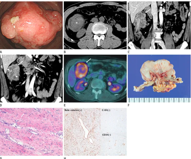
therefore, no specific diagnosis was made by the endoscopic bi- opsy (Fig. 1A). The abdomen-pelvic CT with contrast enhance- ment was performed on the following day for the characteriza- tion of the tumor. The pre-contrast CT scan showed a well- defined bulky mass at the right upper abdomen (Fig. 1B). This mass measured 7.2 cm in the largest diameter and revealed het- erogeneous low density with no hemorrhage or calcifications.
On the contrast-enhanced images, the mass presented as het- erogeneous, relatively less enhanced and causing colo-colic in- tussusceptions (Fig. 1C, D). Colonoscopic findings of endolu- minal leading point showed a lobulating contour, but an overlying inner layer suggested a well-preserved mucosa (Fig.
1C, D). In addition to the absence of bowel obstruction and other extra-intestinal manifestations, there was no pericolic fat infiltration or significantly enlarged lymph nodes adjacent to the lesion. Under the impression of a large bowel lymphoma causing colo-colic intussusception, 18F-fluorodeoxyglucose (FDG) positron emission tomography/CT (PET/CT) was per- formed to evaluate systemic involvement on the 4th day of ad- mission. On the 18F-FDG PET/CT, the mass showed a high FDG uptake (maximum standardized uptake value = 5.8); oth- er FDG uptake lesions were not detected (Fig. 1E). The preop- erative impression was that of a lymphoma or an adenocarci- noma; therefore, the patient underwent laparoscopic right hemicolectomy.
Grossly, a well-defined solid mass measuring 7.5 × 7 cm abutting the ascending colon was observed. Overlying mucosa showed no abnormality. The cut surface of the mass showed a whitish color and contained whirling pattern fibers without ne- crosis or hemorrhage (Fig. 1F). On microscopic evaluation, the tumor invaded the muscle layer of the colonic wall. The tumor was composed of bundles of short spindle cells and collagen fi- ber infiltrating from serosa to the muscle layer with minimal inflammation (Fig. 1G). On immunohistochemistry stain, tu- mor cells were positive for beta catenin and negative for C-kit and CD-34 (Fig. 1H). Based on the immunohistochemistry re- sults, this tumor was confirmed as mesenteric fibromatosis, be- nign fibroblastic proliferation.
The patients was discharged without complication and there
was no recurrence during the 1 year follow-up after surgery.
DISCUSSION
Mesenteric fibromatosis is the most frequent form of intra- abdominal desmoids tumor (1). Thirteen percent of patients with mesenteric fibromatosis have familial adenomatous pol- yposis (FAP), specifically, the Gardner syndrome variant of FAP.
Prior abdominal surgery is an important risk factor in patients with FAP (2).
The CT findings of mesenteric fibromatosis can be generally categorized into the well-circumscribed form or the infiltrative form. The well-circumscribed form has a well-defined margin and shows homogeneous enhancement with similar attenua- tion relative to muscles (3, 4). It is also often characterized by considerable size, which envelops the adjacent structures, espe- cially, the small-bowel loops. In the well-circumscribed form, as in the current case, the tumor can be confused with lympho- ma. The colonic lymphoma usually appears as a polypoid mass that has a circumferential wall thickening and a homogeneous enhancement (5), not very different from benign features of mesenteric fibromatosis. However, lymphoma usually has lymphadenopathy and infiltrates to other abdominal organs, such as liver and spleen, which is absent in the case of mesen- teric fibromatosis (5).
The infiltrative form shows aggressive features such as an ill-de- fined or irregular margin and a rapid growth pattern that looks ma- lignant (4). The imaging findings of this form are similar to those of adenocarcinoma. However, adenocarcinoma often shows met- astatic lymphadenopathy and more intense contrast enhancement (6); furthermore, another key distinguishing point from mesen- teric fibromatosis is the pattern in the bowel wall invasion. A des- moid tumor arises from extra-intestinal mesenteric structures such as the mesocolon, so the invasion of the muscle or the muco- sal layer occurs later than in adenocarcinoma (2).
18F-FDG PET/CT is a good tool to evaluate the malignant po- tential of tumors based on FDG uptake (7). However, it is not the absolute cancer-specific modality (8). It is not clear why mes- enteric fibromatosis could reveal false-positive imaging on 18F- FDG PET/CT like lymphoma or adenocarcinoma (7, 8). Metser and Even-Sapir discussed many similar cases of benign lesions with high FDG uptake and their possible mechanisms. They
suggested possible causes of false positive findings of 18F-FDG PET/CT like that vigorous smooth muscle activity or metaboli- cally active mucosa (7).
In the current case, the tumor was composed of short spindle cells and collagen fiber, but there was a lack of inflammatory cells. The absence of inflammatory cells distinguishes mesen-
Fig. 1. A 47-year-old man presented with mesenteric fibromatosis at the ascending mesocolon forming colo-colic intussusception and mimick- ing malignant tumors.
A. Colonoscopy shows a very large fungating mass with ulceration in the mid ascending colon. The biopsy needle cannot pass through the mass due to its hard-rubber like texture.
B. The axial pre-contrast CT image reveals an approximately 7.2 cm well-defined mass at the right upper quadrant abdomen, which shows some high density area (*).
C, D. The coronal reformatted contrast-enhanced CT image in the portal venous phase shows a lobulating mass forming colocolic intussuscep- tion (arrows). The mass shows a heterogeneous less enhancement feature and thin-enhancing inner layer suggesting that the mucosa is well- preserved (arrowheads). There is no lymph node enlargement or pericolic fat infiltration.
E. Axial 18F-FDG PET/CT image shows an increased FDG-uptake (SUV max = 5.8) around the mid ascending colon (arrow) without evidence of me- tastasis.
F. Gross pathological cut section shows a whitish tumor without necrosis or hemorrhage.
G. The microscopic exam (hematoxylin and eosin stain, × 100) of the mass shows dense bundles of short spindle cells (arrows) and collagen fibers.
H. In immunohistochemistry, the stain is positive for beta catenin (× 100) and negative for C-kit (× 100) and CD34 (× 100), which is very helpful in distinguishing mesenteric fibromatosis from other similar lesions.
FDG = fluorodeoxyglucose, PET = positron emission tomography, SUV max = maximum standardized uptake value A
D
G
B
E
H
C
F
teric fibromatosis from inflammatory myofibroblastic tumor. In addition, positive immunohistochemistry finding for beta catenin supports the diagnosis of mesenteric fibromatosis (1, 9). The high cellularity of the mass composed of fibrotic proliferation may be related to the hardened feature.
Mesenteric fibromatosis is a predisposing factor of intussus- ception (2). The mechanism of intussusception is believed to involve the bowel or lumen that act as irritant and provoke ab- normal peristaltic movement, which may lead to the invagina- tion of one bowel segment into the adjacent segment. Mesen- teric fibromatosis also acts as lead point for an intussusception as other masses (10). Mesenteric fibromatosis can cause not only intussusception but also a small bowel obstruction or fis- tula formation (2). Therefore, the surgical resection is the rec- ommended treatment option. However, the local recurrence is a major issue of mesenteric fibromatosis, even after complete surgical resection. If the resection margin of the tumor is posi- tive or the patient shows functional loss on follow-up, an addi- tional therapy, such as external radiation therapy, non-steroidal anti-inflammatory drug, antiestrogen therapy, or chemothera- py, can be considered (1).
In conclusion, in this study, we described a rare case of mes- enteric fibromatosis involving the mesocolon, which mimicked other malignancies like lymphoma or adenocarcinoma. Such a case can be difficult to diagnose in advance of a surgical resec- tion.
REFERENCES
1. Kasper B, Ströbel P, Hohenberger P. Desmoid tumors: clini- cal features and treatment options for advanced disease.
Oncologist 2011;16:682-693
2. Levy AD, Rimola J, Mehrotra AK, Sobin LH. From the ar-
chives of the AFIP: benign fibrous tumors and tumorlike lesions of the mesentery: radiologic-pathologic correlation.
Radiographics 2006;26:245-264
3. Sheth S, Horton KM, Garland MR, Fishman EK. Mesenteric neoplasms: CT appearances of primary and secondary tu- mors and differential diagnosis. Radiographics 2003;23:
457-473; quiz 535-536
4. Shinagare AB, Ramaiya NH, Jagannathan JP, Krajewski KM, Giardino AA, Butrynski JE, et al. A to Z of desmoid tu- mors. AJR Am J Roentgenol 2011;197:W1008-W1014 5. Ghai S, Pattison J, Ghai S, O’Malley ME, Khalili K, Stephens
M. Primary gastrointestinal lymphoma: spectrum of imag- ing findings with pathologic correlation. Radiographics 2007;
27:1371-1388
6. Horton KM, Abrams RA, Fishman EK. Spiral CT of colon cancer: imaging features and role in management. Radio- graphics 2000;20:419-430
7. Metser U, Even-Sapir E. Increased (18)F-fluorodeoxyglu- cose uptake in benign, nonphysiologic lesions found on whole-body positron emission tomography/computed to- mography (PET/CT): accumulated data from four years of experience with PET/CT. Semin Nucl Med 2007;37:206-222 8. Lo KW. Mesenteric fibromatosis as a potential source of
false-positive interpretation of FDG-PET: report of a case.
Dis Colon Rectum 2007;50:924-926
9. Montgomery E, Torbenson MS, Kaushal M, Fisher C, Abra- ham SC. Beta-catenin immunohistochemistry separates mesenteric fibromatosis from gastrointestinal stromal tu- mor and sclerosing mesenteritis. Am J Surg Pathol 2002;26:
1296-1301
10. Choi SH, Han JK, Kim SH, Lee JM, Lee KH, Kim YJ, et al. In- tussusception in adults: from stomach to rectum. AJR Am J Roentgenol 2004;183:691-698
상행결장의 악성 종양으로 오인될 수 있는 창자간막섬유종증에 의해 유발된 결장장중첩증의 전산화단층촬영과
양전자방출단층촬영술 소견: 증례 보고
이재형 · 하홍일* · 김민정 · 황진호 · 이관섭
창자간막섬유종증은 드문 양성 섬유모세포종양으로 결장간막에 위치한 창자간막섬유종증은 더욱 드물다. 일부 창자간막 섬유종증에서 칩습적인 성장을 보이거나 장중첩증을 유발하는 경우 림프종, 선암종과 같은 악성 종양과 감별이 어렵다.
우리는 상행결장에서 장중첩증을 유발했던 창자간막섬유종증이 림프종, 선암종과 같은 악성 종양으로 오인되었던 증례를 보고하고자 한다.
한림대학교 의료원, 한림대학교 성심병원 영상의학과