INTRODUCTION
Proliferation of vascular smooth muscle cell plays impor- tant roles in atherosclerosis and restenosis after coronary angioplasty (1, 2). However, the exact cellular mechanisms responsible for smooth muscle cell proliferation are unknown.
For many years, reactive oxygen species (ROS), such as superoxide anion (O2._
) and hydrogen peroxide (H2O2), have been thought of as toxic byproducts of aerobic living. But recently, a variety of evidence suggests that ROS are involved in the signal transduction in mammalian cells (3). In vascular smooth muscle cells, ROS were shown to mediate platelet- derived growth factor (PDGF)- or angiotensin II- induced cell proliferation (4-6). The production of ROS in blood vessel is enhanced in experimental models of hypercholes- terolemia, hypertension, diabetes, and balloon injury to the coronary arteries (7-10). These evidences suggest that ROS could mediate the common mechanism of diseases charac- terized by vascular smooth muscle cell proliferation.
The NADH/NADPH oxidase is the major source of ROS induced by various growth factors in vascular smooth muscle cell (11). In non-phagocytic cells, small GTP-binding pro- tein rac1 controls the level of ROS by regulating the NADH/
NADPH oxidase complex (12-16). Recently, treatment with
antioxidants such as pyrrolidinedithiocarbamate (PDTC) or N-acetylcysteine (NAC) and overexpression of catalase have been shown to inhibit proliferation of vascular smooth mus- cle cell by decreasing the level of intracellular ROS (17, 18).
These strategies decrease the preformed ROS.
In this study, we examined whether the inhibition of rac1 could inhibit the proliferation of rat aortic vascular smooth muscle cell (RASMC) stimulated by PDGF via decreasing the production of intracellular ROS. To inhibit NADH/
NADPH oxidase effectively, we used adenoviral-mediated gene transfer of a dominant negative rac1 gene product (N17rac1) as shown in the previous study (12).
MATERIALS AND METHODS Cell Cultures
Primary culture of RASMC was obtained as explants from the thoracic aorta of a 3-month-old Sprague-Dawley rat as previously described (19). Cells were maintained in DMEM (Gibco BRL, Grand Island, NY, U.S.A.) with 10% fetal calf serum (Gibco BRL, Grand Island, NY, U.S.A.) in a humid- ified atmosphere containing 5% CO2at 37℃. For experi-
Gu Kong*, Sahng Lee, Kyung-Soo Kim
Cardiology Division and Pathology Division*, College of Medicine, Hanyang University, Seoul, Korea
Address for correspondence Kyung-Soo Kim, M.D.
Cardiology Division, College of Medicine, Hanyang University, 17 Haengdang-dong, Sungdong-gu, Seoul 133-792, Korea
Tel : +82.2-2290-8312, Fax : +82.2-2298-9183 E-mail: [email protected]
*This work was supported by Korea Research Foundation Grant (KRF-99-015-FP0064).
712
Inhibition of rac1 Reduces PDGF-induced Reactive Oxygen Species and Proliferation in Vascular Smooth Muscle Cells
In vascular smooth muscle cells, reactive oxygen species (ROS) were known to mediate platelet-derived growth factor (PDGF)-induced cell proliferation and NADH/NADPH oxidase is the major source of ROS. NADH/NADPH oxidase is controlled by rac1 in non-phagocytic cells. In this study, we examined whether the inhibition of rac1 by adenoviral-mediated gene transfer of a dominant nega- tive rac1 gene product (Ad.N17rac1) could reduce the proliferation of rat aortic vascular smooth muscle cells (RASMC) stimulated by PDGF via decreasing intra- cellular ROS. RASMC were stimulated by PDGF (80 ng/mL) with or without N- acetylcysteine 1 mM or infected with 100 mutiplicity of infection of Ad.N17rac1.
Intracellular ROS levels were measured at 12 hr using carboxyl-2′,7′-dichlorodi- hydrofluorescein diacetate confocal microscopy. At 72 hr, cellular proliferation was evaluated by cell number counting and XTT assay. Compared with control, ROS levels were increased by 2-folds by PDGF. NAC and Ad.N17rac1 inhibited PDGF-induced increase of ROS by 77% and 65%, respectively. Cell number was increased by PDGF by 1.6-folds compared with control. NAC and Ad.N17rac1 inhibited PDGF-induced cellular growth by 45% and 87%, respectively. XTT assay also showed similar results. We concluded that inhibition of rac1 in RASMCs could reduce intracellular ROS levels and cellular proliferation induced by PDGF.
Key Words : Reactive Oxygen Species; Rac1 GTP-Binding Proteins; Gene Transfer Techniques
Received : 3 May 2001 Accepted : 11 July 2001
ment, RASMCs were pre-cultured on the same constitution of media except 3% fetal bovine serum for 24 hr. All experi- ments were performed on cells between passage 5 and 12 grown to about 80% confluence in 6-well or 96-well plates.
To stimulate proliferation of RASMC, PDGF (BB iso- form) (Upstate Biotechnology, Waltham, MA, U.S.A.) 80 ng/mL was added to the media. The antioxidant, N-acetyl- cysteine (Sigma, St. Louis, MO, U.S.A.) 1 mM was added along with PDGF addition to see the antioxidant effect.
Adenoviruses
One hundred multiplicity of infection (MOI) of Ad.N17- rac1 was infected in 3% fetal calf serum media 24 hr before PDGF stimulation to allow sufficient gene expression in RASMC. Adenovirus Ad.N17rac1 contains the epitope- tagged dominant negative rac1 cDNA, which has a mutation changing amino acid 17 of rac1 from serine to asparagine.
Construction of Ad.N17rac1 was done by homologous recom- binant in 293 cells (13, 20). To assess the effects of adenovi- ral infection alone, a control virus Ad.dl312, which is delet- ed in the E1 region but lacks a recombinant transgene, was used (21). Viruses were amplified, purified, and titrated as previously described (22). All viruses were kindly presented by Dr. Finkel T. at the Laboratory of Molecular Biology, NHLBI.
Gene transfer efficiencies were assessed after infection with Ad.GAL, an adenovirus, which encodes the Escherichia coli LacZ gene product (22). Histochemical X-gal staining of Ad. GAL-infected cells was performed as previously described (22). Western blot analysis of rac1 expression used antibodies directed at the myc-epitope tag (9E10; Santa Cruz Biotech., Heidelberg, Germany), which identified the N17rac1 gene product (13). Twenty g of protein was loaded in each lane and bound immunoglobulins were detected by enhanced chemiluminescence (Tropix, Bedford, MA, U.S.A.). Confir- mation of equal protein loading was assessed using an anti- body directed at -tubulin (Ab-1; Calbiochem-Novabiochem Corp., La Jolla, CA, U.S.A.).
Measuring Intracellular ROS
The level of intracellular ROS was measured at 12 hr after PDGF stimulation with or without NAC and Ad.N17rac1 treatment. Intracellular generation of ROS was detected using 5-(and 6)-carboxyl-2′,7′-dichlorodihydrofluorescein diacetate (carboxyl-DCFH-DA) (Molecular Probes Inc., Eugene, OR, U.S.A.) (23-26). Carboxyl-DCFH-DA fluoresces green when oxidized by O2.
or H2O2. The fluorescence was detected by confocal laser scanning microscope using excitation and emis- sion wavelengths of 488 and 525 nm, respectively, by incu- bating cells for 5 min with 10 g/mL of carboxyl-DCFH- DA as previously described (6). Levels of Carboxyl-DCFH- DA fluorescence represent the values from at least 60 ran-
dom cells (mean±SD), based on an arbitrary scale of fluo- rescence intensity (6).
Assessment of RASMC proliferation
The degrees of RASMC proliferation were evaluated at 72 hr after PDGF stimulation with or without NAC and Ad.N17rac1 treatment. The RASMC proliferation was deter- mined by two methods, cell number count using trypan blue exclusion and XTT assay. In cell number counts, only viable cells by trypan dye are counted in hemocytometer as cells/
mL.‘Cell Proliferation Kit II (XTT)’(Boehringer Mannheim Corp., Mannheim, Germany) was used for XTT assay. Fifty L of XTT labeling mixture was added to each well of 96- well plate and readings were done 24 hr later at 492 nm, corrected against 690 nm.
Statistical analyses
Except when stated otherwise, all results are from tripli- cate cultures (mean±SD) and represent one of at least three similar experiments. Statistical significance was determined by first performing an ANOVA between the experimental groups (p<0.05), and then a two-tailed unpaired t-test to compare N17rac1 with the other experimental groups.
RESULTS
We used primary cultures of RASMC as an in vitro model for growth factor-induced ROS production and cell growth.
Fetal calf serum concentration of 3% was used because it was the minimal concentration that showed linear growth pat- tern. To select the growth factor for RASMC stimulation, we tried angiotensin II, basic FGF and thrombin as well as PDGF. PDGF was chosen because it showed the most promi- nent ROS induction and cell growth. At 80 ng/mL, PDGF showed maximal ROS induction and cell growth, and we used this concentration for this experiment. NAC was used as an antioxidant, because we observed that PDTC and cata- lase induced significant cellular toxicity by trypan blue exclu- sion in the preliminary experiments.
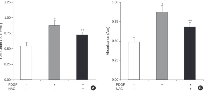
As demonstrated in Fig. 1, compared to the cells without PDGF, the levels of intracellular ROS increased about 2-fold after stimulation with PDGF 80 ng/mL for 12 hr. To see the proliferative effect of PDGF, the cells were cultured with PDGF 80 ng/mL for 72 hr and then cell number count and XTT assay were performed. With PDGF, cell number in- creased about 1.6-fold compared with control (Fig. 2A).
XTT assay showed a similar result with 1.8-fold increase in absorbance (Fig. 2B).
Next, we assessed the effects of treating with the cell per- meant chemical NAC. Consistent with previous studies that demonstrated antiproliferative effects of ROS scavengers by
decreasing intracelluar ROS levels (10, 17), treatment with NAC 1 mM inhibited the increase of intracelluar ROS lev- els (Fig. 1) and the cell proliferation (Fig. 2) induced by PDGF. We have used 1 mM of NAC, because at that con- centration, NAC showed the maximal inhibitory effects to induce ROS and promote cell growth.
Consistent with previous results in vascular smooth mus-
cle cells (27, 28), adenoviral infection at concentration of 100 MOI led to a successful gene transfer in >95% of infected cells (Fig. 3A). Similarly, Western blot analysis of Ad.N17rac1 infected cells demonstrated that the epitope- tagged form of dominant negative rac1 gene product was expressed at a high level (Fig. 3B).
After confirmation of the high efficiency of adenoviral- mediated gene transfer, we examined the effect of inhibition
Relative Fluorescence
80 70 60 50 40 30 20 10
0
PDGF - + +
NAC - - +
Fig. 1. Effects of PDGF and NAC on the levels of reactive oxygen species in rat aortic smooth muscle cells. (A) Representative images obtained by carboxyl-DCFH-DA confocal laser microscopy. Left upper panel: control cells. PDGF: cells incubated with PDGF 80 ng/mL for 12 hr. PDGF+NAC: cells co-incubated with PDGF 80 ng/mL and NAC 1 mM for 12 hr. (B) Levels of carboxyl-DCFH-DA fluorescence measured by confocal laser microscopy. Cells were incubated with PDGF 80 ng/mL (PDGF) or NAC 1 mM (NAC) for 12 hr.
p<0.05, *: PDGF (-) NAC (-) vs. PDGF (+) NAC (-), **: PDGF (+) NAC (-) vs. PDGF (+) NAC (+).
*
**
B
Absorbance (A492)
1.00
0.75
0.50
0.25
0.00
PDGF - + +
NAC - - +
*
**
Cell Count (×105/mL) 1.25
1.00
0.75
0.50
0.25
0.00
PDGF - + +
NAC - - +
Fig. 2. Effects of PDGF and NAC on the proliferations of rat aortic smooth muscle cells. (A) Effects of PDGF and NAC on cell numbers. (B) Effects of PDGF and NAC on relative absorbance at 492 nm on XTT assay. Cells were incubated with PDGF 80 ng/mL (PDGF) or NAC 1 mM (NAC) for 72 hr. p<0.05, *: PDGF (-) NAC (-) vs. PDGF (+) NAC (-), **: PDGF (+) NAC (-) vs. PDGF (+) NAC (+).
*
**
A Control
PDGF+NAC
PDGF
B A
of rac1. To determine whether the inhibition of rac1 could suppress the increase of intracellular ROS levels and cellular proliferation by PDGF stimulation, we used adenoviral-medi- ated gene transfer of a dominant negative rac1 (Ad.N17rac1).
Previous studies have implicated that rac1 plays an impor-
tant role in ligand-stimulated ROS generation in non-phago- cytic cells (13-15, 29, 30). Examination of intracellular ROS levels revealed that in uninfected cells, the stimulation with PDGF produced a significant increase in carboxyl-DCFH- DA fluorescence (Fig. 4). Similar increase in the fluorescence was seen in the cells infected with an E1-deleted adenovirus, which lacked a recombinant transgene (Ad.dl312; Fig. 4).
By contrast, the cells infected with Ad.N17rac1 showed a significant attenuation in the level of ROS, as assessed by carboxyl-DCFH-DA fluorescence, after stimulation with PDGF (Fig. 4).
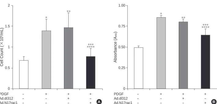
Next, we assessed the effects of N17rac1 expression on cell proliferation after stimulation with PDGF. In the cells treated with PDGF only or PDGF plus Ad.dl312, the num- ber of cells was increased significantly, about 2-folds, com- pared with control (Fig. 5A). By contrast, in the cells infect- ed with Ad.N17rac1, PDGF induced a significant attenua- tion in cell growth by approximately a half of the cell num- ber (Fig. 5A). Similarly, according to the results by the XTT assay, the cells infected with Ad.N17rac1 showed signifi- cant decreases of absorbance compared with the cells treated with PDGF only or PDGF plus Ad.dl312 (Fig. 5B).
DISCUSSION
We found that in RASMCs the inhibition of rac1 by infec-
Fig. 4.Effects of Ad.N17rac1 infection on the levels of reactive oxygen species in rat aortic smooth muscle cells. (A) Representative images obtained by carboxyl-DCFH-DA confocal laser microscopy. Control: uninfected control cells. PDGF cells incubated with PDGF 80 ng/mL. PDGF+Ad.dl312: cells incubated with PDGF 80 ng/mL and infected with 100 mutiplicity of infection (MOI) of Ad.dl312.
PDGF+ Ad.N17rac1: cells incubated with PDGF 80 ng/mL and infected with 100 MOI of Ad.N17rac1. Confocal images were obtained 12 hr after incubation with PDGF and viral infections were performed 24 hr before the start of incubation with PDGF. (B) Levels of car- boxyl-DCFH-DA fluorescence measured by confocal laser microscopy. Cells were incubated with PDGF 80 ng/mL (PDGF) or infected with 100 MOI of Ad.dl312 or Ad.N17rac1 as described in (A).
p<0.05, from left to right, *: 1st vs. 2nd, **: 1st vs. 3rd , ***: 2nd vs. 4th, ****: 3rd vs. 4th bar.
A B
Relative Fluorescence
50
40
30
20
10
0
PDGF - + + +
Ad.dl312 - - + -
Ad.N17rac1 - - - +
*
*******
**
-Tubulin
Fig. 3. Assessment of adenoviral-mediated gene transfer. (A) Histochemical X-gal staining 24 hr after infection with 100 muti- plicity of infection of Ad.GAL, which encodes the Escherichia coli LacZ gene product, showed over 95% gene transfer effi- ciency. (B) Western blots of protein lysates (20 g) obtained from uninfected RASMC (left lane), RASMC infected with Ad.dl312 (middle lane), and Ad.N17rac1 (right lane). Western blotting analysis was proved with -tubulin to confirm equal loading and an antibody (21 kDa) recognizing the myc epitope tag of N17rac1 construct.
c-myc
Control
PDGF+
Ad.dl312
PDGF+
Ad.N17rac1 PDGF
A B
Fig. 5.Effects of Ad.N17rac1 infection on the proliferations of rat aortic smooth muscle cells. (A) Effects of Ad.N17rac1 infection on cell numbers. (B) Effects of Ad.N17rac1 infection on relative absorbance at 492 nm on XTT assay. Cells were incubated with PDGF 80 ng/mL (PDGF) or infected with 100 mutipicity of infection of Ad.dl312 or Ad.N17rac1. Cell number counting and XTT assay were per- formed 72 hr after incubation with PDGF and viral infections were performed 24 hr before the start of incubation with PDGF.
p<0.05, from left to right, *: 1st vs. 2nd, **: 1st vs. 3rd , ***: 2nd vs. 4th, ****: 3rd vs. 4th bar.
B
Absorbance (A492)
1.00
0.75
0.50
0.25
0
PDGF - + + +
Ad.dl312 - - + -
Ad.N17rac1 - - - +
*
*******
**
A Cell Count (×105/mL)
2
1.5
1
0.5
0
PDGF - + + +
Ad.dI312 - - + -
Ad.N17rac1 - - - +
*
*******
**
tion with AdN17rac1, an adenovirus containing gene encod- ing a dominant negative rac1 gene product, resulted in a high expression of N17rac1 in the cells, reduction in the levels of intracellular ROS, and suppression of DNA syn- thesis and cellular proliferation induced by PDGF stimula- tion.
Ligand-stimulated ROS production plays an important role in signal transduction in various cells (3, 31-33) and especially, growth factors, such as PDGF, epidermal growth factor, and angiotensin II trigger the rapid production of ROS.
Also, ROS can stimulate proliferation of vascular smooth muscle cell (5, 34). Furthermore, in RASMC, PDGF stimu- lated H2O2 production and DNA synthesis, and both of these responses are blocked by antioxidant treatment (6).
These reports and our study suggest that endogenously pro- duced ROS have an important role in regulating vascular smooth muscle cell growth.
In phagocytic cells, a variety of stimuli cause burst of super- oxide anion through NADPH oxidase (35). The small GTPase rac2 is one of the essential components of NADPH oxidase complex and controls the activity of the enzyme complex (29, 30). Recent evidences suggest that in non-phagocytic cells, similar enzyme complex, NADPH/NADH oxidase is con- trolled by rac1 (13, 14, 16). Furthermore, NADPH/NADH oxidase is the major source of ROS induced by various growth factors in vascular smooth muscle cell (11). In this experiment, we inhibited NADPH/NADH oxidase in RASMC using rac1 inhibition by adenoviral-mediated gene transfer of a dominant negative rac1 (N17rac1). And we demonstrated
that this resulted in a reduction of intracellular ROS levels and suppression of DNA synthesis and cellular proliferation.
This is the first report to show that the over-expression of N17rac1 could inhibit cellular proliferation induced by PDGF.
Some reports have shown that ROS could induce apopto- sis rather than proliferation in vascular smooth muscle cells (34, 36, 37). These findings are most likely due to the dif- ferences in the amount of ROS applied, but the exact mech- anisms remain to be determined.
Usually to see the effects of growth factors, experiments are performed by serum deprivation or at the minimal serum concentration. In this experiment, we have used fetal calf serum of 3%, which was to observe cell growth on a linear growth phase. We thought this was more physiological than a serum-deprived condition. Due to relatively high serum concentration in controls, PDGF-induced increases in intra- cellular ROS levels, XTT absorbances and cellular prolifera- tions were not so prominent.
Carboxyl-DCFH-DA confocal microscopy to measure the intracellular ROS levels can not discriminate between indi- vidual ROS, that is, superoxide anion (O2._
), hydrogen per- oxide (H2O2), hydroxyl radical (.OH), and nitric oxide (NO).
So, we could not observe the effects of individual ROS.
Nonetheless, our data suggest that under certain condi- tions, inhibition of rac1 in RASMC can reduce intracellular ROS levels and cellular proliferation induced by PDGF. The inhibition of rac1 protein function may be useful in variety of clinical settings, such as restenosis after coronary inter-
vention, in which the proliferation of vascular smooth mus- cle cells plays an important role in pathogenesis.
ACKNOWLEDGMENTS
We thank Dr. Toren Finkel and Ilsa I. Rovira (Lab. of Molec- ular Biology, NHLBI, NIH, U.S.A.) for providing adenovi- ral products and advices for this experiment.
REFERENCES
1. Ross R. The pathogenesis of atherosclerosis: a perspective for the 1990s. Nature 1993; 362: 801-9.
2. Schwartz RS. Pathophysiology of restenosis: interaction of throm- bosis, hyperplasia, and/or remodeling. Am J Cardiol 1998; 81:
14E-7E.
3. Finkel T. Oxygen radicals and signaling. Curr Opin Cell Biol 1998;
10: 248-53.
4. Ushio-Fukai M, Griendling KK, Becker PL, Hilenski L, Halleran S, Alexander RW. Epidermal growth factor receptor transactivation by angiotensin II requires reactive oxygen species in vascular smooth muscle cells. Arterioscler Thromb Vasc Biol 2001; 21: 489- 95.
5. Rao GN, Berk BC. Active oxygen species stimulate vascular smooth muscle cell growth and proto-oncogene expression. Circ Res 1992;
70: 593-9.
6. Sundaresan M, Yu ZX, Ferrans VJ, Irani K, Finkel T. Requirement for generation of H2O2for platelet-derived growth factor signal transduction. Science 1995; 270: 296-9.
7. Ohara Y, Peterson TE, Sayegh HS, Subramanian RR, Wilcox JN, Harrison DG. Dietary correction of hypercholesterolemia in the rabbit normalizes endothelial superoxide anion production. Circu- lation 1995; 92: 898-903.
8. Grunfeld S, Hamilton CA, Mesaros S, McClain SW, Dominiczak AF, Bohr DF, Malinski T. Role of superoxide in the depressed nitric oxide production by the endothelium of genetically hypertensive rats.
Hypertension 1995; 26: 854-7.
9. Langenstroer P, Pieper GM. Regulation of spontaneous EDRF release in diabetic rat aorta by oxygen free radicals. Am J Physiol 1992; 263: H257-65.
10. Nunes GL, Robinson K, Kalynych A, King SB III, Sgoutas DS, Berk BC. Vitamins C and E inhibit O2-production in the pig coronary artery. Circulation 1997; 96: 3593-601.
11. Griendling KK, Minieri CA, Ollerenshaw JD, Alexander RW.
Angiotensin II stimulates NADH and NADPH oxidase activity in cultured vascular smooth muscle cells. Circ Res 1994; 74: 1141-8.
12. Kim KS, Takeda K, Sethi R, Pracyk JB, Tanaka K, Zhou YF, Yu ZX, Ferrans VJ, Bruder JT, Kovesdi I, Irani K, Goldschmidt-Cler- mont P, Finkel T. Protection from reoxygenation injury by inhibi- tion of rac1. J Clin Invest 1998; 101: 1821-6.
13. Sulciner DJ, Irani K, Yu ZX, Ferrans VJ, Goldschmidt-Clermont P, Finkel T. rac1 regulates a cytokine-stimulated, redox-dependent
pathway necessary for NF-kappaB activation. Mol Cell Biol 1996;
16: 7115-21.
14. Sundaresan M, Yu ZX, Ferrans VJ, Sulciner DJ, Gutkind JS, Irani K, Goldschmidt-Clermont PJ, Finkel T. Regulation of reactive-oxy- gen-species generation in fibroblasts by Rac1. Biochem J 1996;
318: 379-82.
15. Irani K, Xia Y, Zweier JL, Sollott SJ, Der CJ, Fearon ER, Sudaresan M, Finkel T, Goldschmidt-Clermont PJ. Mitogenic signaling medi- ated by oxidants in Ras-transformed fibroblasts. Science 1997; 275:
1649-52.
16. Gulbins E, Brenner B, Schlottmann K, Welsch J, Heinle H, Kop- penhoefer U, Linderkamp O, Coggeshall KM, Lang F. Fas-induced programmed cell death is mediated by a Ras-regulated O2-synthe- sis. Immunology 1996; 89: 205-12.
17. Brown MR, Miller FJ, Li WG, Ellingson AN, Mozena JD, Chatterjee P, Engelhardt JF, Zwacka RM, Oberley LW, Fang X, Spector AA, Weintraub NL. Overexpression of human catalase inhibits prolifer- ation and promotes apoptosis in vascular smooth muscle cells. Circ Res 1999; 85: 524-33.
18. Tsai JC, Jain M, Hsieh CM, Lee WS, Yoshizumi M, Patterson C, Perrella MA, Cooke C, Wang H, Haber E, Schlegel R, Lee ME.
Induction of apoptosis by pyrrolidinedithiocarbamate and N-acetyl- cysteine in vascular smooth muscle cells. J Biol Chem 1996; 271:
3667-70.
19. Fang X, Kaduce TL, Weintraub NL, VanRollins M, Spector AA.
Functional implications of a newly characterized pathway of 11,12- epoxyeicosatrienoic acid metabolism in arterial smooth muscle.
Circ Res 1996; 79: 784-93.
20. McGrory WJ, Bautista DS, Graham FL. A simple technique for the rescue of early region I mutations into infectious human adenovirus type 5. Virology 1988; 163: 614-7.
21. Jones N, Shenk T. Isolation of adenovirus type 5 host range dele- tion mutants defective for transformation of rat embryo cells. Cell 1979; 17: 683-9.
22. Guzman RJ, Hirschowitz EA, Brody SL, Crystal RG, Epstein SE, Finkel T. In vivo suppression of injury-induced vascular smooth muscle cell accumulation using adenovirus-mediated transfer of the herpes simplex virus thymidine kinase gene. Proc Natl Acad Sci USA 1994; 91: 10732-6.
23. Speir E, Shibutani T, Yu ZX, Ferrans V, Epstein SE. Role of reac- tive oxygen intermediates in cytomegalovirus gene expression and in the response of human smooth muscle cells to viral infection.
Circ Res 1996; 79: 1143-52.
24. Bass DA, Parce JW, Dechatelet LR, Szejda P, Seeds MC, Thomas M. Flow cytometric studies of oxidative product formation by neu- trophils: a graded response to membrane stimulation. J Immunol 1983; 130: 1910-7.
25. Ohba M, Shibanuma M, Kuroki T, Nose K. Production of hydrogen peroxide by transforming growth factor-beta 1 and its involvement in induction of egr-1 in mouse osteoblastic cells. J. Cell Biol 1994;
126: 1079-88.
26. Zhu H, Bannenberg GL, Moldeus P, Shertzer HG. Oxidation path- ways for the intracellular probe 2′,7′-dichlorofluorescein. Arch Tox- icol 1994; 68: 582-7.
27. Guzman RJ, Lemarchand P, Crystal RG, Epstein SE, Finkel T. Effi- cient and selective adenovirus-mediated gene transfer into vascular neointima. Circulation 1993; 88: 2838-48.
28. Lee S, Trapnell B, Rade J, Virmani R, Dichek D. In vivo adenoviral vector-mediated gene transfer into balloon-injured rat carotid arteries. Circ Res 1993; 73: 797-807.
29. Abo A, Pick E, Hall A, Totty N, Teahan CG, Segal AW. Activation of the NADPH oxidase involves the small GTP-binding protein p21rac1. Nature 1991; 353: 668-70.
30. Knaus UG, Heyworth PG, Evans T, Curnutte JT, Bokoch GM. Reg- ulation of phagocyte oxygen radical production by the GTP-binding protein Rac 2. Science 1991; 254: 1512-5.
31. Rhee SG. Redox signaling: hydrogen peroxide as intracellular mes- senger. Exp Mol Med 1999; 31: 53-9.
32. Finkel T. Redox-dependent signal transduction. FEBS Lett 2000;
476: 52-4.
33. Lander HM. An essential role for free radicals and derived species in signal transduction. FASEB J 1997; 11: 118-24.
34. Fiorani M, Cantoni O, Tasinato A, Boscoboinik D, Azzi A. Hydro- gen peroxide- and fetal bovine serum-induced DNA synthesis in vascular smooth muscle cells: positive and negative regulation by protein kinase C isoforms. Biochim Biophys Acta 1995; 1269: 98- 104.
35. DeLeo FR, Quinn MT. Assembly of the phagocyte NADPH oxi- dase: molecular interaction of oxidase proteins. J Leukoc Biol 1996; 60: 677-91.
36. Li PF, Dietz R, von Harsdorf R. Reactive oxygen species induce apoptosis of vascular smooth muscle cell. FEBS Lett 1997; 404:
249-52.
37. Li P-F, Dietz R, von Harsdorf R. Differential effect of hydrogen peroxide and superoxide anion on apoptosis and proliferation of vascular smooth muscle cells. Circulation 1997; 96: 3602-9.