© 2013 The Korean Academy of Medical Sciences.
This is an Open Access article distributed under the terms of the Creative Commons Attribution Non-Commercial License (http://creativecommons.org/licenses/by-nc/3.0) which permits unrestricted non-commercial use, distribution, and reproduction in any medium, provided the original work is properly cited.
pISSN 1011-8934 eISSN 1598-6357
Caspase-1 Level in Synovial Fluid Is High in Patients with Spondyloarthropathy but Not in Patients with Gout
Activation of caspase-1 by NALP3 inflammasomes has been shown to be important in initiating acute gouty arthritis. The objectives of this study were to measure the levels of caspase-1 in synovial fluid in gout and various arthritides, and to elucidate the clinical significance of caspase-1 levels in synovial fluid. Caspase-1, IL-1β, IL-18, and uric acid were measured in synovial fluid from 112 patients with gout and other arthritides, such as rheumatoid arthritis, osteoarthritis, and spondyloarthropathy. Caspase-1 in synovial fluid from patients with crystal-induced arthritis, inflammatory arthritis, osteoarthritis, and spondyloarthropathy was 35.9 ± 86.7, 49.7 ± 107.7, 2.1 ± 7.0, and 152.6 ± 155.7 pg/
mL, respectively. The mean level and the frequency of high levels (≥125 pg/mL) of caspase-1 in spondyloarthropathy were significantly higher than those in the other arthritides including gout. Caspase-1 was detectible in the synovial fluid of patients with the various arthritides. Contrary to our hypothesis, the caspase-1 level in the synovial fluid of patients with gout was not higher than in that of other arthritides. High levels of caspase-1 may be helpful in differentiating spondyloarthropathy from other arthritides.
Key Words: Caspase-1; Synovial Fluid; Gout; Spondyloarthropathies Chang-Nam Son,1 So-Young Bang,2
Ji Hae Kim,1 Chan-Bum Choi,1 Tae-Hwan Kim,1 and Jae-Bum Jun1
1Department of Rheumatology, Hanyang University Hospital for Rheumatic Diseases, Seoul; 2Division of Rheumatology, Department of Internal Medicine, Hanyang University Guri Hospital, Guri, Korea Received: 14 March 2013
Accepted: 16 July 2013 Address for Correspondence:
Jae-Bum Jun, MD
Department of Rheumatology, Hanyang University Hospital for Rheumatic Diseases, 222 Wangsimni-ro, Sungdong-gu, Seoul 133-792, Korea
Tel: +82.2-2290-9244, Fax: +82.2-2298-8231 E-mail: [email protected]
This work was supported by the research fund of Hanyang University (2010-000-0000-0328).
http://dx.doi.org/10.3346/jkms.2013.28.9.1289 • J Korean Med Sci 2013; 28: 1289-1292
ORIGINAL ARTICLE
Immunology, Allergic Disorders & Rheumatology
INTRODUCTION
Gout is one of the most common types of inflammatory arthri- tis, and its incidence and prevalence is increasing (1). Gouty ar- thritis is characterized by hyperuricemia, which results from overproduction or impaired renal excretion of uric acid (2). It was shown recently that gout-associated monosodium urate (MSU) crystals activate the NALP3 inflammasome (3), leading to processing of procaspase-1 to caspase-1, and the production and secretion of active IL-1β and IL-18 (4).
In a recent study of multiple sclerosis, caspase-1 was mea- sured in biological body fluids, and could be used as a marker of inflammation (5). In that study, the concentration of caspase-1 in synovial fluid (SF) was found to be higher in patients with ju- venile idiopathic arthritis (JIA) than in patients with other in- flammatory diseases.
We hypothesized that caspase-1 would be more active, both within cells and in the synovial fluid, in crystal-induced arthri- tis (CIA) than in other arthritides such as osteoarthritis (OA), spondyloarthropathy (SpA), and inflammatory arthritides in- cluding rheumatoid arthritis (RA). In this study, we evaluated levels of caspase-1 in synovial fluid (SF) which might be useful in the differential diagnosis of CIA from other arthritides.
MATERIALS AND METHODS Subjects
A total of 112 SF samples were obtained from patients with CIA, inflammatory arthritis, OA, and SpA. The patients met the Amer- ican College of Rheumatology criteria for acute gout (6), RA (7), systemic lupus erythematosus (SLE) (8), and knee OA, and the 1984 New York criteria for ankylosing spondylitis (9). Arthro- centesis was carried out to diagnose and/or to attempt to treat arthritis flares such as acute gouty attacks. The presence of MSU crystals was proven in 22 patients with gout, and calcium pyro- phosphate dihydrate (CPPD) crystals were identified in two pa- tients. Only two gout patients were diagnosed clinically. Table 1 gives the diagnoses and current treatments of patients enrolled in the study.
Sampling and assays
Samples of SF were collected from patients requiring knee or ankle arthrocentesis for diagnostic or therapeutic reasons, from September 2010 to March 2012. Plain tubes were used to collect SF for routine examination. Cells were removed by centrifuga- tion at 2,500 RPM for 15 min for ELISAs and the SF was stored at -20°C. Levels of caspase-1, IL-1β, and IL-18 were measured with commercial ELISA kits (caspase-1: Wuhan EIAab Science
Son C-N, et al. • Clinical Significance of Caspase-1 Level in Various Arthritides
1290 http://jkms.org http://dx.doi.org/10.3346/jkms.2013.28.9.1289 Co., Ltd., Wuhan, China; IL-1β and IL-18: Quantikine, R&D Sys-
tems Inc., Minneapolis, MN, USA; IL-1RA: Invitrogen, UK). The detection range for the caspase-1 ELISA kit was 15.6-1,000 pg/
mL. Uric acid in SF was measured with a quantitative colori- metric assay kit as described by the manufacturer (BioAssay Systems, Hayward, CA, USA). C-reactive protein (CRP) and erythrocyte sedimentation rate (ESR) were determined using the data recorded in the medical records within ± 1 day of the study day.
Statistical analysis
All statistical analyses were performed using SAS software, V.9.2 (SAS Institute, Cary, NC, USA). Results are presented as means
± SDs unless specified otherwise. The non-parametric Kruskal- Wallis test was used for between-group comparisons of arthriti- des and between-phase comparisons of gout. Pearson’s and Spearman’s correlation coefficients were used to examine the relationships between concentrations of biomarkers and labo- ratory parameters. The Bonferroni correction was applied to
multiple comparisons within each category of variables. Fish- er’s exact test was used to assess the frequencies of high levels (≥ 125 pg/mL) of caspase-1 in between-group comparisons. P values less than 0.05 was considered significant.
Ethics statement
This study was approved by institutional review board of Han- yang University Hospital (IRB No. 2010-R-22). Written informed consent was obtained from all participants which was confirm- ed by the board.
RESULTS
The clinical and laboratory characteristics of the various patients are summarized in Table 2. As expected, age and gender were disease-specific. CRP was highest in patients with CIA and in- flammatory arthritis. The white blood cell (WBC) count in SF was highest in CIA, followed by inflammatory arthritis, SpA, and OA in descending order.
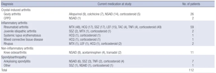
Table 1. Diagnoses and current treatment of patients at study
Diagnosis Current medication at study No. of patients
Crystal-induced arthritis Gouty arthritis CPPD
Allopurinol (9), colchicine (7), NSAID (14), corticosteroid (5) NSAID (1)
26 2 Inflammatory arthritis
Rheumatoid arthritis Juvenile idiopathic arthritis Systemic lupus erythematosus Mixed connective tissue disease Rhupus
MTX (48), HCQ (17), SSZ (17), LEF (15), TAC (4), TNFi (4), corticosteroid (49) SSZ (2), MTX (1), corticosteroid (1)
HCQ (1), corticosteroid (1) HCQ (1), corticosteroid (1)
MTX (1), LEF (1), HCQ (1), corticosteroid (2)
59 2 1 1 2 Non-inflammatory arthritis
Knee osteoarthritis NSAID (8), acetaminophen (4), tramadol (2) 11
Spondyloarthropathy Ankylosing spondylitis Other
NSAID (6), SSZ (3), TNFi (2), corticosteroid (4) SSZ (1), NSAID (1), corticosteroid (1)
7 1
Total 112
No., number; CPPD, calcium pyrophosphate dihydrate; NSAID, nonsteroidal anti-inflammatory drug; TNFi, tumor necrosis factor inhibitor; MTX, methotrexate; LEF, leflunomide;
TAC, tacrolimus; HCQ, hydroxychloroquine; SSZ, sulfasalazine; ( ), Number of patients being taken medication at study.
Table 2. The clinical and laboratory features of patients with crystal-induced arthritis, inflammatory arthritis, osteoarthritis, and spondyloarthropathy Parameters Crystal-induced arthritis Inflammatory arthritis Osteoarthritis Spondyloarthropathy
No. of patients 28 65 11 8 P
Age (yr) 54.9 ± 15.4 51.2 ± 12.6 60.8 ± 6.9 32.9 ± 7.1 < 0.001
Sex (M/F) 23/5 7/58 4/7 4/4
ESR (mm/hr) 40.7 ± 28.1 (n = 21) 41.0 ± 31.2 (n = 27) 43.3 ± 48.7 (n = 4) 37.8 ± 37.9 (n = 4) 0.960 CRP (mg/dL) 7.5 ± 7.2 (n = 25) 1.9 ± 1.6 (n = 27)* 1.0 ± 1.8 (n = 4)* 0.9 ± 0.9 (n = 4)* < 0.001 SF, WBC (103/μL) 24.1 ± 21.9 17.7 ± 15.5 (n = 39) 3.0 ± 7.7 (n = 10)* 7.1 ± 4.3 (n = 3) < 0.001
SF, UA (mg/dL) 7.1 ± 3.8 4.7 ± 3.9* 6.7 ± 4.9 3.9 ± 1.2* < 0.001
SF, Caspase-1 (pg/mL) 35.9 ± 86.7 49.7 ± 107.7 2.1 ± 7.0 152.6 ± 155.7 0.034
SF, IL-1β (pg/mL) 81.1 ± 108.0 11.4 ± 19.0* 0.6 ± 2.1* 24.8 ± 63.0* < 0.001
SF, IL-18 (pg/mL) 351.0 ± 262.7 288.8 ± 255.8 126.5 ± 61.4* 130.2 ± 54.3* < 0.001
SF, IL-1β/caspase-1 1.3 ± 1.8 (n = 10) 0.1 ± 0.1 (n = 23)* 0.0 (n = 1) 0.1 ± 0.2 (n = 5)* 0.003
SF, Caspase-1 > 125 pg/mL† 2/28 = 7.1% 9/65 = 13.9% 0/11 = 0% 5/8 = 62.5% 0.003
SF, synovial fluid; UA, uric acid; P, P value between groups by Kruskal-Wallis test. *Statistically significant compared with crystal-induced arthritis by Bonferroni correction after Kruskal-Wallis test. †The frequency of high levels (≥ 125 pg/mL) of caspase-1 was significantly higher in spondyloarthropathy than in other arthritides by Fisher’s exact test.
Son C-N, et al. • Clinical Significance of Caspase-1 Level in Various Arthritides
http://jkms.org 1291
http://dx.doi.org/10.3346/jkms.2013.28.9.1289
Contrary to our hypothesis, caspase-1 in SF from patients with CIA, mostly with gout, was not higher than in the other ar- thritides (35.9 ± 86.7, 49.7 ± 107.7, 2.1 ± 7.0, and 152.6 ± 155.7 pg/mL for CIA, inflammatory arthritis, OA, and SpA patients, respectively). In fact, caspase-1 in SpA was significantly higher than in the others, even after Bonferroni correction. The frequen- cy of caspase-1 levels ≥ 125 pg/mL was also higher in SpA (62.5%) than in CIA (7.1%), inflammatory arthritis (13.9%), and OA (0%) patients. When the patients with gout were divided into three groups according to the acute gout phases proposed by Scanu et al. (10), the level of caspase-1 in phase I (41.2 ± 48.3 pg/mL, n = 13, 0-48 hr after onset of attack) was higher than in phase II (6.5 ± 11.3 pg/mL, n = 3, days 3-4) or phase III (0 pg/mL, n = 3, days 5-7), but the difference between groups was not statistical- ly significant.
IL-1β and IL-18 were highest in CIA and lowest in OA. More- over, the ratio of IL-1β to caspase-1 differed significantly between groups. Caspase-1 in SF as a whole, and in CIA or inflammatory arthritis in particular, was not correlated with levels of WBC, IL- 1β, or IL-18 in SF (data not shown).
DISCUSSION
We hypothesized that since intracellular caspase-1 is processed from procaspase-1 during MSU-induced acute inflammation and is released into the extracellular environment, as shown by Franciotta et al. (5), there should be a high concentration of caspase-1 in SF in acute gouty arthritis. Unfortunately for this hypothesis, however, caspase-1 was not higher in CIA than in the other arthritides. Moreover, the highest mean level of cas- pase-1 was found, unexpectedly, in the SF of patients with SpA, and the frequency of high level caspase-1 (≥ 125 pg/mL) was also highest in SpA. In the SF of 18 patients with juvenile idio- pathic arthritis (JIA), the level of caspase-1 was 945.5 ± 126.6 pg/mL. It remains to be determined whether caspase-1 is in- herently high in the SF from patients with JIA or whether our findings differ from those of other studies because of differenc- es in the methods used. Meanwhile, the levels of IL-1β and IL- 18 in this study were compatible with previous results in OA and RA (11-14).
Unlike those of caspase-1, levels of IL-1β and IL-18 were high- est in the SF of CIA. This difference between the levels of cas- pase-1 and IL-1β in CIA may be due in part to caspase-1-inde- pendent activation of IL-1β in neutrophil-predominant arthritis (15, 16).
The legitimacy of estimating intracellular pro-inflammatory caspase-1 from measurements of caspase-1 in extracellular body fluids has not been established, but parallelism between the concentration of caspase-1 in cell lysates and in supernatants of THP-1 cells has been demonstrated in vitro (5). In that study the highest mean caspase-1 levels were found in the SF of pa-
tients with JIA, which correlated with ESR. These workers did not mention a correlation between caspase-1 levels in serum and SF, although they measured caspase-1 in paired SF and se- rum samples from patients with JIA. Therefore, they may not have found any correlation.
NALP3 mRNA levels were found to be higher in synovial tis- sue in RA than in OA, but this was thought to be due to a higher number of infiltrating macrophages rather than synovial fibro- blasts in RA synovium (17). Kolly et al. (18) showed that synovi- al fibroblasts lack NALP3, and therefore are unable to activate caspase-1, and that the main sources of inflammasome-associ- ated IL-1β secretion in RA synovium are infiltrating myeloid cells and endothelial cells. With respect to the histopathological fea- tures of the synovial membrane in SpA and RA, the expression of CD163 (a scavenger receptor expressed on mature tissue ma- crophages) on the lining and sublining layers was reported to be significantly higher in SpA than in RA (19). Judging from ob- servations on the activation of NALP3 in infiltrating macropha- ges in RA, and the histopathologic characteristics of SpA syno- vium, the unexpectedly high concentration of caspase-1 in the SF of SpA seems to be associated with the elevated number of infiltrating macrophages. Meanwhile, resident macrophages are very important in initiating MSU crystal-induced inflam- mation in acute gouty arthritis, whereas neutrophils are the main cells recruited in the acute attack phase of gout (20). The fact that macrophages are abundant in the invaded tissues of pa- tients with ankylosing spondylitis, or undifferentiated spondy- loarthropathy (19), not only in synovial tissues (21) but also in sacroiliac joint tissues (22), and even in colonic mucosal tissues (21, 23), has been observed through many studies. Furthermore, in a recent reviews, similar information was mentioned in arti- cles regarding synovial immunopathologic studies for rheuma- toid arthritis and spondyloarthropathy (24). Therefore, the in- creased number of macrophages in peripheral joint lesions in spondyloarthropathy can explain our study results, and caspase-1 is not high in acute gout, even in phase I.
Our study has some limitations. As shown in Table 1, almost all the patients had been taking disease-specific medications.
Although arthrocentesis was carried out during arthritic flares, the medication may have affected the results. Since we were fo- cusing on patients with gout, the number of patients with SpA was too small to draw any conclusions about the issue of cas- pase-1 in SpA.
In conclusion, contrary to our hypothesis, caspase-1 in the SF from patients with gout is not higher than in that from other arthritides. The high level of caspase-1 in SpA could be helpful in differentiating it from other arthritides.
DISCLOSURE
The authors have no conflicts of interest to disclose.
Son C-N, et al. • Clinical Significance of Caspase-1 Level in Various Arthritides
1292 http://jkms.org http://dx.doi.org/10.3346/jkms.2013.28.9.1289
REFERENCES
1. Zhu Y, Pandya BJ, Choi HK. Prevalence of gout and hyperuricemia in the US general population: the National Health and Nutrition Exami- nation Survey 2007-2008. Arthritis Rheum 2011; 63: 3136-41.
2. Agudelo CA, Wise CM. Gout: diagnosis, pathogenesis, and clinical man- ifestations. Curr Opin Rheumatol 2001; 13: 234-9.
3. Martinon F, Pétrilli V, Mayor A, Tardivel A, Tschopp J. Gout-associated uric acid crystals activate the NALP3 inflammasome. Nature 2006; 440:
237-41.
4. Church LD, Cook GP, McDermott MF. Primer: inflammasomes and in- terleukin 1beta in inflammatory disorders. Nat Clin Pract Rheumatol 2008; 4: 34-42.
5. Franciotta D, Martino G, Zardini E, Furlan R, Bergamaschi R, Gironi M, Bergami A, Angelini G, De Benedetti F, Pignatti P, et al. Caspase-1 levels in biological fluids from patients with multiple sclerosis and from pa- tients with other neurological and non-neurological diseases. Eur Cyto- kine Netw 2002; 13: 99-103.
6. Wallace SL, Robinson H, Masi AT, Decker JL, McCarty DJ, Yü TF. Pre- liminary criteria for the classification of the acute arthritis of primary gout. Arthritis Rheum 1977; 20: 895-900.
7. Arnett FC, Edworthy SM, Bloch DA, McShane DJ, Fries JF, Cooper NS, Healey LA, Kaplan SR, Liang MH, Luthra HS, et al. The American Rheu- matism Association 1987 revised criteria for the classification of rheu- matoid arthritis. Arthritis Rheum 1988; 31: 315-24.
8. Tan EM, Cohen AS, Fries JF, Masi AT, McShane DJ, Rothfield NF, Schaller JG, Talal N, Winchester RJ. The 1982 revised criteria for the classification of systemic lupus erythematosus. Arthritis Rheum 1982; 25: 1271-7.
9. Van der Linden S, Valkenburg HA, Cats A. Evaluation of diagnostic cri- teria for ankylosi`ng spondylitis: a proposal for modification of the New York criteria. Arthritis Rheum 1984; 27: 361-8.
10. Scanu A, Oliviero F, Ramonda R, Frallonardo P, Dayer JM, Punzi L. Cy- tokine levels in human synovial fluid during the different stages of acute gout: role of transforming growth factor β1 in the resolution phase. Ann Rheum Dis 2012; 71: 621-4.
11. Scanzello CR, Umoh E, Pessler F, Diaz-Torne C, Miles T, Dicarlo E, Pot- ter HG, Mandl L, Marx R, Rodeo S, et al. Local cytokine profiles in knee osteoarthritis: elevated synovial fluid interleukin-15 differentiates early from end-stage disease. Osteoarthritis Cartilage 2009; 17: 1040-8.
12. Denoble AE, Huffman KM, Stabler TV, Kelly SJ, Hershfield MS, McDan- iel GE, Coleman RE, Kraus VB. Uric acid is a danger signal of increasing risk for osteoarthritis through inflammasome activation. Proc Natl Acad
Sci U S A 2011; 108: 2088-93.
13. Shao XT, Feng L, Gu LJ, Wu LJ, Feng TT, Yang YM, Wu NP, Yao HP. Ex- pression of interleukin-18, IL-18BP, and IL-18R in serum, synovial fluid, and synovial tissue in patients with rheumatoid arthritis. Clin Exp Med 2009; 9: 215-21.
14. Bokarewa M, Hultgren O. Is interleukin-18 useful for monitoring rheu- matoid arthritis? Scand J Rheumatol 2005; 34: 433-6.
15. Guma M, Ronacher L, Liu-Bryan R, Takai S, Karin M, Corr M. Caspase 1-independent activation of interleukin-1beta in neutrophil-predomi- nant inflammation. Arthritis Rheum 2009; 60: 3642-50.
16. Joosten LA, Netea MG, Fantuzzi G, Koenders MI, Helsen MM, Sparrer H, Pham CT, van der Meer JW, Dinarello CA, van den Berg WB. Inflam- matory arthritis in caspase 1 gene-deficient mice: contribution of pro- teinase 3 to caspase 1-independent production of bioactive interleukin- 1beta. Arthritis Rheum 2009; 60: 3651-62.
17. Rosengren S, Hoffman HM, Bugbee W, Boyle DL. Expression and regu- lation of cryopyrin and related proteins in rheumatoid arthritis synovi- um. Ann Rheum Dis 2005; 64: 708-14.
18. Kolly L, Busso N, Palmer G, Talabot-Ayer D, Chobaz V, So A. Expression and function of the NALP3 inflammasome in rheumatoid synovium. Im- munology 2010; 129: 178-85.
19. Baeten D, Kruithof E, De Rycke L, Boots AM, Mielants H, Veys EM, De Keyser F. Infiltration of the synovial membrane with macrophage sub- sets and polymorphonuclear cells reflects global disease activity in spon- dyloarthropathy. Arthritis Res Ther 2005; 7: R359-69.
20. Busso N, So A. Mechanisms of inflammation in gout. Arthritis Res Ther 2010; 12: 206.
21. Baeten D, Demetter P, Cuvelier CA, Kruithof E, Van Damme N, De Vos M, Veys EM, De Keyser F. Macrophages expressing the scavenger recep- tor CD163: a link between immune alterations of the gut and synovial inflammation in spondyloarthropathy. J Pathol 2002; 196: 343-50.
22. François RJ, Gardner DL, Degrave EJ, Bywaters EG. Histopathologic evi- dence that sacroiliitis in ankylosing spondylitis is not merely enthesitis.
Arthritis Rheum 2000; 43: 2011-24.
23. Demetter P, De Vos M, Van Huysse JA, Baeten D, Ferdinande L, Peeters H, Mielants H, Veys EM, De Keyser F, Cuvelier CA. Colon mucosa of pa- tients both with spondyloarthritis and Crohn’s disease is enriched with macrophages expressing the scavenger receptor CD163. Ann Rheum Dis 2005; 64: 321-4.
24. Chou CT. Why is biologic therapy useful in spondyloarthritis? knowl- edge from synovial immunopathologic studies of spondyloarthritis. Int J Rheum Dis 2012; 15: 507-11.