셀룰로스분해 신규 해양미생물 Seonamhaeicola sp. S2-3의
분리 및 동정
김다솜, 지원재*
국립생물자원관생물자원연구부미생물자원과
Received: September 11, 2020 / Revised: October 7, 2020 / Accepted: October 23, 2020
Introduction
식물의세포벽은 cellulose, hemicellulose, pectin, lignin 등으로구성된복잡한구조를이루고있다. 이중 cellulose는 수백에서수천개의 D-glucose 분자가β(1→4) 글리코사이 드결합으로연결되어있는선형의다당류사슬로, 육상식 물뿐만아니라해양식물세포벽의주요한구조골격이다[1− 3]. 육상식물의세포벽에는난분해성 lignin이다수존재하여
미생물이 분비하는 cellulase, hemicellulase, ligninase,
pectinase 등의효소와같은다수의 lignocellulosic 효소처 리에의해서분해될수있다. 반면, 해양식물에는 lignin이 존재하지않아 lignin 제거를위한전처리과정이필요하지 않고, cellulose 분해가보다간단한것으로알려져있다[4− 7]. Cellulase는매개하는반응의형태에따라 endoglucanase (EC 3.2.1.4), cellobiohydrolase (EC 3.2.1.91), β-glucosidase (EC 3.2.1.21) 등으로 구분한다[8]. Endoglucanase는 긴 cellulose 체인의내부 glycosidic 결합을무작위로가수분해 하고, cellobiohydrolase (exocellulase)는 endoglucanase에 의해생성된짧아진 cellulose 체인의끝으로부터 2개혹은 4개단위로당을유리시켜 2탄당이나 4탄당을만든다. 최종 적으로β-glucosidase (cellobiase)는 cellobiohydrolase에의
해생성된올리고당을단당류로분해한다.
Isolation and Characterization of a New Cellulase-producing Marine Bacterium, Seonamhaeicola sp. S2-3
Da Som Kim and Won-Jae Chi*
Microorganism Resources Division, Biological Resources Research Department, National Institute of Biological Resource, Incheon 22689, Republic of Korea
A cellulolytic bacterial strain, S2-3, was isolated from sea water collected in Jeju island, Republic of Korea. The strain was aerobic and gram negative, and formed yellow colored colonies on marine agar medium. S2-3 cells were long rod-shaped, 0.5 × 0.25 µm (width x length) in size, and did not have flagella. The optimal growth conditions for S2-3 were 30-35℃ and pH 6.5-7.0. Analysis of the 16S rRNA gene sequence of S2-3 revealed that it had the highest identity with those of Seonamhaeicola algicola Gy8 (97.08%), Hyunsoon-leella udonensis JG48 (95.01%), and Aestuariibaculum scopimerae I-15 (94.86%). In phylogenetic analysis, S2-3 formed the same clade as S. algicola Gy8, implying that S2-3 belongs to the genus Seonamhaeicola. The major fatty acids (>10%) comprised C15:1 iso G (22.29%), C15:0 iso (17.71%), C17:0 iso 3OH (16.06%), and C15:0 iso 3OH (10.7%), resulting in quite different ratio of the component from those of S. algicola Gy8. Moreover, its biochemical characteristics, including acid production and enzyme activities, were different from those of S. algicola Gy8. Therefore, putting all these results together, we concluded S2-3 is distinct species from S. algicola Gy8, and thus named it Seonamhaeicola sp. S2-3. In liquid culture, S2-3 produced extracellular cel-lulases that can hydrolyze cellulose or cellooligosaccharides into cellobiose, which is a good enzyme resource that deserves further research.
Keywords: Celluase, Seonamhaeicola sp. S2-3, KACC 19205, cellooligosaccharide
*Corresponding author
Tel: +82-32-590-7371, Fax: +82-32-590-7230 E-mail: [email protected]
해양식물중에는녹조류(green algae)와같이 cellulose를 주요구성성분으로하는 해조류가다수 존재하며, 이들은 해양세균이분비하는 cellulase, hemicellulase 등의효소에 의해서 분해될 수 있다. Cellulose는 분해 단계 중 endoglucanase에의해서선택적이고특이적으로부분분해 되어 cellooligosaccharide가생산되는데, 이것은각종생리 활성이알려져건강기능성식품과화장품등의다양한바이 오제품의소재로도사용되고있다. 미생물효소를이용한 다당체의가수분해법은유해부산물을생산하는화학적분 해법의대안으로인식되고있어[9], 친환경적바이오분해공 정에적합한효소를생산하는신규한미생물을찾는많은연 구가진행되고있다[10−13]. 현재까지의 보고에 의하면, Bacillus spp., Clostridium spp., Alteromonas spp., Vibrio spp., Brevibacillus spp., Streptomyces spp., Pseudomonas spp., Cellulomonas spp.
등이식물바이오매스를분해할수있는 cellulase 생산세 균으로잘알려져있으며, Anoxybacillus spp.와 Geobacillus spp. 등의고온성세균도 cellulase 생산균주로주목받고있 다. 이러한 cellulose 분해세균탐색은대부분토양이나장 내에서식하는미생물을대상으로이루어졌다. 최근에는해 양생태계가새로운효소나미생물을탐색할수있는거대한 소재로서각광받고있으며, 실제로해양시료로부터 cellulase 등의다당체분해효소를분비하는많은해양세균들이발 견되고있다[10, 14]. 본연구에서는해수로부터육상및해양식물을분해할수 있는신규의세균을발굴하였고, 이세균이생산하는 cellulase 효소가 cellulose로부터고부가가치의 cellooligosaccharide를 생산할수있음을검증하였다.
Materials and Methods
Isolation of microorganisms
제주도성산연안해수를채취하여멸균수에 10-1−10-5으
로 serial dilution 법으로희석하였다. 희석액을각각 200 μl 씩 Marine agar (MA) 배지에도말한후, 30℃에서 48시간 배양하였다. 생성된 콜로니를 0.2% azurine-crosslinked (AZCL)-cellulose (Megazyme, USA)가포함된 MA 배지에
도말한후같은온도조건에서 48시간배양하였다. AZCL-cellulose 기질은효소에의해분해되면푸른색을나타내어 분해효소생산여부를육안으로식별이가능함으로, 콜로니 주변에푸른색을형성하는균만을회수하여새로운고체배 지에계대한후배양하였다. 3차례이상의계대배양을통해 순수한콜로니를회수하였다. 순수분리된콜로니는 0.3% carboxylmethylcelluose (CMC)가포함된 MA 배지에배양 후 congo red로 염색하여 효소활성을 다시 확인하였다. Cellulose 분해활성을갖는콜로니들중에서 S2-3 균주를선 택하여선택하여다음실험에사용하였다. Phylogenetic analysis 미생물균주의콜로니를 Marine broth (MB) 배지에접종 하여 48시간 진탕배양(150 rpm, 30℃) 한 후, 배양액을 10,000 rpm에서 10분간원심분리하여상등액을제거하고균 체를회수하였다. 회수된균체는 genomic DNA Extraction kit (DyneBio, Korea)를사용하여 genomic DNA를추출하 는데사용하였다. 16S rRNA 유전자는 genomic DNA를주 형으로하는 polymerase chain reaction (PCR)법으로증폭 하였고[15], 프라이머는 bacterial universal primer (27F와 1492R)를사용하였다[16]. 증폭된유전자단편의염기서열
분석은바이오닉스(Korea)에의뢰하여수행하였다. 16S rRNA
유전자염기서열의상동성검사는 BLAST program을사용
하였고, 표준균주(type strain)들과의유사성(similarity) 조 사 및 16S rRNA 유전자 염기서열의 확보는 Ezbiocloud server (http://www.ezbiocloud.net/eztaxon)에서수행하였다 [17]. Multialignment, 5’ 및 3’의 gap의편집및 phylogenetic tree 제작은 Mega 6 프로그램(https://www.megasoftware. net/)을사용하여수행하였으며, bootstrap value는 1,000회의
재구성된 자료로부터 계산되었다. evolutionary distance
matrices는 Kimura’s two-parameter model의 algorithm에따
라서계산하였다[18].
Analysis of morphological and physiological characteristics
선발된균주는 Gram stain kit (BD, USA)를사용하여염
색후현미경으로관찰하였다. MA 배지에서 24시간배양된
균체를 1% phosphotungstic acid (PTA)로염색한후투과전 자현미경(JEM1010, Jeol, Japan)을사용하여균체크기, 모
양, 편모의유무를관찰하였다. 선발된균주는 pH 4.5-11.0 (pH 0.5 간격)의고체배지에 접종하여 30℃에서 48시간동안배양하면서 pH 변화에따 른균체의성장을관찰하였다. 균주의성장에필요한온도 조건을 알아보기 위해 4, 10, 15, 20, 25, 30, 35, 40, 45, 50℃의조건에서 48시간동안배양하면서성장유무를관찰 하였다.
API ZYM kit, Staph kit (Biomérieux, France)를사용하
여, 제조사의제시된방법에따라미생물균주가생산하는
세포외효소생산능력, 기질이용능력, 유기산생산능력에관
한생리적특성을판별하였다.
Analysis of biochemical characteristics
학적 특성 분석은 한국미생물보존센터(Korean Culture Center of Microorganisms)에의뢰하여수행하였다. S2-3과 16S rRNA 유전자염기서열상동성및 N-J tree 로부터계
통분석발생적유연관계가가장높은표준균주를선발하여
비교 분석에 사용하였다. 표준균주는 Seonamhaeicola
algicola KCTC 42396, Aestuariibaculum scopimarae KCTC 32459, Seonamhaeicola aphaedonensis KCTC 42396, Gaetbulibacter aestuarii KACC 17489, Aestuariibaculum suncheonense KACC 16186을선정하였다. 모든균주는 MA
배지에서동일조건으로배양하였고, 지방산분석은 Miller
등[19]의방법에준하여 methyl ester화시킨 fatty acid methyl esters (FAME) mixture를제작한후, microbial identification system (MIDI)의지침에따라 gas chromatography (GC)를 사용하였다[20]. Polar lipid 분석은 Minikin 등[21]의방법 에 따라 시료 준비 및 high performance TLC (HPTLC plate, 10 cm × 10 cm, Merck)를수행하였다. DNA-DNA hybridization S2-3과선발된 5종의표준균주와의 DNA-DNA hybridization (DDH) 분석을한국미생물보존센터(KCCM)에의뢰하여수 행하였다. S2-3 및표준균주는 MA 배지에서배양하고각균 체로부터 genomic DNA를 DNA Extraction Kit로추출하여 준비하였다. 탐침용 DNA 준비 및 hybridization 반응은 DIG High Prime DNA Labeling and Detection Starter Kit II (Roche Applied Science, Germany)를사용하여수행 하였다. Hybridization signal은 Quantity One Program (Bio-rad, USA)으로 측정하였고, S2-3의 signal을 100%로 하여 hybridization 정도를 환산하였다. 각균주들에대한 hybridization 값은세번의반복실험을통하여얻은평균 값으로표시하였다. Negative standard 균주는 Lactobacillus acidophilus KCCM 32820 균주를사용하였다.
Determination of cell growth and enzyme production S2-3을 MB 액체배지에서 24시간진탕배양한후 100 ml MB 액체배지가들어있는 500 ml flask에 1%가되도록접 종하여 96시간동안추가배양하였다. 접종후 24시간까지 는 6시간과 12시간에샘플링하였으며, 24시간이후부터는 24시간간격으로샘플링하였다. 각샘플의세포농도는분광 광도계를이용하여 600 nm에서측정하였고, cellulase 활성 은 carboxylmethylcellulose (Sigma, USA)를 기질로하여 DNS 법으로측정하였다. 효소반응은 50 mM Tris-Cl (pH 7.5) 완충액을 37℃에서 30분간수행하였다.
Thin layer chromatography
S2-3을 200 ml의 MB 액체배지에 접종하여 37℃에서
48시간진탕배양한후 15,000 rpm에서 30분간원심분리하
여균체를제거한상등액만을취하였다. 상등액은 0.22 µm
syringe filter (Millipore, USA)를 이용하여 여과한 후 amicon ultracentrifugal filter (Millipore)로농축하여최종
2 ml의배양여액을준비하였다. S2-3이세포외부로분비하
는 cellulase에의한분해산물을분석하기위해서, 준비된 배양여액과 carboxylmethylcellulose (Sigma), cellobiose, cellotriose, cellotetraose, cellopentaose, cellohexaose (Megazyme, USA)를기질로하여효소반응을수행하였다. 농 축액 100 µl를 동량의 기질액(0.3% CMC solution in 50 mM Tris-Cl, pH 7.5)에섞은후 37℃에서 24시간반응
시켰다. 각기질에대한효소반응액의산물은TLC(박층크로
마토그래피) 방법으로분석하였다. 각반응액 10 µl를 Silica gel 60 plate (Merck, USA)에스폿팅한후이동상(n-부탄올: 아 세트산:증류수= 2:1:2) 용액으로전개하였다. 분리된 spot은 발색시약(에탄올:황산= 9:1)을스프레이로 plate 위에골고
루뿌린후 120℃에서색이나타날때까지가열하여확인하
였다.
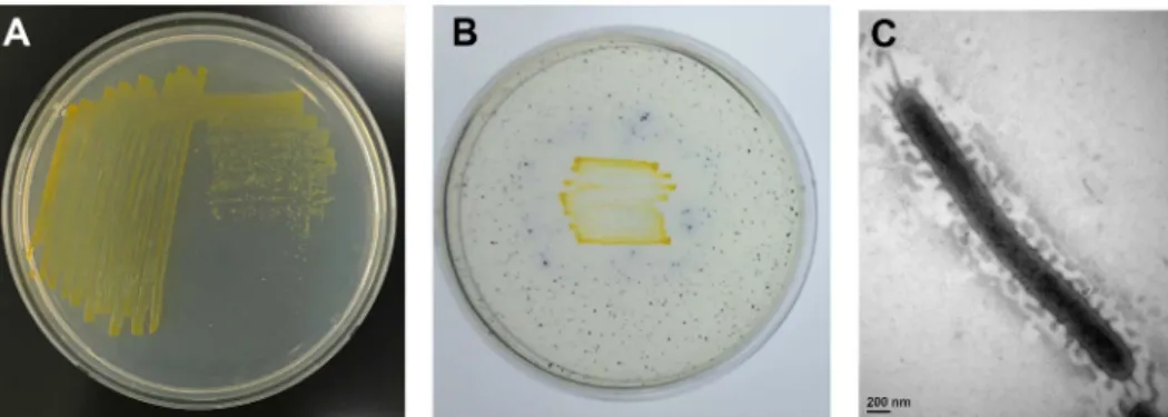
Fig. 1. Phenotypic characteristics of S2-3. (A) Formation of yellow colored colonies of S2-3 grown on marine agar medium at 30℃.
(B) Detection of cellulase activity based on blue color appearing around colonies due to decomposition of AZCL-cellulose by S2-3 on marine agar medium. (C) Transmission electron microscopy of S2-3 cell grown on marine agar medium at 30℃. Scale bar is presented at the bottom.
Results
Identification of strain S2-3
제주도연안해수로부터 cellulose 분해활성을갖는해양
미생물 균주 S2-3을 분리하여, Korean Agricultural Culture collection (KACC)에기탁(기탁번호: KACC 19205)
하였다. S2-3은그람음성균으로서콜로니에짙은노란색을 띄고, 매끈한표면의콜로니를형성하였다(Fig. 1A). S2-3의 cellulose 분해활성은고체배지에함유된 AZCL-cellulose의 분해로콜로니주위에푸른색을띄는색상의변화로확인하 였다(Fig. 1B). 전자현미경분석결과, S2-3 세포는긴막대 기형태이며균체의크기는 2.85 × 0.25 um (가로 × 세로)이
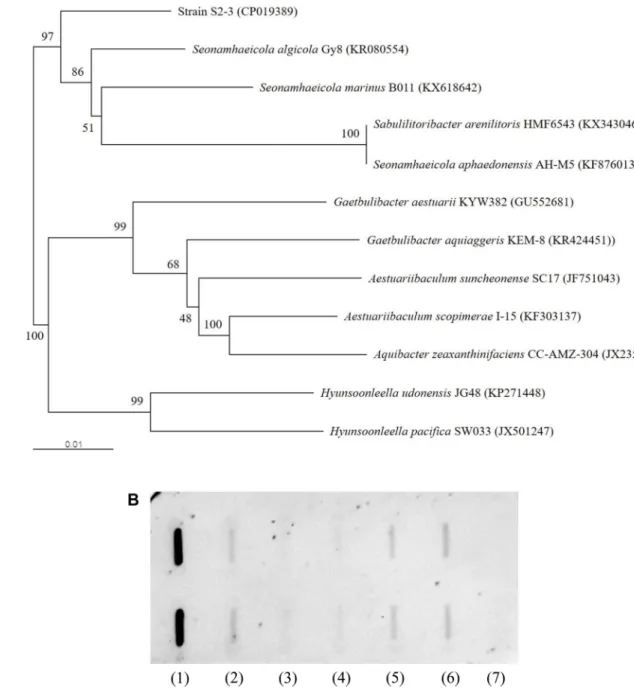
Fig. 2. Genetic analysis of S2-3. (A) Phylogenetic tree of S2-3 constructed from the nucleotide sequence of 16S rRNA gene using the
neighbor-joining method. The scale bar indicates a genetic distance of 0.01. The number shown next to each node indicates the percentage bootstrap value of 1,000 replicates. Accession nos. of the 16S rRNA sequences are given in parentheses. (B) DNA-DNA hybridization of S2-3 with the five phylogenetically close type strains. (1) S2-3, (2) Seonamheaicola algicola KCTC 42396, (3) Seonamheaicola aphaedonensis KCTC 32578, (4) Aestuariibaculum scopimerae KCTC 32459, (5) Aestuariibaculum suncheonense KACC 16186, (6) Gaetbulibacter aestuarii KACC 17489, (7) Lactobacillus acidophilus KCCM 32820.
고, 편모를갖지않는것으로관찰되었다(Fig. 1C). S2-3은 10−45℃의범위에서성장이가능하고 30−35℃에서가장잘 자랐으며, pH 5.5−8.5의범위에서성장이가능하고 pH 6.5− 7.0에서가장잘자랐다. S2-3은 API ZYM kit 및 Staph kit 테스트에서 alkaline phosphatase, esterase (C4), esterase lipase (C8), leucine arylamidase, valine arylamidase, acid phosphatase, naphtol-AS-BI-phosphohydrolase, β -glucosidase 효소가 양성반응을 보였으며, lipase (C14), cystine arylamidase, Trypsin protease, α-chymotrypsin protease, α-galactosidase, β-galactosidase, β-glucuronidase, α -glucosidase, N-acetyl-β-glucosaminidase, α-mannosidase, α-fucosidase, nitrate reduction, arginine dihydrolase, urease 등의효소는음성반응을보였다. S2-3은 Staph kit를 이용한 당성분의 acidification 테스트에서 glucose, D-fructose (약함), D-maltose (약함), D-mannitol (약함), D-xylose에서 양성반응을 보였고 mannose, lactose, D-trehalose, xylitol, D-melibiose, D-raffinose, D-sucrose, methyl-α-D-glucopyranoside, N-acetylglucosamine에서음
성반응을보였다.
S2-3의 16S rRNA 유전자의 염기서열 정보를 토대로 NCBI와 Ezbiocloud 서버에서 상동성 검색을 수행하여
Seonamhaeicola algicola Gy8, Hyunsoonleella udonensis JG48, Aestuariibaculum scopimerae I-15, Aestuariibaculum suncheonense SC17, Seonamhaeicola aphaedonensis AH-M5, Gaetbulibacter aestuarii KYW382, Hyunsoonleella pacifica SW033, Gaetbulibacter aquiaggeris KEM-8 등과 각각 97.08, 95.01, 94.86, 94.778, 94.72, 94.72, 94.60, 94.58%의높은상동성을보였다.
16S rRNA 유전자염기서열상동성검색결과를토대로하 는 N-J 계통수제작을통한계통발생적연관성분석결과, 균 주 S2-3은 S. algicola Gy8와 S. aphaedonensis AH-M5와
가장가까운연관관계를형성하고있는것으로분석되었다 (Fig. 2A). S2-3을탐침으로하는 DDH 결과에서 S. algicola Gy8와 가장 높은 32%의 hybridization 수치를 보였으며, S. algicola Gy8을 탐침으로하는 DDH 실험에서는 33%의 hybridization 수치를 보였다. 그 외의표준균들과는 24− 26%의 hybridization 수치를보였다(Fig. 2B). 이러한결과 를토대로 S2-3은 Seonamhaeicola 속의한종으로서신종 일 것으로 판단되었다. 이후 본 논문에서는 균주 S2-3을 Seonamhaeicola sp. S2-3으로명명하였다. Seonamhaeicola sp. S2-3의세포내주요지방산(>5%)은
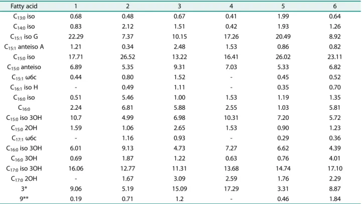
Table 1. Cellular fatty acid compositions (>1%) of strain S2-3 and its closest members.
Strains: 1, S2-3; 2, Seonamhaeicola algicola; 3, Aestuariibaculum scopimarae; 4, Seonamhaeicola aphaedonensis; 5, Gaetbulibacter aestuarii; 6, Aestuariibaculum suncheonense Fatty acid 1 2 3 4 5 6 C13:0iso 0.68 0.48 0.67 0.41 1.99 0.64 C14:0iso 0.83 2.12 1.51 0.42 1.93 1.26 C15:1 iso G 22.29 7.37 10.15 17.26 20.49 8.92 C15:1anteiso A 1.21 0.34 2.48 1.53 0.86 0.82 C15:0 iso 17.71 26.52 13.22 16.41 26.02 23.11 C15:0anteiso 6.89 5.35 9.31 7.03 5.33 6.82 C15:1 ω6c 0.44 0.80 1.52 - 0.45 0.52 C16:1 iso H - 0.49 1.11 - 0.35 0.70 C16:0iso 0.51 5.46 1.00 1.53 1.19 1.35 C16:0 2.24 6.81 5.88 2.55 1.03 5.81 C15:0 iso 3OH 10.7 4.99 6.98 10.31 7.20 5.72 C15:02OH 1.59 1.06 2.65 1.53 0.90 1.23 C17:1 ω6c - 1.16 0.93 - 0.29 0.36 C16:0iso 3OH 6.01 9.13 4.73 7.27 6.62 4.39 C16:03OH 0.69 1.87 1.22 0.63 0.76 4.01 C17:0 iso 3OH 16.06 12.77 11.31 13.68 14.74 17.10 C17:0 2OH - 1.67 3.09 2.59 1.76 2.29 3* 9.06 5.19 15.09 17.29 3.31 8.87 9** 0.19 0.71 1.2 - 0.46 1.84 3*, C16:1 ω7c/C16:1 ω6c or C16:1 ω6c/C16:1 ω7c; 9**, C16:0 10-mehtyl or C17:1 iso ω9c.
C15:1 iso G (22.29%), C15:0 iso (17.71%), C17:0 iso 3OH
(16.06%), C15:0 iso 3OH (10.7%), summed feature 3 (9.06%),
C15:0 anteiso (6.89%), C16:0 iso 3OH (6.01%) 등으로분석
되었는데, 이들주요지방산은다른표준균주들에서도주요
지방산으로나타났으나, 그함량및비율등에서큰차이를
보이고있다(Table 1).
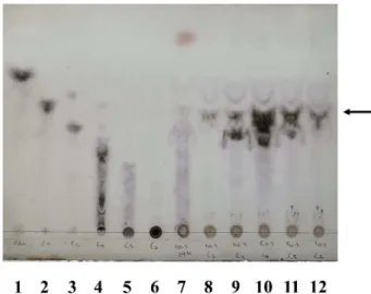
Seonamhaeicola sp. S2-3의 polar lipid 성분을분석한결 과, 1개의 phosphatidylethanolamine, 2개의 unidentified aminolipid와 5개의 unidentified lipid를 확인하였다(Fig. 3). Seonamhaeicola sp. S2-3의 셀룰로스 분해 활성 Seonamhaeicola sp. S2-3은 AZCL-cellulose 가 함유된 배지에서기질을분해하여푸른색을띄었고, 액체배지배양 액의균체를제거한상등액에서 cellulose 분해능력을확인 할수있었다. 이러한결과로부터, Seonamhaeicola sp. S2-3가세포의외부로 cellulose 분해효소를분비하고있음을예 상하였다. 실제액체배양에서, Seonamhaeicola sp. S2-3은 액체배지접종후 24시간에 stationary phase에진입하여 48시간에최대성장을보였고, 72시간이후급격히사멸하는 것으로관찰되었는데, 이러한현상은배지성분내특정영양 성분의고갈에따른영향으로알려져있다[22]. Seonamhaeicola sp. S2-3의 cellulase 생산능력역시배양 24시간까지급격히 증가하여 48시간에최대생산을보인후급격히감소하였다 (Fig. 4). 액체배양 48시간배양액으부터배양여액을제조하 여, cellulose 및 cellooligosaccharides 기질과반응시킨후분 해산물의 TLC 분석을실시하였다. 그결과, Seonamhaeicola sp. S2-3가세포외부로분비하는 cellulase는 polysaccharide 인 cellulose를분해하여다양한길이의 cellooligosaccharides를 생산하며, triose 이상의 cellooligosaccharide를 biose까지분 해하는것을확인하였다(Fig. 5). 이러한 TLC pattern은일 반적인 endo-type cellulase의가수분해활성특성과일치한 다. 또한 Seonamhaeicola sp. S2-3의배양여액은기질로사 용된 다른 cellooligosaccharide에 대한 활성과 비교하여 CMC에대한활성이비교적약한것으로관찰되었는데, 이 는 crystalline cellulose의복잡한삼차구조가효소의접근을 방해하기때문으로추측된다.
Fig. 3. Polar lipid analysis of S2-3 cells by molybdophosphoric acid staining (A) and ninhydrin staining (B). PE,
phosphatidy-lethanolamine; AL, unidentified aminolipids; L, unidentified lipid.
Fig. 4. Determination of cell growth (filled circle) and cellu-lase activity (filled square) depending on cultivation time in marine broth. Cell growth was measured at 600 nm and
cellu-lase activity was measured by DNS method using carboxylmeth-ylcellulose as a substrate at 540 nm.
Fig. 5. Thin layer chromatographic analysis of the hydrolysates of various substrates by crude cellulase prepared from S2-3 culture broth. 1, D-glucose; 2, cellobiose; 3, cellotriose; 4,
cello-tetraose; 5, cellopentaose; 6, cellohexaose; 7, carboxy-methylcellulose + S2-3 cellulase; 8, cellobiose + S2-3 cellulase; 9, cellotriose + S2-3 cellulase; 10, cellotetraose + S2-3 cellulase; 11, cellopentaose + S2-3 cellulase; 12, cellohexaose + S2-3 cellulase. Spots corresponding to cellobiose are indicated by arrow.
Discussion
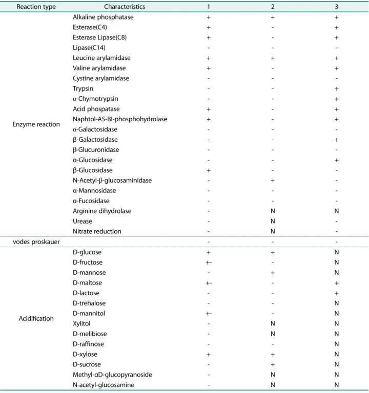
Seonamhaeicola 속은 2014년 Park 등[23]에의해서처음 소개된후현재까지 6개의종이알려져있으며, 모든종은 해양시료로부터유래되었다. Seonamhaeicola 속균주는그 람음성균으로주요퀴논으로 menaquinone-6 (MK-6)을포 함하고있고주요세포지방산으로 iso-C15:0으로구성되어 있으며, 유전체 DNA G+C 농도는약 33−36 mol%로알려져 있다. 이번연구로부터분리된균주 S2-3은형태적, 생리적, 유전적특성분석을통해 Seonamhaeicola 속에속하는새Table 2. Biochemical characteristics of strain S2-3 and its closest members.
Strains: 1, S2-3; 2, Seonamhaeicola aphaedonensis[23]; 3, Seonamhaeicola algicola[24] Symbol: N, no data
Reaction type Characteristics 1 2 3
Enzyme reaction Alkaline phosphatase + + + Esterase(C4) + - + Esterase Lipase(C8) + - + Lipase(C14) - - -Leucine arylamidase + + + Valine arylamidase + - + Cystine arylamidase - - -Trypsin - - + α-Chymotrypsin - - + Acid phospatase + - + Naphtol-AS-BI-phosphohydrolase + - + α-Galactosidase - - -β-Galactosidase - - + β-Glucuronidase - - -α-Glucosidase - - + β-Glucosidase + - -N-Acetyl-β-glucosaminidase - + -α-Mannosidase - - -α-Fucosidase - - -Arginine dihydrolase - N N Urease - N -Nitrate reduction - N -vodes proskauer - - -Acidification D-glucose + + N D-fructose +- - N D-mannose - + N D-maltose +- - + D-lactose - - + D-trehalose - - N D-mannitol +- - N Xylitol - N N D-melibiose - N N D-raffinose - - N D-xylose + + N D-sucrose - + N Methyl-αD-glucopyranoside - N N N-acetyl-glucosamine - N N
로운종으로분류되었다.
Seonamhaeicola 속에속하는 6개의종에관한문헌은모
두 분류학상의 특징만을 기술하고 있고, 그 중에서 S.
algicola Gy86가 cellulose를분해하는것으로언급이되어 있을뿐이다. 이번에분리한균주 Seonamhaeicola sp. S2-3는 cellulose 분해활성을갖는신종이며, 이균주의배양여 액은 cellulose를분해하여다양한길이의 cellooligosaccharides 를만들고, 최종적으로 cellobiose까지분해할수있음을확 인하였다. 따라서, Seonamhaeicola sp. S2-3은세포외부로 cellulase를 분비하여 다당체인 cellulose를 최소단위인 cellobiose까지분해하여세포내로흡수한후, β-glucosidase 등과같은효소를이용하여단당인 glucose로완전분해하여 에너지원으로사용할것으로추측된다. Seonamhaeicola sp. S2-3 유래의 cellulase가어떤생화학적특성을갖는지는추 후연구가필요하며, 이러한연구를통해지구상의탄소재 순환을위한해결책이마련되길기대한다.
Acknowledgments
This work was supported by a grant from the National Institute of Biological Resources (NIBR), funded by the Ministry of Environment (MOE) of the Republic of Korea (NIBR202020101).
Conflict of Interest
The authors have no financial conflicts of interest to declare.
References
1. Beguin P, Aubert JP. 1994. The biological degradation of cellulose.
FEMS Microbiol. Rev. 13: 25-58.
2. Carpita NC. 1996. Structure and biogenesis of the cell walls of grasses. Annu. Rev. Plant Physiol. Plant Mol. Biol. 47: 445-476. 3. Warren RA. 1996. Microbial hydrolysis of polysaccharides. Annu.
Rev. Microbiol. 50: 183-212.
4. Studer MH, DeMartini JD, Davis MF, Sykes RW, Davison B, Keller M, et al. 2011. Lignin content in natural Populus variants affects sugar release. Proc. Natl. Acad. Sci. USA 108: 6300-6305. 5. Si S, Chem Y, Fan C, Hu H, Li Y, Huang J, et al. 2015. Lignin
extraction distinctively enhances biomass enzymatic saccharifi-cation in hemicelluloses-rich Miscanthus species under various alkali and acid pretreatments. Bioresour. Technol. 183: 248-254. 6. Vermaas JV, Petridis L, Qi X, Schulz R, Lindner B, Smith JC. 2015.
Mechanism of lignin inhibition of enzymatic biomass decon-struction. Biotechnol. Biofuels 8: 1-16.
7. Patyshakuliyeva A, Falkoski DL, Wiebenga A, Timmermans K, Vries RP. 2019. Macroalgae derived fungi have high abilities to degrade algal polymers. Microorganisms 8: 52.
8. Bhat MK. 2000. Cellulases and related enzymes in
biotechnol-ogy. Adv. Biotechnol. 1: 355-383.
9. Biely PJ, Schneider H. 1985. Acetyl xylan esterases in fungal cel-lulolytic systems. FEBS Lett. 186: 80-84.
10. Yin LJ, Huang PS, Lin HH. 2010. Isolation of cellulase-producing bacteria and characterization of the cellulase from the isolated bacterium Cellulomonas sp. YJ5. J. Agric. Food Chem. 58: 9833-9837. 11. Fan C, Li S, Li C, Ma S, Zou L, Wu Q. 2012. Isolation, identification and cellulase production of a cellulolytic bacterium from intes-tines of giant panda. Wei Sheng Wu Xue Bao 52: 1113-1121. 12. Gaur R, Tiwari S. 2015. Isolation, production, purification and
characterization of an organic-solvent-thermostable alkalophilic cellulase from Bacillus vallismortis RG-07. BMC Biotechnol. 15: 19. 13. Kim DS, Chi WJ, Hong SK. 2019. Molecular characterization of an endo-β-1,4-glucanase, CelAJ93, from the recently isolated marine bacterium, Cellulophaga sp. J9-3. Appl. Sci. 9: 4061-4073. 14. Dos Santos YQ, de Veras BO, de Franca AFJ, Gorlach-Lira K,
Velasques J, Migliolo L, et al. 2018. A new salt-tolerant thermo-stable cellulase from a marine Bacillus sp. Strain. J. Microbiol.
Biotechnol. 28: 1078-1085.
15. Baker GC, Smith JJ, Cowan DA. 2003. Review and re-analysis of domain-specific 16S primers. J. Microbiol. Methods 55: 541-555. 16. Galkiewicz JP, Kellogg CA. 2008. Cross-kingdom amplification
using bacteria-specific primers: complications for studies of coral microbial ecology. Appl. Environ. Microbiol. 74: 7828-7831. 17. Chun J, Lee JH, Jung YY, Kim MJ, Kim SI, Kim BK, et al. 2007.
EzTaxon: a web-based tool for the identification of prokaryotes based on 16S ribosomal RNA gene sequences. Int. J. Syst. Evol.
Microbiol. 57: 2259-2261.
18. Kimura M. 1980. A simple method for estimating evolutionary rates of base substitutions through comparative studies of nucleotide sequences. J. Mol. Evol. 16: 111-120.
19. Miller L, Berger T. 1985. Bacterial identification by gas chroma-tography of whole cell fatty acids. Hewlett-Packard Application
note pp. 228-241. Hewlett-Packard Co, Avondale, Pa.
20. Sasser M. 1990. Identification of bacteria by gas chromatography of cellular fatty acids. MIDI Technical Note 101. pp. 1-7. Newark, DE:MIDI Inc.
21. Minikin DE, O'Donnell AG, Goodfellow M, Alderson G, Athalye M, Schaal A, et al. 1984. An integrated procedure for the extraction of bacterial isoprenoid quinones and polar lipids. J. Microbiol.
Methods 2: 233-241.
22. Zhang Z, Chen Y, Wang R, Cai R, Fu Y, Jiao N. 2015. The fate of marine bacterial exopolysaccharide in natural marine microbial communities. PLoS One 10: e0142690.
23. Park S, Won SM, Park DS, Yoon JH. 2014. Seonamhaeicola
aphae-donensis gen. nov., sp. nov., a member of the family Flavobacteri-aceae isolated from a tidal flat sediment. Int. J. Syst. Evol. Microbiol. 64: 1867-1881.
24. Zhou YX, Du ZJ, Chen GJ. 2016. Seonamhaeicola algicola sp. nov., a complex polysaccharide-degrading bacterium isolated from
Gracilaria blodgettii, and emended description of the genus Seonamhaeicola. Int. J. Syst. Microbiol. 66: 2064-2068.