• 검색 결과가 없습니다.

Comparisons of Development Potential in Bovine SCNT Embryos using Donor Cells treated with Different Demethylating Inhibitors

N/A
N/A
Protected

Academic year: 2021

Share "Comparisons of Development Potential in Bovine SCNT Embryos using Donor Cells treated with Different Demethylating Inhibitors"

Copied!
9
0
0

로드 중.... (전체 텍스트 보기)

전체 글

(1)

Comparisons of Development Potential in Bovine SCNT Embryos using Donor Cells treated with Different Demethylating Inhibitors

Byeong-Gyun Jeon

1,3†

, Gie-Joon Jeong

1

and Gyu-Jin Rho

2

1

Dept. of Biology Education, College of Education, Gyeongsang National University, Jinju 660-701, Korea

2

OBS/Theriogenology and Biotechnology, College of Veterinary Medicine, Gyeongsang National University, Jinju 660-701, Korea

3

Research Institute of Education, Gyeongsang National University, Jinju 660-701, Korea

ABSTRACT

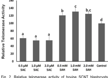
To improve the developmental potential of bovine somatic cell nuclear transfer (SCNT) embryos, this study compared the developmental rates to blastocyst stage in the SCNT embryos using donor fibroblasts treated with 5-azacytidine (5AC) and S-adenosylhomocysteine (SAH) at different concentrations. Their reprogramming efficiency level was investigated with level of telomerase activity. Donor fibroblasts isolated from adult ear skin of a cow were exposed to 5AC and SAH at different concentrations during 2 passages. After nuclear transfer into enucleated recipient oocytes, the cleavage and developmental rates were significantly (p<0.05) decreased in the SCNT embryos using 5AC-treated fibroblasts (5AC-SCNT embryos), compared with those of non-treated control (control-SCNT embryos) and SAH-treated fibroblasts (SAH-SCNT embryos). The developmental rates to blastocyst stage tended to be slightly increased in the SAH-SCNT embryos at each of the concentrations, and especially, the developmental rates in the SCNT embryos using 1.0 mM SAH-treated fibroblasts were significantly (p<0.05) higher than that of control SCNT embryos. The mean numbers of total and ICM cell in blastocysts were also significantly (p<0.05) decreased in the 5AC-SCNT embryos, compared with those of other SCNT blastocysts. Further, the level of telomerase activity was also significantly (p<

0.05) decreased in the 5AC-SCNT embryos than those of control and SAH-SCNT embryos. Whereas, a significantly (p<0.05) up-regulated telomerase activity was observed in SAH-SCNT embryos, compare with that of control-SCNT embryos. In conclusion, SCNT embryos using hypomethylated donor cells with SAH, not 5AC, may improve the developmental potential and reprogramming efficiency.

(Key words: somatic cell nuclear transfer, reprogramming, hypomethylation, telomerase activity, bovine)

Correspondence : [email protected]

INTRODUCTION

The matured oocytes can induce reprogramming of the nuclei of differentiated somatic cells into embryo and fetus status.

Thus, nuclear transfer technique (SCNT), inserting donor so- matic cell into a host recipient oocyte, should be regarded as a powerful tool for the mass production of cloned animals.

The SCNT technique has also shown some advantages in parts of the production of endangered animals, epigenetics control in developmental biology and others. However, although cloned animals in various species have been successfully produced by SCNT technique up to now, low pregnancy rates of cloned animals, high stillborn and early neonatal mortality with mor- phologic abnormalities have frequently been observed in animal clones. These are the main obstacles to applications for animal

cloning with SCNT technique (Cibelli et al., 2002; Rideout et al., 2001).

It has been now suggested that one of the main reasons on the low production efficiency of cloned embryos and animals by SCNT technique is tightly related with the incomplete re- programming of differentiated somatic cells inserted into the recipient oocytes (Han et al., 2003; Niemann et al., 2008).

Under normal fertilization, paternal and maternal genomes from

sperm and oocytes are fully demethylated by active and passive

mechanisms at an early developmental stage, respectively. Both

genomes are subsequently remethylated in an orderly way,

according to the procedure of embryo development (Guo et al.,

2014; Morgan et al. 2005). However, the donor nuclei inserted

into the recipient oocyte is not fully demethylated during early

development of SCNT embryos, and the incomplete demethy-

(2)

lation leads to abnormal and incorrect remethylation patterns of donor nuclei. The precise expression timing of genes along with developmental stage of embryos is essential for the nor- mal development of embryos and fetus (Guo et al., 2014;

Morgan et al. 2005). However, it has been reported that SCNT embryos are displayed to abnormal expression patterns of genes for embryonic development by incomplete reprogramming effi- ciency of the inserted donor nuclei (Han et al., 2003; Niemann et al., 2008). By considering these findings, several previous studies have attempted that hypomethylated or demethylated somatic donor cells are often used for improving development potential of SCNT embryos (Jafari et al., 2011; Jeon et al., 2008; Kumar et al., 2013). Both 5-azacytidine (5AC) and S- adenosylhomocysteine (SAH) are the most commonly used for inducing DNA hypomethylation/demethylation. In previous stu- dies, the bovine and porcine adult fibroblasts treated with 5AC and SAH have been shown to be easily induced to DNA hypo- methylation (Jeon et al., 2008; Kumar et al., 2007). 5AC, an analogue of cytidine that is present in DNA, can induce hy- pomethylation by incorporation into newly synthesized DNA instead of methyl-cytosine (Christman, 2002). Otherwise, S- adenosylhomocysteine (SAM) plays an important role as a methyl donor that catalyzes methylation of DNA and proteins, and is converted into SAH, but SAH strongly react with DNA methyltransferase at higher concentration than SAM, and induce DNA hypomethylation (Castro et al., 2005; Caudill et al., 2001;

Yi et al., 2000). Further, several reports have shown beneficial effects on the developmental potential of the SCNT embryos using somatic donor cells hypomethylated with 5AC or SAH (Ding et al., 2008; Jafari et al., 2011; Jeon et al., 2008; Kumar et al., 2013). Whereas, it has been shown that 5AC or SAH treatment at a high dose is induced to cell cytotoxicity, such as cell cycle arrest, apoptosis and cell death (Issa et al., 2004;

Wang et al., 2015), and the hypomethylated donor cells with 5AC is ineffective in the developmental potential of the porcine SCNT embryos (Das et al., 2010; Ning et al., 2012; Ysuji et al., 2009). Up to now, the influences on the developmental potential of the SCNT embryos using donor cells hypomethylated with various demethylating inhibitors remain confused and suitable demethylating inhibitors should be precisely investigated for improving the production efficiency of bovine SCNT embryos.

Therefore, the present study investigated the influences of donor fibroblasts hypomethylated with 5AC and SAH at diffe- rent concentrations on the developmental potential of the bovine

SCNT embryos, and their reprogramming efficiency was asse- ssed with the level of telomerase activity.

MATERIALS AND METHODS

1. Media and Chemicals

All chemicals and media were purchased from Sigma (USA) and Gibco (USA), unless otherwise specified. TCM-199 medium supplemented with 2.5 mM Na-pyruvate, 1 mM L-glutamine, 1% (v/v) penicillin-streptomycin (10,000 IU and 10,000 μg/ml, respectively, Pen-Strep), 2% steer serum (Cansera, Canada), 1 μg/ml estradiol-17 β, 0.5 μg/ml FSH, and 1 μg/ml LH was used for in vitro maturation (IVM) of recipient oocytes. Culture me- dium for SCNT embryo was modified synthetic oviduct fluid (mSOF) supplemented with 2.9 μg/ml EDTA and 8 mg/ml BSA (essentially fatty acid free). Donor fibroblasts were cultured in Dulbecco’s modified Eagle’s medium (DMEM) containing 10%

(v/v) fetal bovine serum (FBS). Tyrode’s albumin lactate py- ruvate medium containing 2 mg/ml bovine serum albumin (BSA, essentially fatty acid free) and 10 mM Hepes (HEPES-TALP) was used for oocyte micromanipulation and handling. For all the media, the pH was adjusted to 7.4 and osmolality to 280 mOsm/kg.

2. Treatments of Donor Cells

The donor fibroblasts for SCNT embryos were isolated from ear skin tissues of a 4-year-old cow as described previously by Jeon et al. (2008). Briefly, the fibroblast pellets were extracted from pieces of ear’s skin tissue by treating 0.5% collagenase at 38.5 ℃ for 3∼6 h with agitation. The extracted fibroblasts were washed once by centrifugation at 300 ×g for 10 min, and were subcultured in DMEM plus 10% FBS and 1% Pen-Strep at 38.5 ℃ in a humidified atmosphere of 5% until fully conflu- ence. For each experiment, the exposure concentrations and durations to 5-azacytidine (5AC) and S-adenosylhomocysteine (SAH) were adapted from Kumar et al. (2013) and Jeon et al.

(2008). Briefly, the fibroblasts at passages 3 were treated in

DMEM plus 10% FBS and 1% Pen-Strep containing 0 (control),

5AC (0.5, 1.0 and 2.0 μM) and SAH (0.5, 1.0 and 2.0 mM)

in 4-well dishes (Nunc, Denmark) for an additional 2 passages

until confluence. The cell culture media was changed daily with

fresh DMEM containing respective concentrations of 5AC and

SAH. After treatment, fully confluent fibroblasts were used as

donor cells for SCNT embryos.

(3)

3. Preparation of Recipient Cytoplasts

The SCNT procedure was performed according to the pre- viously described protocol (Jeon et al., 2011a). Briefly, ovaries of cows were acquired from a local abattoir at 25 ℃ in PBS and cumulus-oocyte complexes (COCs) were collected from small antral follicles (2 ∼8 mm in diameter) in Ham’s F10.

Groups of 10 grade I COCs with at least several layers of cu- mulus cells and morphologically homogeneous cytoplasm were selected and in vitro matured in 50 μl droplets of IVM medium at 38.5 ℃ in a humidified atmosphere of 5% CO

2

in air. Cu- mulus cells were removed by vortexing for 2 min in 3% (v/v) sodium citrate solution at 12 h after maturation. After being cultured in IVM medium for additional 6 h, the denuded oocytes were enucleated by removing a small amount of cytoplasm along with first polar body under an inverted microscope equi- pped with micromanipulator. Enucleation of recipient oocytes was confirmed by staining with 5 µg/ml bisbenzimide (Hoechst 33342) under a fluorescence inverted microscope equipped with a UV light.

4. Nuclear Transfer and Oocyte Activation

Small and morphologically normal donor fibroblast treated with 5AC and SAH at different concentrations was inserted into the perivitelline space of each enucleated recipient oocyte. Mem- brane fusion of recipient oocyte and inserted donor cell was induced by two electric pulses at 1.0 kV/cm for 15 μsec in 0.26 M mannitol solution containing 50 μM MgCl

2

with an ECM 2001 BTX cell manipulator (VWR Laboratories, USA) equipped with electrode needles. Successful cell fusion was checked at 2 h after electrical stimulation. Only fused eggs were induced to oocyte activation in HEPES-TALP containing 5 μM ionomycin for 5 min, sequential exposure to mSOF containing 30 mg/ml BSA for 4 min, and further incubated in mSOF containing 10 μg/ml cycloheximide for 5 h at 38.5 ℃ and 5% CO

2

in air.

5. In vitro Culture of SCNT Embryos

Groups of 30 SCNT eggs were cultured in 30 μl droplets of mSOF for 3 days, subsequently transferred to 30 μl droplets of mSOF supplemented with MEM amino acids, and further cul- tured for 6 days at 38.5 ℃ in a humidified atmosphere of 5%

CO

2

, and 5% O

2

. Cleavage and blastocyst rates were assessed on day 2 and 9, respectively. A differential staining method was employed for mean numbers of total and ICM cells in SCNT blastocysts as previously represented by Thouas et al. (2001).

6. Relative-Quantitative Telomerase Repeat Amplification Protocol (RQ-TRAP)

To assess reprogramming efficiency by the level of telomerase activity in SCNT embryos, RQ-TRAP assay using LightCycler 4.0 (Roche, USA) modified from a conventional EILZA-TRAP assay was used as previously described by Jeon et al. (2008).

Briefly, SCNT embryos developed to blastocyst stage were harvested on day 9 and immediately frozen at —80℃ for fu- ture analysis or lysed in 0.5% CHAPS lysis buffer (pH 7.5, Chemicon, USA) at a concentration of 5 blastocysts/20 μl for 30 min on ice. Samples were then centrifuged for 20 min at 12,000 ×g at 4 ℃. Eighty percent of the lysed supernatant ex- cluding cell debris and DNA was transferred to a new Eppen- dorf centrifuge tube and analyzed by RQ-TRAP assay using the PCR reagent LightCycler FastStart DNA Master SYBR Green 1 (Roche, USA). A reaction volume of 20 µl contained 2 µl of lysed sample buffer, 2.5 mM MgCl

2

, 0.02 µg of primer TS (5’- AAT CCG TCG GAG CAG AGT T-3’) and 0.04 µg of primer ACX (5’-GCG CGG CTT ACC CTT ACC CTT ACC CTA ACC- 3’). The assay program for amplification consisted of a 20 min incubation at 25 ℃, followed by a 10 min incubation at 94℃, and 40 cycles of PCR at 94 ℃ for 30 s and 60℃ for 90 s. A second derivative method of crossing point determination with LightCycler Quantification Software (Roche, Canada) was used for quantification of samples, and the level of telomerase activity of 5AC and SAH-SCNT blastocysts was calculated to ratio based on the level of telomerase activity in the control-SCNT blastocysts.

7. Statistical Analysis

Differences among treatments were analyzed by using one- way analysis of variance (ANOVA). Differences in the percen- tage of in vitro developmental rate were analyzed using a Chi- square test. Differences in the telomerase activity and the total cell number of blastocyst were analyzed using a Student’s t-test.

A five percent probability (P<0.05) was used as the level of significance.

RESULTS

1. Developmental Potential of SCNT Embryos

The donor fibroblasts were treated with 5AC and SAH at

different concentrations, and their represented growth appea-

rance are showed in Fig. 1. Compared with non-treated control

(4)

Table 1. In vitro developmental potential of bovine somatic cell nuclear transfer (SCNT) using donor fibroblasts treated with 5-azacytidine (5AC) and s-adenosylhomocysteine (SAH) at different concentrations

Treatment No. (%) of oocytes fused

No. (%) of embryos developed to No. of mean cells**

2-cell Blastocyst H-Blastocysts* Total ICM

5AC

0.5 μM 156/168 (92.8) 91/150 (60.6)

a

29/150 (19.3)

a

10/150 ( 6.6)

a

147.9±30.2

a

23.3±6.40

a

1.0 μM 152/170 (89.4) 74/138 (53.6)

a,b

16/138 (11.6)

a

5/138 ( 3.6)

a

139.1±28.6

a

25.2±5.60

a

2.0 μM 181/205 (88.3) 84/165 (50.9)

b

20/165 (12.1)

a

7/165 ( 4.2)

a

128.3±23.5

a

20.4±7.70

a

SAH

0.5 mM 123/135 (91.1) 105/121 (87.6)

d

49/121 (40.5)

b,c

36/121 (29.7)

b,c

190.9±29.5

b

43.6±8.60

b

1.0 mM 166/180 (92.2) 118/141 (83.7)

d

62/141 (43.9)

b

49/141 (34.7)

b,c

195.4±25.6

b

47.2±12.5

b

2.0 mM 140/158 (88.6) 111/130 (85.3)

d

50/130 (38.5)

b,c

38/130 (29.2)

b,c

188.4±35.2

b

40.3±9.80

b

Control 355/389 (91.3) 283/329 (86.0)

d

113/329 (34.3)

c

84/329 (25.4)

c

179.2±30.8

b

45.3±8.60

b

* Hatched blastocysts.

** Values indicate the mean cell number (mean±SEM). Different superscripts within columns indicate significant differences (P<0.05).

Control 1.0 μm 5AC 1.0 mM SAH

Fig. 1. Morphological features of bovine ear skin donor fibroblasts treated with control, 1.0 μM 5-azacytidine (5AC) and 1.0 mM s- adenosylhomocysteine (SAH). Fibroblasts at passages 3 were cultured for an additional 2 passages in media containing each of concentrations 5AC and SAH. The control and 1.0 mM SAH-treated fibroblasts were reached to fully confluent status at 8 days during treatment of additional 2 passages, but delayed cell growth with enlarged and star-shaped cell morphology was observed in the fibroblasts exposed with 1.0 μM 5AC, scale bar, 50 μm.

and SAH-treated fibroblasts, the rate of cell growth was obser- ved to remarkably be delayed in the 5AC-treated fibroblasts, and the cells is gradually changed to enlarged and star-shaped morphology, reaching at senescent status.

The rates of fusion, cleavage and development to blastocyst stage in the SCNT embryos using hypomethylated with 5AC and SAH at different concentrations are tabulated in Table 1.

The fusion rates between recipient cytoplasm and donor cell were 91.3% in the control-SCNT embryos. The fusion rates were 92.8, 89.4 and 88.3% in the SCNT embryos using 0.5, 1.0 and 2.0 μM 5AC-treated fibroblasts, and 91.1, 92.2 and 88.6% in the SCNT embryos using 0.5, 1.0 and 2.0 mM SAH-treated fi- broblasts, respectively. The fusion rates were not significantly (p<0.05) different in the 5AC and SAH-SCNT embryos, com- pared with that of control-SCNT embryos. The cleavage rates were 86.0% in the control-SCNT embryos. The cleavage rates

were 60.6, 53.6 and 50.9% in the SCNT embryos using 0.5, 1.0 and 2.0 μM 5AC-treated fibroblasts, respectively. Whereas, the cleavage rates were 87.6, 83.7 and 85.3% in the SCNT embryos using 0.5, 1.0 and 2.0 mM SAH-treated fibroblasts, respectively, and the cleavage rate in the SAH-SCNT embryos are similar with control-SCNT embryos. However, the cleavage rate was significantly (P<0.05) decreased in the 5AC-SCNT embryos, compared with those of control and SAH-SCNT embryos.

The rates of embryos developed to blastocyst stage on day

9 were 34.3%, and the rates of hatched and hatching blastocysts

among developed blastocysts 25.4% in the control-SCNT em-

bryos. Whereas, the developmental rates to blastocyst stage and

hatched blastocyst stage were 19.3 and 6.6, 11.6 and 3.6, and

12.1 and 4.2% in the SCNT embryos using 0.5, 1.0 and 2.0 μM

5AC-treated fibroblasts, respectively. The developmental rates

to blastocyst stage in the 5AC-SCNT embryos was signifi-

(5)

cantly (P<0.05) decreased than that of control-SCNT embryos.

However, the developmental rates of SCNT embryos using 0.5, 1.0 and 2.0 mM SAH-treated fibroblasts to blastocyst stage and hatched blastocyst stage were 40.5 and 29.7, 43.9 and 34.7, and 38.5 and 29.2%, respectively. The developmental rates were slightly increased in the SAH-SCNT embryos at treated all concentrations. Particularly, the developmental rates to blasto- cyst and hatched blastocyst stage in the SCNT embryos using 1.0 mM SAH-treated fibroblasts were significantly (P<0.05) increased than that of control-SCNT embryos.

The mean numbers of total and ICM cells were investigated by a different staining method in the SCNT embryos. The mean total (ICM) cell numbers were 179.2±30.8 (45.3±8.6) in the control-SCNT blastocysts. The mean total (ICM) cell numbers in the SCNT blastocysts using 0.5, 1.0 and 2.0 mM SAH-treated fibroblasts were 190.9±29.5 (43.6±8.6), 195.4±25.6 (47.2±12.5) and 188.4±35.2 (40.3±9.8), respectively, and there was no signi- ficant (P<0.05) difference, compared with that of control-SCNT blastocysts. However, the mean total (ICM) cell numbers were 147.9±30.2 (23.3±6.4), 139.1±28.6 (25.2±5.6) and 128.3±23.5 (20.4±7.7) in the SCNT embryos using 0.5, 1.0 and 2.0 μM 5AC-treated fibroblasts, respectively. The mean total (ICM) cell numbers were significantly (P<0.05) decreased in the 5AC-SCNT embryos than those of control and SAH-SCNT embryos.

2. Relative Telomerase Activity of SCNT Embryos

The relative telomerase activity (RTA) by RQ-TRAP was investigated the control, 5AC and SAH-SCNT blastocysts, as summarized in Fig. 2. The telomerase activity in the control- SCNT blastocysts was counted as 100% for comparison with 5AC and SAH-SCNT blastocysts. The RTA in the SCNT blas- tocysts using 0.5, 1.0 and 2.0 μM 5AC-treated fibroblasts was 61±5.3, 55±5.2, 54±9.8%, respectively. The RTA in the 5AC- SCNT blastocysts was significantly (P<0.05) decreased than that of control-SCNT blastocysts. Whereas, the RTA was 121±6.3, 129±8.8, 125±3.3% in the SCNT blastocysts using 0.5, 1.0 and 2.0 mM SAH-treated fibroblasts, respectively. The RTA in the SAH-SCNT blastocysts was significantly (P<0.05) up-regulated, compared with that of control-SCNT blastocysts.

DISCUSSION

In the present study, we compared the developmental po- tential along with the developmental rates to blastocyst stage

Fig. 2. Relative telomerase activity of bovine SCNT blastocysts using donor fibroblasts treated with 5AC and SAH at di- fferent concentrations. Telomerase activity in control-SCNT blastocysts was regarded as 100% for comparison with 5AC and SAH-SCNT blastocysts. Different superscripts within columns indicate significant differences (P<0.05).

and mean numbers of total and ICM cells in the bovine SCNT embryos using donor fibroblasts hypomethylated with 5-azacy- tidine (5AC) and S-adenosylhomocysteine (SAH) at different concentrations, and their reprogramming efficiency was subse- quently assessed by measuring level of telomerase activity.

The findings obtained in this study have demonstrated that donor fibroblasts treated with SAH, not 5AC, outstandingly exhibit beneficial effects on the development potential and reprogra- mming efficiency of the SCNT embryos in bovine.

Methylation covalently adds or substitutes a methyl ( —CH

3

)

group to a biomolecule, including DNA, RNA, protein and

others instead of a hydrogen atom, and the methylated bio-

molecules exhibit slightly different biological activity. DNA

methylation also substitutes methyl groups to cytosine and

adenine nucleotide among four DNA nucleotides. Particularly,

methylated cytosine nucleotides were modified by the covalent

addition of a methyl group to the cytosine pyrimidine ring at

CpG islands near or in promoter of the genes. The binding

transcription factors for the gene expression were efficiently

inhibited by the methylated cytosine nucleotides of the CpG

islands at the major groove of DNA. Thus, DNA methylation

can stably induce alternation or down-regulation of the genes

(Breiling and Lyko, 2015). Embryonic and fetal development

is controlled by the precise timing of expression and silence

of each of the genes in the developmental stage of embryo and

fetus, called epigenetics control that implies long-term changes

(6)

of gene expression without alteration of the DNA sequence (Bird, 2002). The poor outcome of SCNT embryos and fetus by error and incompleteness of DNA methylation pattern along with developmental stage have fully identified (Han et al., 2003; Niemann et al., 2008). Further, the epigenetics controls are clearly related with the process of cellular differentiation of stem cells, X-chromosome inactivation in the female embryo and genomic imprinting (Payer et al., 2011). DNA methylation in epigenetics is most important factor, followed by modification (acetylation and methylation) of histone lysine tail that packed the DNA into nucleosome.

Generally, differentiated cells, such as fibroblasts and cu- mulus cells and others with highly methylated CpG islands were used as a donor cell in the SCNT procedure. In the de- velopmental procedure of normal embryos, the DNA methylation of each of the genes is nearly removed during the early zygote stage and re-established with timely and cell-specific patterns during additional embryo development (Morgan et al., 2005;

Whitworth and Prather, 2010; Ju et al., 2010). Even though the differentiated donor cells are reprogrammed in the recipient oocytes into embryonic state, it has been shown that DNA me- thylation is not fully removed in zygotic stage of SCNT embryos, and DNA re-methylation subsequently exhibits abnormal pattern (Han et al., 2003; Niemann et al., 2008). Thus, it has been tried that donor cells treated with demethylating agents, such as 5AC and SAH are used for SCNT embryos (Ding et al., 2008; Jafari et al., 2011; Jeon et al., 2008; Kumar et al., 2013).

The cytosine of newly synthesized DNA stands are immedia- tely methylated by DNA methyltransferases (DNMTs) during DNA replication. 5AC, a methyl-cytosine analogs that is most commonly used for inducing demethylation of the cells is incorporated into the replicating DNA strands. The 5AC co- valently combine with DNA methyltransferases (DNMTs) and subsequently inhibits DNA methylation of the newly replicating DNA strands (Christman, 2002275). The porcine fibroblasts derived from ear skin tissues have shown to be efficiently hypomethylated by the treatment of 5AC (Kumar et al., 2006).

However, the 5AC covalently combined with DNMTs interfere elongation of the newly replicating DNA strands and can po- tentially lead to cellular cytotoxicity, including cell cycle arrest at S phase, apoptosis and chromosomal abnormalities (Funayama and Ishikawa, 2007; Kumar et al., 2006; Lavelle et al., 2003;

Momparler, 2005). Otherwise, S-adenosylmethionine (SAM) is an important methyl donor for methylation of DNA, RNA and

others, and converted to S-adenosylhomocysteine (SAH) after transfer of the methyl group by methyltransferase. However, DNMTs exhibit higher affinity to SAH than SAM and DNA methylation is efficiently inhibited at a high SAH concen- tration by inhibiting transfer of the methyl groups at DNMTs active s291) ite after DNA replication (De Cabo et al. 1994).

Previous studies have demonstrated that any cytotoxic effects are not displayed in SAH-treated cells (De Cabo et al. 1994, Jeon et al., 2008; Nieto et al. 2004). Further, our previous study has shown that SAH treatment induces global DNA hy- pomethylation and re-activation of in-activated X-chromosome with highly hypermethylated state in bovine adult fibroblast 1s (Jeon et al., 2008).

Previous several studies have shown that the 5AC-SCNT embryos exhibit the enhanced developmental potential (Gómez et al., 2012; Kumar et al., 2013). However, our study has shown that the 5AC treatment is induced to the decreased developmental potential of SCNT embryos, and other studies have also de- monstrated the decreased developmental potential (Das et al., 2010; Ning et al., 2012; Tsuji et al., 2009). One of the main reasons for the decreased developmental potential in the 5AC- SCNT embryos may be related with cell cycle stage of the treated donor cells. It has been strongly suggested that the efficiency of SCNT embryos is enhanced with donor cells at the G

0

/G

1

phase of cell cycle than S phase (Cibelli et al., 1998;

Wakayama et al., 1998; Onishi et al., 2000). Present results

have shown that the delayed cell growth with morphological

alterations into star-shaped and enlarged cells is observed in

the 5AC-treated fibroblasts, probably suggesting that fibroblasts

are reached at senescent status. It has been demonstrated that

star-shaped, flat and enlarged cells are the main characterizations

showing in the cells reached at senescent status and their cell

cycle is arrested at S-phase with high level of apoptotic rate

(Funayama and Ishikawa, 2007; Kumar et al., 2006; Lavelle et

al., 2003; Momparler, 2005). Further, we have attempted the

high level of DNA hypomethylation by prolonged 5AC exposure

during 2 passages (up to 10 days) and daily changes of media

containing 5AC, but prolonged exposure and daily media

changes probably leads to more cellular cytotoxicity, such as

apoptosis and senescence. Even though stage of cell cycle,

apoptosis and senescence rates of the donor fibroblasts treated

with 5AC was not examined in the present study, the decrea-

sed developmental potential in the 5AC-SCNT embryos might

be tightly related with stage of cell cycle and apoptotic state

(7)

of the donor fibroblasts treated with 5AC, as pointed out earlier studies. Further, in a previous report, the level of DNA methylation was dramatically decreased in the 5AC-treated fibroblasts, but the decreased developmental potential of the SCNT embryos derived from the fibroblasts were observed by cellular cytotoxic effects (Enright et al., 2005). Whereas, it has been demonstrated that SAH-treated fibroblasts exposed during 2 passages are exhibited to hi1gh level of DNA hypomethy- lation and the increased developmental rates of the SCNT embryos (Jeon et al., 2008; Jafari et al., 2011). In the present study, the increased developmental potential was also observed in the SCNT embryos using SAH-treated donor fibroblasts.

Moreover, our results demonstrated that the level of telo- merase activity is up-regulated in the SAH-SCNT embryos, compared with that of control-SCNT embryos. Although telo- merase enzyme consists of several proteins and molecules, telomerase activity is tightly related with the expression of two genes, telomerase reverse transcriptase (TERT) protein and telomerase RNA component (TERC) (Cao et al., 2008). The high level of telomerase activity generally means up-regulated expression of TERT and TERC by DNA demethylation of CpG islands at their promoter (Guilleret et al., 2003; Hoare et al., 2001). It has been reported that most differentiated somatic cells, such as donor fibroblasts are undetectably exhibited at a very low level of telomerase activity (Betts, et al., 2001; Jeon et al., 2011b). The high level of telomerase activity in the SCNT embryos imply to be reprogrammed the somatic donor fibroblasts with undetectable telomeres activity by re-expression of TERT and TERC genes. Therefore, many studies have used as an index of reprogramming efficiency by measuring the level of telomerase activity in the SCNT embryos (Betts et al. 2001;

Jeon et al. 2008; Jeon et al., 2012). The up-regulated level of telomerase activity in the SAH-SCNT embryos, not 5AC-SCNT embryos, is probably related with modifications of DNA me- thylation patterns, suggesting that SAH treatment is more efficient in reprogramming of donor cells than that of 5AC treatment. Further, thelevel of telomerase activity was signi- ficantly down-regulated in the 5AC-treated SCNT embryos. In previous studies, it has been suggested that level of telomerase activity calculated per embryos is tightly related with the cell numbers of embryos at blastocyst stage (Betts et al., 2001;

Jeon et al., 2008). As shown in present results, the SCNT em- bryos cloned with donor fibroblasts by 5AC treatment were exhibited to decrease of the developmental potential and less

cell numbers of the blastocysts, and the down-regulated telo- merase activity may be exhibited in the 5AC -SCNT embryos.

In conclusion, we clearly demonstrated that SAH treatment, not 5AC, should be provided for the improved developmental potential and reprogramming efficiency of SCNT embryos pro- bably by epigenetic modification of donor fibroblasts. However, SAH treatment may be induced to random DNA demethylation of each of the individual genes. Successive expression of the genes in the developmental stage is positively necessary for successful development and differentiation of embryos to term.

Thus further understandings on expression of the genes in SAH- treated fibroblasts and SCNT embryos are required for post- implantation development.

REFERENCES

Betts DH, Perrault S, Harrington L and King WA. 2006. Quan- titative analysis of telomerase activity and telomere length in domestic animal clones. Methods in Molecular Biology.

325:149-180.

Bird A. 2002. DNA methylation patterns and epigenetic memory.

Genes and Development. 16:6-21.

Breiling A and Lyko F. 2015. Epigenetic regulatory functions of DNA modifications: 5-methylcytosine and beyond. Epi- genetics Chromatin. 8:24.

Castro R, Rivera I, Martins C, Struys EA, Jansen EE, Clode N, Graca LM, Blom HJ, Jakobs C and de Almeida IT. 2005.

Intracellular S-adenosylhomocysteine increased levels are associated with DNA hypomethylation in HUVEC. Journal of Molecular Medicine. 83:831-836.

Cao Y, Bryan TM and Reddel RR. 2008. Increased copy num- ber of the TERT and TERC telomerase subunit genes in cancer cells. Cancer Sci. 99:1092-1099.

Caudill MA, Wang JC, Melnyk S, Pogribny IP, Jernigan S, Collins MD, Santos-Guzman J, Swendseid ME, Cogger EA and James SJ. 2001. Intracellular S-adenosylhomocysteine concentrations predict global DNA hypomethylation in ti- ssues of methyl-deficient cystathionine beta-synthase hete- rozygous mice. Journal of Nutrition. 131:2811-2818.

Christman JK. 2002. 5-Azacytidine and 5-aza-2’-deoxycytidine as inhibitors of DNA methylation: mechanistic studies and their implications for cancer therapy. Oncogene. 21:5483- 5495.

Cibelli JB, Campbell KH, Seidel GE, West MD and Lanza RP.

(8)

2002. The health profile of cloned animals. Nat. Biotechnol.

20:13-14.

Cibelli JB, Stice SL, Golueke PJ, Kane JJ, Jerry J, Blackwell C, Ponce de León FA and Robl JM. 1998. Transgenic bo- vine chimeric offspring produced from somatic cell-derived stem-like cells. Nat. Biotechnol. 16:642-646.

Das ZC, Gupta MK, Uhm SJ and Lee HT. 2010. Increasing histone acetylation of cloned embryos, but not donor cells, by sodium butyrate improves their in vitro development in pigs. Cell. Reprogram. 12:95-104.

De Cabo SF, Hazen MJ, Molero ML and Fernandez-Piqueras J. 1994. S adenosyl-L-homocysteine: A non-cytotoxic hypo- methylating agent. Experientia. 50:658-659.

Ding X, Wang Y, Zhang D, Wang Y, Guo Z and Zhang Y.

2008. Increased pre-implantation development of cloned bovine embryos treated with 5-aza-2'-deoxycytidine and tri- chostatin A. Theriogenology. 70:622-630.

Enright BP, Kubota C, Yang X, Tian XC. 2003. Epigenetic characteristics and development of embryos cloned from donor cells treated by trichostatin A or 5-aza-2’-deoxycy- tidine. Biol. Reprod. 69:896-901.

Funayama R and Ishikawa F. 2007. Cellular senescence and chromatin structure. Chromosoma. 116:431-440.

Gómez MC, Biancardi MN, Jenkins JA, Dumas C, Galiguis J, Wang G and Earle Pope C. 2012. Scriptaid and 5-aza-2’de- oxycytidine enhanced expression of pluripotent genes and in vitro developmental competence in interspecies black- footed cat cloned embryos. Reprod. Domest. Anim. 47 Suppl.

6:130-135.

Guilleret I and Benhattar J. 2003. Demethylation of the human telomerase catalytic subunit (hTERT) gene promoter redu- ced hTERT expression and telomerase activity and shortened telomeres. Experimental Cell Research. 289:326-334.

Guo F, Li X, Liang D, Li T, Zhu P, Guo H, Wu X, Wen L, Gu TP, Hu B, Walsh CP, Li J, Tang F and Xu GL. 2014.

Active and passive demethylation of male and female pro- nuclear DNA in the mammalian zygote. Cell Stem Cell.

15:447-458.

Han YM, Kang YK, Koo DB and Lee KK. 2003. Nuclear reprogramming of cloned embryos produced in vitro. The- riogenology. 59:33 –44.

Hoare SF, Bryce LA, Wisman GB, Burns S, Going JJ, van der Zee AG and Keith WN. 2001. Lack of telomerase RNA gene hTERC expression in alternative lengthening of telo-

meres cells is associated with methylation of the hTERC promoter. Cancer Res. 61:27-32.

Issa JP, Garcia-Manero G, Giles FJ, Mannari R, Thomas D, Faderl S, Bayar E, Lyons J, Rosenfeld CS, Cortes J and Kantarjian HM. 2004. Phase 1 study of low-dose prolonged exposure schedules of the hypomethylating agent 5-aza-2’- deoxycytidine (decitabine) in hematopoietic malignancies.

Blood. 103:1635-1640.

Jafari S, Hosseini MS, Hajian M, Forouzanfar M, Jafarpour F, Abedi P, Ostadhosseini S, Abbasi H, Gourabi H, Shahverdi AH, Dizaj AV, Anjomshoaa M, Haron W, Noorshariza N, Yakub H and Nasr-Esfahani MH. 2011. Improved in vitro development of cloned bovine embryos using S-adenosyl- homocysteine, a non-toxic epigenetic modifying reagent.

Mol. Reprod. Dev. 78:576-584.

Jeon BG, Betts DH, King WA, Rho GJ. 2011a. In vitro de- velopmental potential of nuclear transfer embryos cloned with enucleation methods using pre-denuded bovine oocytes.

Reprod. Domest. Anim. 46:1035-1042.

Jeon BG, Coppola G, Perrault SD, Rho GJ, Betts DH and King WA. 2008. S-adenosylhomocysteine treatment of adult female fibroblasts alters X-chromosome inactivation and improves in vitro embryo development after somatic cell nuclear transfer. Reproduction. 135:815-828.

Jeon BG, Kwack DO and Rho GJ. 2011b. Variation of telo- merase activity and morphology in porcine mesenchymal stem cells and fibroblasts during prolonged in vitro culture.

Anim. Biotechnol. 22:197-210.

Jeon BG, Rho GJ, Betts DH, Petrik JJ, Favetta LA and King WA. 2012. Low levels of X-inactive specific transcript in somatic cell nuclear transfer embryos derived from female bovine freemartin donor cells. Sex Dev. 6:151-159.

Ju S1, Rui R, Lu Q, Lin P and Guo H. 2010. Analysis of apop- tosis and methyltransferase mRNA expression in porcine cloned embryos cultured in vitro. J. Assist. Reprod. Genet.

27:49-59.

Kumar BM, Jin HF, Kim JG, Song HJ, Hong YG, Balasubra- manian S, Choe SY and Rho GJ. 2006. DNA methylation levels in porcine fetal fibroblasts induced by an inhibitor of methylation, 5-azacytidine. Cell Tissue Res. 325:445-454 Kumar BM, Maeng GH, Lee YM, Lee JH, Jeon BG, Ock SA,

Kang T and Rho GJ. 2013. Epigenetic modification of fetal

fibroblasts improves developmental competency and gene

expression in porcine cloned embryos. Vet. Res. Commun.

(9)

37:19-28.

Lavelle D, DeSimone J, Hankewych M, Kousnetzova T, Chen YH. 2003. Decitabine induces cell cycle arrest at the G1 phase via p21(WAF1) and the G2/M phase via the p38 MAP kinase pathway. Leuk. Res. 27:999-1007. Momparler RL. 2005. Epigenetic therapy of cancer with 5-aza-2’-de- oxycytidine (decitabine). Semin. Oncol. 32:443-451.

Morgan HD, Santos F, Green K, Dean W and Reik W. 2005.

Epigenetic reprogramming in mammals. Human Molecular Genetics. 1:R47-R58.

Niemann H, Tian XC, King WA and Lee RS. 2008. Epige- netic reprogramming in embryonic and foetal development upon somatic cell nuclear transfer cloning. Reproduction.

135:151-163.

Nieto M, Samper E, Fraga MF, Gonzalez de Buitrago G, Esteller M and Serrano M. 2004. The absence of p53 is critical for the induction of apoptosis by 5-aza-20-deoxy- cytidine. Oncogene. 23:735-743.

Ning SF, Li QY, Liang MM, Yang XG, Xu HY, Lu YQ, Lu SS and Lu KH. 2013. Methylation characteristics and de- velopmental potential of Guangxi Bama minipig (Sus scrofa domestica) cloned embryos from donor cells treated with trichostatin A and 5-aza-2'-deoxycytidine. Zygote. 21:178- 86.

Onishi A, Iwamoto M, Akita T, Mikawa S, Takeda K, Awata T, Hanada H and Perry AC. 2000. Pig cloning by micro- injection of fetal fibroblast nuclei. Science. 289:1188-1190.

Payer B, Lee JT and Namekawa SH. 2011. X-inactivation and

X-reactivation: Epigenetic hallmarks of mammalian repro- duction and pluripotent stem cells. Hum. Genet. 130:265- 280.

Thouas GA, Korfiatis NA, French AJ, Jones GM and Troun- son AO. 2001. Simplified technique for differential staining of inner cell mass and trophectoderm cells of mouse and bovine blastocysts. Reprod. Biomed. Online. 3:25-29.

Tsuji Y1, Kato Y and Tsunoda Y. 2009. The developmental potential of mouse somatic cell nuclear-transferred oocytes treated with trichostatin A and 5-aza-2’-deoxycytidine. Zygote.

17:109-115.

Wakayama T, Perry AC, Zuccotti M, Johnson KR and Yanagi- machi R. 1998. Full-term development of mice from enu- cleated oocytes injected with cumulus cell nuclei. Nature.

394:369-374.

Whitworth KM and Prather RS. 2010. Somatic cell nuclear transfer efficiency: How can it be improved through nu- clear remodeling and reprogramming? Mol. Reprod. Dev.

77:1001-1015.

Yi P, Melnyk S, Pogribna M, Pogribny IP, Hine RJ and James SJ. 2000. Increase in plasma homocysteine associated with parallel increases in plasma Sadenosylhomocysteine and lymphocyte DNA hypomethylation. J. Biological Chemistry.

275:29318-29323.

Received August 19, 2015, Revised September 2, 2015, Accepted

September 22, 2015

수치

Fig.  1.  Morphological  features  of  bovine  ear  skin  donor  fibroblasts  treated  with  control,  1.0  μM  5-azacytidine  (5AC)  and  1.0  mM  s-  adenosylhomocysteine  (SAH)
Fig.  2.  Relative  telomerase  activity  of  bovine  SCNT  blastocysts  using  donor  fibroblasts  treated  with  5AC  and  SAH  at  di-  fferent  concentrations

참조

관련 문서

JVS Original Article J Vet Sci 2019, 20(1), 2 9ㆍhttps //doi org/10 4142/jvs 2019 20 1 2 Expression of polo like kinase 1 in pre implantation stage murine somatic cell nuclear

In this study, we examined the feasibility of using subzonal cell injection with electrofusion for interspecies somatic cell nuclear transfer (iSCNT) to produce sei

Our results suggest that a supplement of TUDCA during vitrification improves blastocyst formation rates of vitrified mouse 8-cell stage embryos by attenuating ER

In vitro development of cloned embryos produced by interspecies nuclear transfer of porcine oocytes with goat fetal fibroblast cells, porcine nuclear transfer