원 원 저저
INTRODUCTION
Mushroom poisoning is an increasingly common medical emergency in many countries because out- door leisure activities increase these days. The over- whelming majority of lethal mushroom poisonings
알파 아마니틴에 의한 간독성에 대한 녹차 추출물의 보호 효과
조선대학교 의과대학 응급의학교실1, 병리학교실2, 생화학교실3
안수환1∙선경훈1∙홍홍란2∙이병래3∙박용진1
The Protective Effect of Green Tea Extract on Alpha-amanitin Induced Hepatotoxicity
Su Hwan An, M.D.1, Kyung Hoon Sun, M.D., Ph.D.1, Ran Hong, M.D., Ph.D.2, Byoung Rai Lee, M.D., Ph.D.3, Yongjin Park, M.D., Ph.D.1
Department of Emergency Medicine1, Department of Pathology2,
Department of Biochemistry3, School of Medicine, Chosun University, Gwangju, Korea
Purpose: Alpha-amanitin induces potent oxidative stress and apoptosis, and may play a significant role in the pathogenesis of hepatotoxicity. This study examined the mechanisms ofα-amanitin-induced apoptosis in vitro, and whether green tea extract (GTE) offers protection against hepatic damage caused byα-amanitin (AMA) induced apoptosis in vivo.
Methods: The effects of GTE and SIL on the cell viability of cultured murine hepatocytes induced by AMA were evaluated using an MTT assay. Apoptosis was assessed by an analysis of DNA fragmentation and caspase-3. In the in vivo protocol, mice were divided into the following four groups: control group (0.9% saline injection), AMA group (α-amanitin 0.6 mg/kg), AMA+SIL group (α-amanitin and silibinin 50 mg/kg), and AMA+GTE group (α-amanitin and green tea extract 25 mg/kg). After 48 hours of treatment, the hepatic aminotransferase and the extent of hepa- tonecrosis of each subject was evaluated.
Results: In the hepatocytes exposed to AMA and the tested antidotes, the cell viability was significantly lower than the AMA only group. An analysis of DNA fragmentation showed distinctive cleavage of hepatocyte nuclear DNA in the cells exposed to AMA. In addition, the AMA and GTE or SIL groups showed more relief of the cleavage of the nuclear DNA ladder. Similarly, values of caspase-3 in the AMA+GTE and AMA+SIL groups were significantly lower than in the AMA group. The serum AST and ALT levels were significantly higher in the AMA group than in the con- trol and significantly lower in the AMA+GTE group. In addition, AMA+GTE induced a significant decrease in hepa- tonecrosis compared to the controls when a histologic grading scale was used.
Conclusion: GTE is effective against AMA-induced hepatotoxicity with its apoptosis regulatory properties under in vitro and in vivo conditions.
Key Words: Alpha-amanitin, Green tea extract, Liver toxicity, Antidotes, Apoptosis
책임저자: 박 용 진
광주광역시 동구 필문대로 365 조선대학교 의과대학 응급의학교실 Tel: 062) 220-3285 Fax: 062) 224-3501, E-mail: eryongjin@chosun.ac.kr
투고일: 2019년 10월 4일 1차 심사일: 2019년 10월 5일 게재 승인일: 2019년 11월 25일
are attributable to the genus Amanita. Of the three common Amanita species-A. phalloides, A. verna, and A. virosa-A. phalloides has been held account- able for more than 90% of fatalities
1). Alpha-amanitin (α-amanitin) is the most potent of the toxins occur- ring in poisonous mushrooms in the genus Amanita.
These differ from the other amatoxins in that they are heat resistant, alcohol and lipid soluble and indi- gestible in gastric and small intestinal enzymes
2). In addition, they can be absorbed rapidly by both gas- tric and duodenal tissues. Alpha-amanitin does not cause a direct cytolytic effect but blocks a RNA poly- merase II. This results in inhibition of transcription of DNA and protein synthesis processes and leads to cell death
3,4). We reported the effect that these find- ings support that α-amanitin generates free radicals, which may contribute to its severe hepatotoxicity
5). Alpha-amanitin is also a strong apoptosis inductor
6-8). Experiments performed on canine hepatocyte cultures suggest that apoptosis may play a significant role in pathogenesis of hepatic damage in course of aman- itin intoxication
9). Green tea catechins are involved in many biological activities and have antioxidative, antivi- ral, and antitumor properties
10). And anti-proliferation and apoptosis controlling effects of green tea polyphe- nols were reported by many previous studies. This study aims to examine the protective effect of green tea on the disturbances in apoptosis related factors pro- duced due to α-amanitin exposure that subsequently induces liver cell damage in both in vitro and in vivo models and to compare this with silibinin, a known antidote.
MATERIALS AND METHODS
1. In vitro studies1) Chemicals and materials
3-(4,5-Dimethylthiazol-2-yl)-2,5-diphenyl tetrazoli- um bromide (MTT), penicillin, streptomycin, silibinin (SIL) andα-amanitin (AMA) were purchased from Sigma Chemical (St. Louis, MO, USA). Fetal bovine serum (FBS), Dulbecco’s Modified Eagle Medium (DMEM), trypan blue, trypsin and Hank’s buffer were obtained
from Hyclone Laboratories, INC. (Logan, UT, USA).
All other reagents used were of analytical grade.
2) Preparation of extract
The green tea extract (GTE) was prepared from green tea (Thea sinensis L., Theaceae) cultivated in Bosung area in Chonnam province, Republic of Korea. The GTE was prepared according to Maity et al.
11)by soaking 15 gram of green tea powder in 100 ml of boiling dis- tilled water for 5 minutes. The GTE contained epigal- locatechin gallate (337 mg/l), epigallocatechin (268 mg/l), epicatechin (90 mg/l), epicatechingallate (60 mg/l), and coffeic acid (35 mg/l) as determined by the HPLC method
12).
3) Cell culture
The protocol of the experiment was approved by Animal Care and Use ethical Committee of Chosun University (CIACUC2016-A0038). Hepatocytes were iso- lated from white male laboratory mice with weight 20- 30 gram by collagenase perfusion
13). The viability of the isolated hepatocytes was over 90% as determined by trypan blue exclusion test. The cells were cultured overnight in a humidified atmosphere at 37。C. This medium was renewed the next day with Hank’s buffer with or without AMA, GTE and SIL. AMA (60μmol/L), GTE (2μM), SIL (500μmol/L) were dosed at different final concentrations by previous study
14,15). The incu- bation buffer was changed to Hank’s buffer because this did not interfere with other factors in this experi- ment. The green tea extract and tea polyphenols were dissolved in ethanol (final concentration of ethanol was 0.25%), before being added to the incubation medi- um. The control consisted of the medium, ethanol and DMSO. Viability and apoptosis evaluation of cul- tured cells were performed after 24 hours of expo- sure to α-amanitin and/or tested antidotes.
4) Analytical methods
Retained functional integrity and viability of cultured hepatocytes were assessed using the MTT assay.
Reduction in yellow salt MTT by mitochondrial dehy-
drogenases in viable cells to a purple formazan pre-
cipitate was determined by measuring the absorbance
at 570 nm on a plate reader (Bio-TEK Instruments, Winooski, VT, USA). Detection of apoptosis in murine hepatocyte cultures was performed by analysis of DNA fragmentation (DNA laddering). For analysis of DNA fragmentation, hepatocyte DNAs was extracted and purified using theApopLadderEx
TMKit (Takara Bio Inc., Otsu, Shiga, Japan). For analysis of DNA fragmen- tation by agarose gel electrophoresis, total DNA was extracted and purified using the ApopLadderEx
TMKit.
The individual DNA extracts were loaded into the wells of a 1.5% agarose gel containing 1 μg/ml of ethidium bromide and the bands were visualized by the Gel- DocXR (BioRad, USA) using the QuantityOne4.6.1 soft- ware. Intensity and mechanisms of apoptosis process- es were evaluated by determination of caspase-3 activ- ity (Caspase-3 Colorimetric Assay Kit, BioVision Research Products, USA).
2. In vivo studies
1) Animals and laboratory
After getting approval for the experimental proto- col as already described, male BALB/c mice weighing 20-30 gram were used (n=40). These were housed in a 12 hours light/12 hours dark cycle in the mouse accommodation room. Mice were fed with standard chow and water and were fasted for 12 hours before the experiment. All efforts were made to minimize animal suffering.
2) Experimental protocol
The mice were randomly assigned to one of four groups (n=10 for the control group; CTL, n=10 for the α-amanitin treated group; AMA, n=10 for the AMA and silibinin treated group; AMA+SIL, and n=10 for the AMA and GTE treated group; AMA+GTE). After dissolving it in distilled water, α-amanitin was admin- istered to all animals except those in the CTL group in doses of 0.6 mg/kg intraperitoneally (i.p.) (in 0.2 mL distilled water) because it represents 50% lethal dose value (LD50) of α-amanitin in mice according to previous study. During the experimental procedure, GTE (25 mg/kg) and silibinin (50 mg/kg) at 12-hours intervals were given for 48 hours, while normal saline
(NS) was given every 12 hours as previous study pro- tocol
16). NS was also administered between the GTE treat- ments in order to reduce the potential stress factor asso- ciated with the number of injections and to ensure standardization among the mice in terms of injury.
All injections were performed intraperitoneally (i.p.) to lower abdominal quadrant.
3) Data collection and processing
At the end of the 48-hours experimental protocol, the mice were killed by cervical dislocation under ether anesthesia, and blood was collected by cardiac punc- ture. Immediately afterwards, the right hepatic lobe was fixed in 10% formalin buffer and embedded in paraf- fin for examination under light microscopy (Olympus BX-50, Japan).
4) Biochemical analysis
Blood specimens (0.3 ml) were centrifuged to sep- arate the sera, and aspartate aminotransferase (AST) and alanine aminotransferase (ALT) values were then measured using the methods recommended by the International Federation of Clinical Chemistry (IFCC).
5) Histopathological study
Five-micron sections were taken from liver tissues and stained with hematoxylin-eosin. Specimens were assessed by independent board-certified pathologist under a blind fashion in terms of congestion, necrosis, cytoplasmic vacuolization, eosinophilia, nuclear pykno- sis and inflammatory cell density. A Hepatic Histological Damage Score (HHDS) was used during evaluation 0:
no or minimal damage, 1: mild damage, 2: moderate damage, and 3: severe damage.
3. Statistical analysis
Differences between MTT values and biochemical parameters were analyzed by the one-way ANOVA with Tukey test, whereas caspase-3 values and histopatho- logical parameters were analyzed by the Kruskal-Wallis test. All data were expressed as the form of mean (±
SEM) and analyzed by SPSS software version 19.0 (SPSS
Inc., Chicago, IL, USA). p<0.05 was regarded as sta-
tistically significant.
RESULTS
In vitro the cytotoxicity of α-amanitin and the protec- tive effect of GTE were evaluated by the MTT assay in murine hepatocytes. As shown in Fig. 1, the cell via- bility decreased remarkably (p<0.001), after incuba- tion with α-amanitin. In hepatocytes exposed simulta- neously to AMA and tested antidotes (AMA+GTE and AMA+SIL groups), cell viability was significantly lower compared to the control, but remained signifi- cantly higher compared to the AMA group.
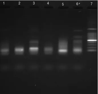
Analysis of DNA fragmentation by agarose gel elec- trophoresis showed changes in the characteristic of apoptosis with a distinctive cleavage of hepatocyte nuclear DNA after 24 hours of exposition to AMA. But in AMA+GTE and AMA+SIL groups showed the reduc-
tion of cleavage. In control, GTE and SIL groups no cleavage of hepatocyte nuclear DNA typical for apop- tosis was revealed (Fig. 2).
Similarly, values of apoptosis marker (caspase-3), in AMA+GTE and AMA+SIL groups were higher com- pared to the control, but significantly lower compared to the AMA group (Fig. 3).
In vivo study, aminotransferase values increased sig- nificantly in the AMA group compared to the CTL group (p<0.01, respectively). However, the AST and ALT val- ues in the AMA+SIL group were lower than those of the AMA group. AMA+GTE group showed a marked decrease in the serum AST and ALT levels as compared with the AMA and AMA+SIL groups. However, they were still higher than those of the CTL group. The biochemical analysis results of the liver function tests of all groups were given in Table 1.
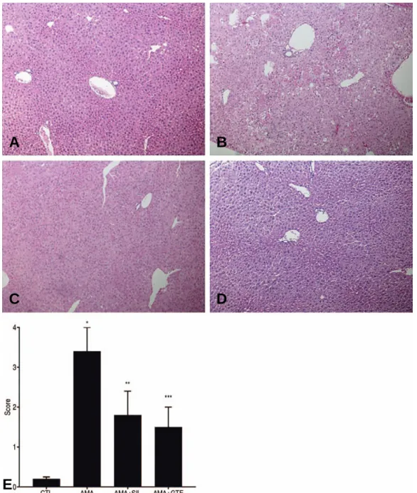
In the AMA group, light microscopy revealed liver cell injury, periportal mononuclear cell (MNC) infil- tration, focal necrosis, and also diffuse liver cell necro- sis and sinusoidal dilatation in specimens from some mice (Fig. 4B). Less liver cell injury, periportal MNC infiltration, focal necrosis and sinusoidal dilatation were observed in the AMA+SIL and AMA+GTE group
Fig. 1.MTT activity in control (CTL) and experimental groups (AMA; GTE, SIL; AMA+GTE; and AMA+SIL). The number of viable hepatocytes (one representative hepato- cyte preparation) is proportional to the MTT reaction product, as determined by the optical density. Each value represents the mean±SEM.
Fig. 1.* p<0.001 AMA vs. CTL; GTE; SIL; AMA+GTE;
AMA+SIL,
Fig. 1.** p<0.05 AMA+GTE vs. CTL; GTE; SIL, Fig. 1.*** p<0.05 AMA+SIL vs. CTL; GTE; and SIL.
Fig. 1.CTL: control; AMA:α-amanitin treated; GTE: green tea extract treated; SIL: silibinin treated; AMA+GTE:α-amanitin and green tea extract treated; AMA+SIL:α-amanitin and silibinin treated
Fig. 2. Analysis of fragmented DNA by electrophoresis on 1.5%
agarose gel after 24 hour exposure to AMA in groups:
Control (lane 1); AMA (lane 3); GTE (lane 2); SIL (lane 4), AMA+GTE (lane 5) and AMA+SIL (lane 6). Marker 500 kbp (lanes 7).
(Fig. 4C, D). As shown in Fig. 4E, significantly lower hepatic injury scores compared to those in the AMA group (3.4±0.6) were observed in the CTL group (0.2±0.05), the AMA+SIL group (1.8±0.6), and in the AMA+GTE group (1.5±0.5) (p<0.01 respectively).
DISCUSSION
This study investigated the efficacy of green tea extract in an experimental in vitro and in vivo model of hepatotoxicity induced in mice with AMA and compared it with that of silibinin, used as an antidote in the treatment of poisoning. In vitro part of present experiment, both GTE and SIL efficiently protected murine hepatocytes against AMA-induced viability loss and cell death. While hepatocyte viabilities in AMA+GTE and AMA+SIL groups were lower com- pared to the control, it was significantly higher than in AMA group. Similar results were obtained, assess- ing values of apoptosis marker (caspase-3). Thus, probably protective effect of GTE and SIL against AMA-
induced apoptosis and viability loss resulted from an antidote-associated inhibition of AMA uptake by murine hepatocytes.
In vivo part of this study, we primarily determined the hepatotoxic dose of AMA and GTE and SIL solvent on the liver. We employed an increase in aminotrans- ferase levels as a supporting biochemical parameter in addition to histopathological analysis in confirm- ing AMA-induced hepatotoxicity. AMA+GTE group resulted in a marked decrease in the serum AST and ALT levels as compared with the AMA (p<0.001 for AST and ALT) and AMA+SIL groups (p<0.002 for AST and ALT). And histopathologic finding of AMA+anti- dotes groups the Hepatic Histological Damage Score (HHDS) was significantly lower compared to the AMA group, but remained significantly higher com- pared to the CTL group.
AMA has been shown to cause toxicity in hepato- cytes cell culture via necrosis and apoptosis. AMA directly interacts with the enzyme RNA polymerase II in eukaryotic cells and inhibits the transcription, caus- ing a progressive decrease in mRNA, deficient pro- tein synthesis, and cell death. For this reason, meta- bolically active tissues dependent on high rates of protein synthesis, such as the cells of the gastroin- testinal tract, hepatocytes, and the proximal tubules of kidney, are disproportionately affected. But, recent in vitro and in vivo studies have suggested oxidative stress might contribute to powerful amatoxin hepa- totoxicity in studies
17-19). And among other potential toxic mechanisms, it has been proposed that AMA acts in synergy with endogenous cytokines (e.g., tumor
Fig. 3. Caspase-3 activity in cell lysates in control (CTL) andexperimental groups (AMA, GTE, SIL, AMA+GTE, and AMA+SIL). The caspase-3 activity is proportional to the concentration of chromophore p-nitroanilide, as determined by the optical density. Each value represents the mean±
SEM.
Fig. 3. * p< 0.001 AMA vs. CTL; GTE; SIL, Fig. 3. # p< 0.05 AMA vs. AMA+GTE; AMA+SIL, Fig. 3. ** p< 0.05 AMA+GTE vs. CTL; GTE; SIL, Fig. 3. *** p< 0.05 AMA+SIL vs. CTL; GTE; and SIL.
Table 1. Comparison of biochemical liver function test results.
Biochemical Parameters AST ALT
CTL group 61.3±4.200. 26.7±2.700.
AMA group 3723±40*00. 4378±321*0.
AMA+SIL group 1659±129*,# 2199±215*,#
AMA+GTE group 1068±60*,** 1079±19*,**
AST, ALT: IU/L. Values are expressed as means±SEM. n=10 all groups. Statistical analysis was done by One way ANOVA followed by Bonferroni test.
* p<0.01 difference from CTL;
# p<0.01 difference from the AMA group;
** p<0.01 difference from AMA and AMA+SIL groups.
necrosis factor) and that this might cause cell damage through the induction of apoptosis
20). Apoptosis is a form of programmed cell death that occurs in multicel- lular organisms. Biochemical events lead to character- istic cell changes and death. These changes include
zeiosis, cell shrinkage, nuclear fragmentation, chro- matin condensation, chromosomal DNA fragmenta- tion, and global mRNA decay. Because apoptosis can- not stop once it has begun, it is a highly regulated process. Apoptosis can be initiated through one of
Fig. 4. Represents the micrographs related to histopathologic evaluation (×100 enlargement after hematoxylin-eosin staining) of the liver tissues. Section (A) shows normal liver architecture. Section (B) shows severe hepatocellular necrosis after AMA administration, which is mainly centrilobular in nature. Sections (C) and (D) show a marked decrease in the severity of hepatocellular necrosis.(E): The degree of liver necrosis was further characterized by using Hepatic Histological Damage Score (HHDS) analysis. Each value represents the mean±SEM.
Fig. 4. * p<0.001 AMA vs. CTL; AMA+SIL; AMA+GTE, Fig. 4. ** p<0.001 AMA+SIL vs. CTL; AMA+ GTE, Fig. 4. *** p<0.05 AMA+GTE vs. AMA+SIL.
A B
C
E
D
two pathways. In the intrinsic pathway the cell kills itself because it senses cell stress, while in the extrin- sic pathway the cell kills itself because of signals from other cells. Weak external signals may also activate the intrinsic pathway of apoptosis. Both pathways induce cell death by activating caspases, which are proteases, or enzymes that degrade proteins. The two pathways both activate initiator caspases, which then activate exe- cutioner caspases, which then kill the cell by degrading proteins indiscriminately. Caspases play the central role in the transduction of apoptotic signals. Caspases are enzymes regulating numerous intracellular process- es and play a crucial role in programmed cell death.
Caspase-3 activity is established as an apoptotic mark-
er
21,22). AMA is a toxin that inhibits global transcription
by different mechanisms and induces programmed cell death.
Green tea made from Camellia sinensis (Theaceae family) is a widely consumed beverage which provides a dietary source of biologically active compounds con- sidered to be beneficial to human health. Green tea extract contains polyphenols, tannin and caffeine, and catechins have been most widely studied to prove the pharmacological action of green tea. They have demon- strated significant antioxidant, anticarcinogenic, anti- inflammatory, thermogenic, probiotic, and antimicro- bial properties
23-25).
Although the researches concerning GTE are still on the road accompanied with quite a few controversies, GTE is more likely to be beneficial to health. The results of the present analysis, combined with the previously published report from Dekant et al.
26)suggest that the composition of green tea preparations that most close- ly reflects that of a traditional infusion is safe
27). But, preparations based on concentrated extracts, contain- ing high levels of individual constituents, such as EGCG, and consumed in solid dosage form, may require health-based guidance values to assure their safe use. Because the catechins of green tea are syn- ergistic with each other, it is more effective than tak- ing EGCG alone.
There are various limitations to our experimentation, particularly their applicability to clinical practice. We could not measure the effects of each component of
GTE catechins separately. We were only able to sup- port the thesis that GTE prevents AMA-related hepa- totoxicity through its apoptosis controlling effect by means of biochemical and histopathological data, and we did not measure various anti-inflammatory and apoptosis parameters. In addition, the pharmacody- namics of orally ingested α-amanitin may differ from injected intraperitoneally. Also, intravenous adminis- tration of silibinin is ideal, but it was administered intraperitoneally in this experiment.
CONCLUSION
Summing up, AMA-induced apoptosis in murine hepa- tocyte cultures is caspase-3-dependent. Murine hepa- tocyte cultures exposed simultaneously to AMA and tested antidotes (GTE or SIL) showed significantly higher cell viability and significantly lower values of apoptosis markers compared to the cultures exposed to AMA, only. And in murine model, green tea extract was effective in limiting hepatic injury after α-aman- itin poisoning by decreases of aminotransferases and pathologic degrees of hepatonecrosis.
ORCID
Su Hwan An (https://orcid.org/0000-0001-5968-6148) Yongjin Park (https://orcid.org/0000-0001-8033-1704)
ACKNOWLEDGEMENTS
This study was supported by research fund from Chosun University, 2018.
REFERENCES
01. Vetter J. Toxins of Amanita phalloides. Toxicon 1998;36:
13-24.
02. Mas A. Mushrooms, amatoxins and the liver. Journal of hepatology 2005;42:166-9.
03. Bushnell DA, Cramer P, Kornberg RD. Structural basis of transcription:α-amanitin-RNA polymerase II cocrystal at 2.8 Å resolution. Proceedings of the National Academy of Sciences 2002;99:1218-22.
04. Nguyen VT, Giannoni F, Dubois M-F, et al. In vivo degra-
dation of RNA polymerase II largest subunit triggered by α-amanitin. Nucleic acids research 1996;24:2924-9.
05. Kim BH, Sun KH, Kim SP, et al. In vitro Protective Effects of Glehnia Littoralis on Alpha-amanitin Induced Hepatotoxicity.
J Korean Soc Clin Toxicol 2017;15(2):107-15.
06. Andera L, Wasylyk B. Transcription abnormalities poten- tiate apoptosis of normal human fibroblasts. Molecular Medicine 1997;3:852-63.
07. Arima Y, Nitta M, Kuninaka S, et al. Transcriptional block- ade induces p53-dependent apoptosis associated with translocation of p53 to mitochondria. Journal of Biological Chemistry 2005;280:19166-76.
08. Gartel AL. Transcriptional inhibitors, p53 and apoptosis.
Biochimica et Biophysica Acta (BBA)-Reviews on Cancer 2008;1786:83-6.
09. Magdalan J, Ostrowska A, Podhorska-Oko
ł
ów M, et al.Early morphological and functional alterations in canine hepatocytes due toα-amanitin, a major toxin of Amanita phalloides. Archives of toxicology 2009;83:55.
10. Mohamadin A, El-Beshbishy H, El-Mahdy M. Green tea extract attenuates cyclosporine A-induced oxidative stress in rats. Pharmacological research 2005;51:51-7.
11. Maity S, Vedasiromoni JR, Ganguly DK. Role of glutathione in the antiulcer effect of hot water extract of black tea (Camellia sinensis). Jpn J Pharmacol 1998;78:285-92.
12. Maiani G, Serafini M, Salucci M, et al. Application of a new high-performance liquid chromatographic method for measuring selected polyphenols in human plasma. Journal of Chromatography B: Biomedical Sciences and Applications 1997;692:311-7.
13. Li W-C, Ralphs KL, Tosh D. Isolation and culture of adult mouse hepatocytes. In: Mouse Cell Culture: Springer; 2010.
p.185-96.
14. Ryu CY, Sun KH, Hong R, et al. The Effect of Glehnia Littoralis on Alpha-amanitin Induced Hepatotoxicity in a Murine Model. J Korean Soc Clin Toxicol 2018;16:108.
15. Surai P. Silymarin as a natural antioxidant: an overview of the current evidence and perspectives. Antioxidants 2015;
4:204-47.
16. Tong TC, Hernandez M, Richardson III WH, et al.
Comparative treatment ofα-amanitin poisoning with N- acetylcysteine, benzylpenicillin, cimetidine, thioctic acid,
and silybin in a murine model. Annals of emergency medi- cine 2007;50:282-8.
17. Marciniak B,
Ł
opaczy´nska D, Kowalczyk E, et al.Evaluation of micronuclei in mice bone marrow and antiox- idant systems in erythrocytes exposed toα-amanitin.
Toxicon 2013;63:147-53.
18. Nikolova G, Karamalakova Y, Hadjibojeva P, et al. Severe mushroom toxin alpha amanitin causes generation of reac- tive oxygen species in liver tissues of mice-a comparative study by two different instrumental methods. Trakia Journal of Sciences 2010;8:149-54.
19. Wu X, Zeng J, Hu J, et al. Hepatoprotective effects of aqueous extract from lingzhi or reishi medicinal mush- room Ganoderma lucidum (higher basidiomycetes) onα- Amanitin-induced liver injury in mice. Int J Med Mushrooms 2013;15(4):383-91.
20. Leist M, Gantner F, Naumann H, et al. Tumor necrosis fac- tor-induced apoptosis during the poisoning of mice with hepatotoxins. Gastroenterology 1997;112:923-34.
21. Cohen GM. Caspases: the executioners of apoptosis.
Biochemical Journal. 1997;326:1-16.
22. Wolf BB, Green DR. Suicidal tendencies: apoptotic cell death by caspase family proteinases. Journal of Biological Chemistry 1999;274:20049-52.
23. Balentine DA, Wiseman SA, Bouwens LC. The chemistry of tea flavonoids. Critical Reviews in Food Science & Nutrition 1997;37:693-704.
24. Roedig-Penman A, Gordon MH. Antioxidant properties of catechins and green tea extracts in model food emulsions.
Journal of Agricultural and Food Chemistry 1997;45:
4267-70.
25. Chu C, Deng J, Man Y, et al. Green tea extracts epigallo- catechin-3-gallate for different treatments. BioMed research international 2017;2017:5615647.
26. Dekant W, Fujii K, Shibata E, et al. Safety assessment of green tea based beverages and dried green tea extracts as nutritional supplements. Toxicol Lett 2017;277:104-8.
27. Hu J, Webster D, Cao J, et al. The safety of green tea and green tea extract consumption in adults-Results of a sys- tematic review. Regulatory toxicology and pharmacology 2018;95:412-33.