Edwardsiella tarda의 특이 Bacteriophage와 Bacillus subtilis KM-1혼합액이 Edwardsiella tarda 에 미치는 항균효과
백민석⋅황요셉⋅최상훈†6) 군산대학교 해양과학대학 수산생명의학과
Mixture of Edwardsiella tarda specific Bacteriophage and Bacillus subtilis KM-1enhanced bactericidal activity against Edwardsiella tarda
Min Suk Baek, Yo Sep Hwang, and Sanghoon Choi*†
Department of Aquatic Life Medicine, Kunsan National University, Gunsan 573-400, Korea
The present study was performed to investigate an antibacterial activity of specific bacteriophage (phage) and Bacillus subtilis KM-1 (B. subtilis) mixture against Edwardsiella tarda (E. tarda). An appropriate number of phage showing the most effective antibacterial activity was 2×10⁵ PFU/㎖ with 1×10⁷ CFU/㎖ of B. subtilis 36 h post incubation. On the other hand, B. subtilis showed a dose dependant manner in inducing antibacterial activity in the presence of phage (2×10⁵ PFU/㎖). The phage and B. subtilis mixture showed higher antibacterial activity against E. tarda than phage or B. subtilis only. These results suggest that the phage and B. subtilis mixture could be utilized as an alternative to antibiotics in the control of fish diseases caused by E. tarda.
Key words :Phage, B. subtilis, E. tarda, antibacterial activity
에드와드증의 원인균인 Edwardsiella tarde (E.
tarda)는 수중 및 정상어의 장내에 상재해 있지만 어 떤 stress 조건하에서는 기회성 감염균으로서 각종 양식 어류에 감염되어 경제적으로 많은 손실을 초래 하고 있다(Kanai et al., 1988; Bang et al., 1992). 특히 넙치 양식에서 가장 많은 피해를 주는 병원성균으로 서 에드와드증의 발병 증상으로는 복부팽창, 탈장 등의 증상이 있으며 주로 고수온기에 많이 나타나는 질병이다(Kusuda and Kawai, 1998). 현재 본 질병은 화학적 치료와 관련된 오염 및 다양한 항생제 내성균
†Corresponding author: Sanghoon Choi
Tel: 063-469-1886, Fax: 063-63-9493 E-mail: [email protected]
의 출현 때문에 어류양식에 있어서 예방 및 치료가 더욱 어려워지고 있다(Chinabut and Puttinaowarat, 2005). 따라서 기존의 질병예방 및 치료의 문제점에 대한 대책으로 친환경적인 bacteriophage (phage)나 Bacillus subtilis (B. subtilis)와 같은 probiotics에 대한 관심과 연구가 점차 증가되고 있는 실정이다(Salminen et al., 1999; Park et al., 2000).
Phage는 숙주세포인 세균에 감염되어 숙주세포의 생체 기능을 이용하여 증식하는 바이러스로서 자연 계에 널리 분포하고 있다(Carlton, 1999). Phage는 1915년 Twort와 1917년 d’Herelle에 의해서 최초로 발견되었으며(Sulakvelidze et al., 2001) 1980년도에 는 phage의 임상실험이 성공하였다(Smith and
Huggins, 1980; Smith and Huggins, 1982). 이후 동물의 세균성 질병에 phage를 이용한 치료가 가능하다는 연구가 많이 보고되었으며(Soothill, 1992; Soothill, 1994; Merril et al., 1996; Barrow et al., 1998) 또한 어류의 세균성 질병에 대한 phage치료연구도 상당수 보고되었다(Rodgers et al., 1981; Stevenson and Airdrie, 1984; Park et al., 2000).
Probiotics는 숙주의 건강에 유익한 효과를 나타내 는 살아있는 미생물로서 다양한 종류의 정상 장내 세균총에서 병원성균에 대해 증식을 억제하는 항균 력이 있는 것으로 알려져 있으므로 정상 장내 세균총 의 균형을 유지하고 병원성 세균의 증식을 억제하는 데 중요한 역할을 담당한다(Shahani and Ayebo, 1980;
Fuller, 1989). 이러한 probiotics의 유용한 효과는 주로 사람과 가축에 대한 실험보고 내용이 대부분을 차지 하고 있다(Tournut, 1989). 그러나 probiotics는 어류와 다른 수생생물에서도 선천적 면역반응 향상과 병원 성 미생물을 억제시키는 것으로 연구보고 되고 있다 (Burr et al., 2005; Wang et al., 2008; Nayak, 2010).
수산양식에서 사용되는 probiotics는 Lactobacillus sp., Bacillus sp., Saccharomyces sp. 및 Enterococcus sp 등 이 있다(Kumar et al., 2008). Probiotics의 일종인 B.
subtilis가 첨가된 사료공급 시 어류에서 다양한 유익 한 효과들이 보고 되어있다(Nayak et al., 2007; Kumar et al., 2008; Aly et al., 2008).
이에 본 연구에서는 항생제 대체제를 개발하기 위한 기초적 자료를 수집하고자 E. tarda의 특이 phage와 B. subtilis의 혼합액이 in vitro 상의 E. tarda에 어떠한 항균효과를 나타내는 지 조사하였다.
재료 및 방법
Bacillus subtilis KM-1분리 및 동정
숙성된 김치를 Luria Bertani (LB) broth에 넣고 3 7℃에서 24시간 배양한 후 멸균된 생리식염수
(Phosphate Buffered Saline, pH 9.2, PBS)로 1:10 단계 희석하고 LB agar에 도말하여 B. subtilis와 유사한 형태 집락 5가지를 분리하였다. 분리된 균들은 E.
tarda에 대한 항균효과를 측정한 후 가장 효과가 좋은 균주를 16S rRNA gene을 이용하여 B. subtilis임을 증명하였다. DNA sequencing에 사용된 B. subtilis의 16S rRNA gene에 대한 universal primer는 Table 1에 나타내었다.
Primer
Name Object Primer Sequence 5′
to 3′
16S-27F 16S rRNA sequence amplification
AGAGTTTGATC MTGGCTCAG 16S-1492
R
16S rRNA sequence amplification
TACGGYTACCT TGTTACGAC Table 1. Universal primers for B. subtilis KM-1 16S rRNA
병원성 균주
본 연구에서 사용한 E. tarda (KCTC 12267)는 Korean Collection for Type Culture (KCTC)에서 분리 보존하고 있던 균주를 분양받았다. 분양 받은 균주는 brain heart infusion (BHI) broth 와 Salmonella shigella (SS) agar를 이용하여 2차 계대배양 한 후 사용하였다.
Phage
서해안 및 남해안에 위치한 100여 군데의 양식장 에서 확보한 물 sample 20 ㎖를 기준으로 이 등(2001) 이 분리한 방법을 이용하였다. E. tarda 2 ㎕와 Brain Heart Infusion (BHI) 2 ㎖를 첨가하여 32 ℃에서 배양 하였다. 약 12시간 후 박테리아가 증식되지 않는 것이 관찰되면 더 많은 양의 phage를 증폭시키기 위해 박 테리아 5 ㎕를 더 첨가하여 32 ℃에 배양하였다. 이후 E. tarda가 더 이상 자라지 않는 것을 확인한 후 phage 가 있는 것으로 추정되는 배양액을 800 x g로 원심 분리하였으며 상청액을 0.2 ㎛ 실린지 필터로 여과하
였다. 여과된 phage sample을 E. tarda 500 ㎕가 도말되 어 있는 BHI agar에 접종하여 나타나는 monolayer spot plaque을 관찰하였다. Phage를 순수 정제하기 위 해 멸균된 Top agar를 E. tarda와 phage가 손상을 받지 않도록 45∼50 ℃의 적정온도를 유지한 상태에서 10⁷ CFU/㎖의 E. tarda 500 ㎕을 첨가하고 100 ㎕의 phage 를 첨가하여 중층배지를 만들었다. Plaque assay를 통해 10¹¹ PFU/㎖ 정도까지 증폭된 phage를 확인한 후 0.2 ㎛ 실린지 필터로 여과하였다. 여과된 phage는 50 % 글리세롤을 1:1로 혼합한 후 사용하기 전까지 4 ℃에 보관 하였다.
In vitro 항균효과 실험
E. tarda에 대한 B. subtilis와 phage의 in vitro상 항균 효과를 조사하기 위해 3가지 방법으로 실험을 수행 하였다. 1차 실험은 E. tarda (2×10⁸ CFU/㎖)와 B.
subtilis (1×10⁷ CFU/㎖)의 농도는 변함없이 phage농 도만을 2×10¹, 10³, 10⁵10⁷ 및 10⁹ PFU/㎖ 씩 변화를 주었다. 이 때 BHI broth 5 ㎖에 E. tarda, B. subtilis 및 각기 다른 농도의 phage를 각각 100 ㎕를 분주하였 으며 대조군으로는 phage를 첨가하지 것으로 하였다.
2차 실험은 E. tarda (2×10⁸ CFU/㎖)와 phage (2×10³ PFU/㎖)의 농도는 변함없이 B. subtilis농도만을 2×10³, 10⁵, 10⁷ CFU/㎖ 씩 변화를 주어 1차 실험과 같은 방법으로 측정하였으며 대조군으로는 B.
subtilis를 적용하지 않은 것으로 하였다. 3차 실험은 4개의 sample로 나누어 실험을 수행하였다. 첫 번째 sample은 BHI broth 5 ㎖에 E. tarda (2×10⁸ CFU/㎖) 100 ㎕, B. subtilis (1×10⁷ CFU/㎖) 100 ㎕, phage (2×10⁵ PFU/㎖) 100 ㎕를 분주하였다. 두 번째 sample은 BHI broth 5 ㎖에 E. tarda (2×10⁸ CFU/㎖) 100 ㎕, B. subtilis (1×10⁷ CFU/㎖) 100 ㎕를 각각 분주 하였고 세 번째 sample은 BHI broth 5 ㎖에 E. tarda (2×10⁸ CFU/㎖) 100 ㎕, phage (2×10⁵ PFU/㎖) 100 ㎕를 분주하였다.
마지막으로 네 번째 대조군 sample은 BHI broth 5
㎖에 E. tarda (2×10⁸ CFU/㎖) 100 ㎕만 분주한 후 모든 sample을 25℃에서 배양하였다. 배양 0, 6, 12, 18, 24, 36시간 및 48시간째에 각각의 sample에서 SS agar에 평판도말법을 이용하여 E. tarda의 생균수를 측정하였다.
결과 및 고찰
본 연구에서는 수산양식산업에 있어서 다양한 항 생제 내성균의 출현으로 기존의 항생제 대체제로서 의 이용가능성이 높아지고 있는 친환경적인 박테리 아 특이 phage와 생균제의 일종인 B. subtilis 혼합제재 를 이용하여 에드와드증의 원인균인 E. tarda에 대한 항균효과를 조사하였다. 분리된 phage는 여러 종이 었으나 그 중 가장 활성이 강한 phage를 선별하였으 며 담수보다는 해수에서 안정성이 높게 나타났으며 수온 약 50oC까지 그 활성이 유지됨을 관찰하였다(결 과 미 제시). 김치에서 분리된 균주를 16S rRNA universal primer를 이용 한 염기서열 분석 결과 Genbank에 등록된 accession number JF496383.1인 B.
subtilis의 16S ribosomal RNA gene과 100% 상동성을 나타내었다.
B. subtilis와 phage의 E. tarda에 대한 in vitro 항균효 과 1차 실험 결과는 Fig. 1에 나타내었다. 즉 phage (2×10¹, 10³, 10⁵, 10⁷, 10⁹ PFU/㎖)농도 차에 따른 B.
subtilis와 phage의 E. tarda에 대한 in vitro 항균효과 실험을 통해 2×10¹ PFU/㎖ 만큼의 phage만 존재하더 라도 B. subtilis만 첨가한 대조군보다 높은 항균효과 가 확인되었다. 그러나 phage의 수가 10⁵ PFU/㎖ 이후 부터는 농도에 상관없이 배양 36시간 째 가장 높은 항균효과가 나타났다. Fig. 2는 일정한 phage (10⁵ PFU/㎖)의 존재 하에 B. subtilis의 수에 따른 항균력의 차이를 나타낸 결과로서 생균제 첨가 양에 비례하는 양상을 보이고 있으며 배양 48시간 째 1×10⁷ CFU/㎖
의 B. subtilis에서 가장 높은 항균력을 나타냈다. E.
Incubation time (h)
0 6 12 18 24 36 48
Viable cells (log10n CFU/ml) 0 2 4 6 8 10 12
E. tarda+B. subtilis
E. tarda+B. subtilis+phage (2x101 PFU/ml) E. tarda+B. subtilis+phage (2x10³ PFU/ml) E. tarda+B. subtilis+phage (2x10⁵ PFU/ml) E. tarda+B. subtilis+phage (2x10⁷ PFU/ml) E. tarda+B. subtilis+phage (2x10⁹ PFU/ml)
Fig. 1. Antibacterial effect of B. subtilis (1×10⁷ CFU/㎖) and different numbers of phage (2×10¹, 10³, 10⁵, 10⁷, 10⁹ PFU/㎖) mixture on E. tarda (2×10⁸ CFU/㎖). The result is a representative of three different experiments.
Incubation time (h)
0 6 12 18 24 36 48
Viable cells (log10n CFU/ml)
0 2 4 6 8 10 12
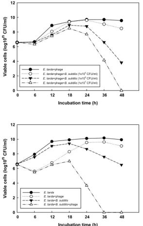
E. tarda+phage
E. tarda+phage+B. subtilis (1x10³ CFU/ml) E. tarda+phage+B. subtilis (1x10⁵ CFU/ml) E. tarda+phage+B. subtilis (1x10⁷ CFU/ml)
Fig. 2. Antibacterial effect of three different numbers of B. subtilis (1×10³, 10⁵, 10⁷ CFU/㎖) and phage (2×10³ PFU/
㎖) mixture on E. tarda (2×10⁸ CFU/㎖). The result is a representative of three different experiments.
Incubation time (h)
0 6 12 18 24 36 48
Viable cells (log10n CFU/ml) 0 2 4 6 8 10 12
E. tarda E. tarda+phage E. tarda+B. subtilis E. tarda+B. subtilis+phage
Fig. 3. Antibacterial effect of B. subtilis (1×10⁷ CFU/㎖) and phage (2×10⁵ PFU/㎖) mixture on E. tarda (2×10⁸ CFU/
㎖). The result is a representative of four different experiments.
tarda에 대한 항균력에 있어서 B. subtilis와 phage의 혼합제가 각각을 첨가한 실험군과 어떠한 차이를 나 타내는 지에 대한 결과는 Fig. 3에서 보여주고 있다.
B. subtilis만을 첨가한 군은 배양 24시간 이후부터 대조군과 phage만을 첨가한 군에 비해 E. tarda의 생 균수가 감소되는 것이 확인되었다. 이전 연구에서 다른 종류의 probiotics를 이용한 어류의 병원성 세균 에 대한 항균효과 보고(백 등, 2001)와 본 연구의 결과 는 유사하였다. 또한 phage만을 첨가한 sample에서는 배양 6시간째에 B. subtilis와 phage를 함께 첨가한 sample과 유사한 결과를 보였지만 그 이후부터는 E.
tarda의 생균수가 증가 되는 것이 확인되었다(Fig.
3). 이러한 결과는 phage에 대한 내성균주가 배양 6시 간째에 형성되어 E. tarda 생균수가 증가하기 시작하 였지만 그 이전 배양시간까지 배지 내에 B. subtilis가 존재한다면 B. subtilis가 배지 내에서 우점하기 더 좋은 환경이 되었기 때문일 것으로 추측 된다. 사실 phage나 B. subtilis 만을 첨가한 sample보다 함께 복합 투여 한 그룹에서 배양 18시간부터 E. tarda에 대한 더 좋은 항균효과가 확인되었다. 또한 지금까지 많이 보고된 B. subtilis가 만들어 내는 다양한 항균성 peptide (Zheng et al., 1999; Bizani and Brandelli, 2002) 와 phage의 상호작용에 의해 항균효과가 향상되었을 것이라고 추측된다. 그러나 E. tarda에 대한 B. subtilis 와 phage의 상호작용에 의한 항균효과에 관한 추가적 인 보충연구를 통하여 이에 대한 정확한 메커니즘을 규명할 필요성이 있다.
요 약
본 연구는 E. tarda에 대한 특이박테리오phage와 생균제의 일종인 B. subtilis KM-1의 혼합제가 E. tarda 에 대한 항균활성에 미치는 효과를 알아보고자 수행 되었다. 가장 효과적인 혼합제의 항균활성을 보인 phage의 적정 수는 B. subtilis 가 2×10⁵ CFU/㎖의 농도
로 존재할 때 배양 36시간째 2×10⁵ PFU/㎖로 나타났 다. B. subtilis의 경우 2×10⁵ PFU/㎖의 phage가 존재할 때 첨가량에 비례하는 항균활성을 보여주었다. phage 와 B. subtilis의 혼합투여는 phage나 B. subtilis 각각을 투여한 그룹보다 훨씬 높은 항균활성을 나타냈다. 본 연구의 결과는 수산양식에서 대두되고 있는 양식 어류의 E. tarda 감염성 질병 치료를 위한 항생제 대체 제로서의 사료첨가제 개발 가능성을 제시하고 있다.
감사의 글
이 논문은 2013년도 군산대학교 수산과학연구소 학술연구비 지원으로 연구되었음.
참고문헌
Ackermann, H.W. and Nguyen, T.M. 1983. Sewage coliphages studied by electron microscopy.
Appl. Environ. Microbiol. 45(3): 1049-1059.
Aly, S. M., Abdel-Galil Ahmed, Y., Abdel-Aziz Ghareeb, A. and Mohamed, M. F. 2008. Studies on Bacillus subtilis and Lactobacillus acidophilus, as potential probiotics, on the immune response and resistance of Tilapia nilotica (Oreochromis niloticus) to challenge infections. Fish Shellfish Immunol. 25: 128-136.
Bang, J. D., Chun, S. K., Park, S. I. and Choi, Y. J.
1992. Studies on the biochemical and serological characteristics of Edwardsiella tarda isolated from cultured flounder, Paralichthys olivaceus.
Fish Pathol. 5(1): 29-35.
Barrow, P., Lovell, M. and Berchieri, A. Jr. 1998. Use of lytic bacteriophage for control of experimental Escherichia coli septicemia and meningitis in
chickens and calves. Clin. Diagn. Lab. Immunol.
5: 294-298.
Bizani, D. and Brandelli, A. 2002. Characterization of a bacteriocin produced by a newly isolated Bacillus sp. strain 8A. J. Appl. Microbiol. 93:
512-519.
Bradley, D. E. 1967. Ultrastructure of bacteriophages and bacteriocins. Bacteriol. Rev. 31(4): 230-314.
Burr, G., Gatlin, D. and Ricke, S. 2005. Microbial ecology of the gastrointestinal tract of fish and the potential application of prebiotics and probiotics in finfish aquaculture. J. World Aquacul. Soc.
36: 425-436.
Carlton, R. M. 1999. Phage therapy: past history and future prospects. Immunol. Ther. Exp. 47:
267-274.
Chinabut, S. and Puttinaowarat, S. 2005. The choice of disease control strategies to secure international market access for aquaculture products. Dev.
Biol. 121: 255-261.
Fuller, R. 1989. probiotics in man and animals. J. Appl.
Bacteriol. 66: 365-378.
Kanai, K., Tawaki, S. and Uchida, Y. 1988. An ecological study of Edwardsiella tarda in flounder farm.
Fish Pathol. 22: 41-47.
Kumar, R., Mukherjee, S. C., Ranjan, R. and Nayak, S. K. 2008. Enhanced innate immune parameters in Labeo rohita (Ham.) following oral administration of Bacillus subtilis. Fish Shellfish Immunol.
24: 168-172.
Kusuda, R. and Kawai, K. 1988. Bacterial diseases of cultured marine fish in Japan. Fish Pathol. 33:
221-227.
Lee, C. H., Heo, Y. J., Baek, M. S., Lee, J. U., Kang, J. Y., Han, M. J., Kyoung, S. B. and Choi, S. H. 2011. Characterization of Edwardsiella tarda specific phage isolated from fish farms on west coast of Korea. J. Fish Pathol. 24(2):
85-93.
Merril, C. R., Biswas, B., Carlton, R., Jensen, N. C., Creed, G. J., Zullo, S. and Adhya, S. 1996.
Long-circulating bacteriophage as antibacterial agents. Proc. Natl. Acad. Sci. USA. 93:
3188-3192.
Nayak, S. K. 2010. Probiotics and immunity: a fish perspective. Fish Shellfish Immunol. 29: 2-14.
Nayak, S. K., Swain, P. and Mukherjee, S. C. 2007.
Effect of dietary supplementation of probiotic and vitamin C on the immune response of Indian major carp. Fish Shellfish Immunol. 23:
892-896.
Paek, N. S., Lim, Y. B. and Kim, Y. M. 2001. Antibacterial Activity and Growth Promotion in Aquacultured Fish by Probiotics. Kor. J. Appl, Microbiol.
Biotechnol. 29(1): 56-61.
Park, S. C., Ichiro, S., Minoru, F., Koh-Ichiro, M. and Toshihiro, N. 2000. Isolation of Bacteriophage Specific to a Fish Pathogen, Pseudomonas plecoglossicida, as a Candidate for Disease Control. Applied and Environmental Microbiology.
1416-1422.
Rodgers, C. J., Pringle, J. H., McCarthy D. H. and Austin, B. 1981. Quantitative and qualitative studies of Aeromonas salmonicida bacteriophage. J.
Gen. Microbiol. 125: 335-345.
Salminen, S., Ouwehand, A., Benno, Y. and Lee, Y.
K. 1999. Probiotics: How should they be defined Trends. Food Sci. Technol. 10: 107-110.
Shahani, K. M. and Ayebo, A. D. 1980. Role of dietary lactobacilli in gastrointestinal microccology.
Am. J. Clin. Nutr. 33: 2448-2457.
Smith, H. W. and Huggins, M. B. 1980. The association of the O18, K1 and H7 antigens and the Co1V plasmid of a strain of Escherichia coli with its virulence and immunogenecity. J. Gen.
Microbiol. 128: 387-400.
Smith, H. W. and Huggins, M. B. 1982. Successful treatment of experimental Escherichia coli infections in mice using phage: its general superiority over antibiotics. J. Gen. Microbiol.
128: 307-318.
Soothill, J. S. 1992. Treatment of experimental infections of mice with bacteriophages. J. Med. Microbiol.
37: 258-261.
Soothill, J. S. 1994. Bacteriophage prevents destruction of skin grafts by Pseudomonas aeruginosa.
Burns. 20: 209-211.
Stevenson, R. M. W. and Airdrie, D. W. 1984. Isolation of Yersinia ruckeri bacteriophages. Appl.
Environ. Microbiol. 47: 1201-1205.
Sulakvelidze, A., Alavidze, Z. and Morris, J. G., Jr.
2001. Bacteriophage therapy. Antimicrob.
Agents Chemother. 45: 649-659.
Tournut, J. 1989. Application of probiotics to animal husbundary. Rev. Sci. Tech. Off. Int. Epiz. 8:
551-566.
Wang, Y., Tian, Z., Yao, j. and Li, W. 2008. Effect of probiotics, Enterococcus faecium, on tilapia (Oreochromis niloticus) growth performance
and immune response. Aquaculture. 277:
203-207.
Zheng, G., Yan, L. Z., Vederas, J. C. and Zuber, P., 1999. Genes of the sbo-alb locus of Bacillus subtilis are required for production of the
antilisterial bacteriocin subtilosin. J. Bacteriol.
181: 7346-7355.
Manuscript Received : October 10, 2013 Revised : November 05, 2013 Accepted : November 19, 2013