Use of Cellulose and Recent Research into Butyrate
Tae Jong Yeo
1, In Soon Choi
2* and Kwang Keun Cho
1*
1
Department of Animal Resources Technology, Gyeongnam National University of Science and Technology, Gyeongnam 660-758, Korea
2
Department of Biological Science, Silla University, Busan 617-736, Korea
Received October 4, 2012 /Revised October 15, 2012 /Accepted October 19, 2012
On earth, there are about 5,400 kinds of mammals, of which about 1,000 kinds are herbivores. Among herbivores, about 250 kinds are known to be ruminants. As for cattle and sheep, which are ruminants, fermentation takes places mainly in their rumen; in contrast, for pigs and men, which are non-ru- minants, fermentation takes place mainly in their caecum, colon, and rectum. As for the kind and dominance of rumen microorganisms, Bacteroidetes account for 51% and Firmicutes for 43%. As for the dominance of the large intestine microorganisms in men, Firmicutes account for 65% and Bacteroidetes for 25%. Cell wall components are decomposed by microorganisms, and short chain fatty acids (SCFAs) are generated through fermentation; the ratio of acetate, propionate, and butyrate generate is 60:25:15. Butyrate absorbed through the primary butyrate transporter MCT1 (mono carboxylate transports-1) in the intestines activates such SCFA receptors as GPR43 and GPR41. Butyrate has a strong anti-tumorigenic function. Butyrate is characterized by the fact that it has an effect on many cancer cells, contributes to the coordination of functions in the cells, and induces cancer apoptosis.
Butyrate activates caspase but inhibits the activity of HDAC (histone deacetylase), so as to induce apoptosis. In addition, it increases p53 expression, so as to induce cell cycle arrest and apoptosis.
Anti-inflammation actions of SCFA include the reduction of IL-8 expression in intestinal epithelial cells, the inhibition of NO synthesis, and the restraint of the activity of NF-kB (nuclear factor kB), so as to suppress the occurrence of cancers caused by inflammation. Butyrate plays an important role in maintaining physiological functions of intestinal mucous membranes and is used as a cure for in- flammatory bowel disease (IBD).
Key words : Cellulose, microorganism, butyrate, apoptosis, cancer
*Corresponding author (Kwang Keun Cho)
*Tel:+82-55-751-3286; Fax:+82-55-751-3689
*E-mail: [email protected]
*Co-corresponding author (In Soon Choi)
*Tel:+82-51-999-5647, Fax:+82-51-999-5644
*E-mail: [email protected]
ISSN (Print) 1225-9918 ISSN (Online) 2287-3406 Journal of Life Science 2012 Vol. 22. No. 11. 1571~1586 DOI : http://dx.doi.org/10.5352/JLS.2012.22.11.1571
Digestive organ of ruminant and mono-gastric animals
Ruminants are referred to as so because of their rumina- tion or special digestive organ (the stomach), in which cud can be chewed. Ruminants are the most advanced animals of ungulates and the most multiplied and prevalent through- out the world. They have a rumination, which is a very pe- culiar digestive organ and possibly enables them to have survived poor vegetation conditions, allowing them to flour- ish greatly. The stomach of all ruminants is divided into four compartments including the rumen, reticulum, omasum, and abomasums (of which rumen and reticulum are in- volved in rumination and called rumen). Rumination is a process in which food, once swallowed, is emitted back unto the mouth for re-chewing; of feeds returned into reticulum
via rumen, those of which over a certain size will be re- turned again into rumen, accumulated, and regurgitated to the mouth. The regurgitated feeds will be smashed finely between molar teeth and mixed with saliva in the mouth, then returned to rumen for fermentation. Ruminants include cattle, sheep, goats, deer, musk deer, giraffes, and impalas.
Pseudo-ruminants have a similar stomach to rumen in which microorganism fermentation takes place, but their stomach is composed of three compartments since omasum and abomasums are not specialized. Pseudo-ruminants in- clude animals that belong to the family of camel (camel, la- ma, and alpaca) and the family of chevrotain (chevrotain and mouse deer). Non-ruminant herbivores are animals that are herbivores and have not developed rumen (but the cae- cum and others have), with which fiber materials are di- gested and fermented for gaining nutriments. They include horse, rabbit, elephant and hippopotamus [94].
When ruminants eat food they chew the cud, which is
a process for earning energy from cell wall substances
(cellulose) of plants. In other words, cellulose is finely crush-
ed and decomposed through microorganisms in rumen, and
- Review -
Table 1. Relative area of digestive intestines where fermentation takes place in each animal (%) [12]
Animal Rumen Caecum Colon/rectum Total
Cattle 64 5 5~8 75
Sheep 71 8 4 83
Pig - 15 54 69
Man - 32 29 61
it is metabolized to be used as an energy source. Until the present, a focus has been placed only on the fermentation taking place in rumen of ruminants in relation to the diges- tion process. However, as can be seen in Table 1, much of the fermentation takes place not only in rumen of cattle but also in the caecum, colon, and rectum of pigs and men. An investigation was made into the ratio of intestines versus all digestive intestines where fermentation takes place, and it was found that the ratio is 83% for sheep, 75% for cattle, and 69% and 61% for pig and man, respectively, which are mono-gastric animals. The fermentation takes place mainly in rumen of ruminants like cattle and sheep, while it takes place mainly in the caecum, colon, and rectum of mono-gas- tric animals such as pigs and men. More than we have con- ceived, the fermentation takes place in digestive intestines of pig and man. Since a lot of fermentation takes place in men, the concept of dietary cellulose for adjusting the fer- mentation in intestines appeared long ago to maintain the health of the digestive organs of men, and the importance of the concept is steadily increasing. Many microorganisms exist in each digestive intestine and they have an effect on the health of the intestines including the damage, develop- ment, and digesting & absorbing abilities of intestinal epi- thelial cells; therefore, it is very important to maintain the health of the intestines with the help of intestinal micro- organisms [90]. Moreover, obesity may be induced by such microorganisms that are dominant in the intestines.
Checking of the kinds of microorganisms existing in the in- testines of obese patients, Firmicutes accounts for more than 90% and Bacteroidetes no more than 3%; Bacteroidetes accounts for 30% in men whose body weight is normal. As obese men lose their weight by means of dietary adjustment, intestinal Bacteroidetes increases and Firmicutes decreases while the in- testines of the men resemble those of slender men [55].
Ruminant microorganisms
It is known that more than 200 kinds of rumen bacteria number 10
10~10
11per ml and account for more than 50%
of the total quantity of microorganisms. Rumen bacteria pri-
marily secrete such enzymes that can digest the cellulose of grass feed, and their life is 20 minutes~3 hours. Protozoa are larger than bacteria and exist in gastric juice in the num- ber of 10
6per ml, accounting for 50% of the total quantity of microorganisms and 2% of the contents in the stomach.
The rumination can be operated without protozoan but func- tion more smoothly with protozoa. Protozoa undertake about 20~40% of starch digestion and are thoroughly anaero- bic, with a lifespan of 8~36 hours. Fungi account for 8% of the total quantity of microorganisms and are important mi- croorganisms that stick to plants and decompose cellulose.
They disintegrate a small quantity of lignin into small par- ticles, so that bacteria may digest them and have high activ- ity in cellulose and hemicellulose; their germination cycle is 24 hours [94].
In order to identify the kind and dominance of rumen microorganisms, 16 Holstein cows were bred in the same conditions, then DNA was extracted from rumen micro- organisms and applied with a pyrosequencing technique in which V2 and V3 regions of 16s rRNA were amplified. As result, it was found that Bacteroidetes existed in the ratio of 51%, Firmicutes 43%, Proteobacteria 5.21%, Actinobacteria 0.87%, and Tenericutes 0.68% [44].
Microorganisms in the intestines of men
500~1,000 kinds of microorganisms live in the intestines
of adult men. From the stomach down to the large intestine,
increasingly more anaerobic microorganisms live; from the
stomach upwards, increasingly more aerobic micro-
organisms live. Microorganisms exist in the stomach in the
amount of 10² cfu/ml, in the duodenum 10
1-3cfu/ml, in the
jejunum 10
3-4cfu/ml, in the ileum 10
7-9cfu/ml, and in the
large intestine 10
10-12cfu/ml. Fiber material of food taken
into the body is fermented in the large intestine and pro-
duces SCFAs (Fig. 1) [18,21,28]. As can be seen in Fig. 1,
compared to the other digestive organs, microorganisms ex-
ist in the large intestine the most. According to a recent
study that conducted a biopsy into the fecal and the large
intestine of men, 9 kinds of microorganisms (Firmicutes,
Journal of Life Science 2012, Vol. 22. No. 11 1573
Fig. 1. Key physiologic and microbiological features of the gut [21]. Relative concentrations of bacteria and the pH at various locations within the adult gut are also noted. cfu: colony forming unit.
Bacteroidetes, Actinobacteria, Fusobacteria, Proteobacteria, Verrucomicrobia, Cyanobacteria, Spirochaetes, and Vadin BE 97) exist in terms of the phyla [3,18]; and as for the dominance of microorganisms, Firmicutes accounts for 65%, Bacteroidetes 25%, and Proteobacteria, Actinobacteria, and Fusobacteria ac- count for the remaining 10% [1,17,24,85].
Microorganisms that decompose polysaccharide
In omnivorous mammals (i.e., men), less than 10% of en- ergy is generated through the fermentation in the large intes- tine, but microorganisms in the large intestine play a very important role for the health of omnivorous animals.
Microorganisms generate butyrate, which is a SCFA and pre- vents colon cancer in mice and men [60,68]. Bacteria in the large intestine bring on various metabolic changes and many mutual actions, which involve immune actions of the host [23,38]. In rumen, fungi and protozoa play an important role in decomposing the cellulose of plants; in omnivorous mam- mals, intestinal microorganisms perform this role. The diver- sity of microorganisms can be identified against the nucleo- tide sequence of 16s rRNA. A comparison was made of the
number of phylum of microorganisms in the large intestine
and rumen of men, pig, and horse; it was found that
Firmicutes and gram-negative Bacteroidetes exist in more than
90% of the phylum [19,79]. A recent study compared in-
testinal microorganisms in obese men and normal men, and
found that the dominance ratio of 2 kinds of bacteria
(Firmicutes and Bacteroidetes) is different; also, when a com-
parison is made of mice, microorganisms (Firmicutes) domi-
nant in obese mice generate more energy from taken feed
than microorganisms (Bacteroidetes) dominant in normal
mice do [55]. Intestinal microorganisms directly decompose
non-digested cell walls of plants. Cell walls of plants consist
of cellulose, hemicellulose, pectinaceous material, and
protein. Bacteria that can decompose cellulose decompose
cellulose and bacteria that cannot do so decompose other
substances. When feed is taken in, various kinds of poly-
saccharide move into the intestines and rumen. There are
various polysaccharides including oligosaccharide, inulin
which is a storage polysaccharide, resistant starch (RS)
which is a polymer protecting cell walls, and xylan and pec-
tin which are cell wall polysaccharides. Therefore, there are
a greatly increasing number of microorganisms that can de-
compose polysaccharides. As seen in Fig. 2, it was found
Fig. 2. Polysaccharide-degrading bacteria in the ruminant and human gastrointestinal tracts [27]. The major sites of microbial break- down of dietary polysaccharides, which also support the highest densities of bacteria, are the rumen in ruminant animals and the large intestine in humans. Examples of cultured polysaccharide-degrading species are shown for these sites, together with the phylum to which they belong ( Firmicutes or Bacteroidetes ) and their characteristic polysaccharide-utilizing abilities.
Much of the diversity remains undefined, however, and new species of polysaccharide-utilizing bacteria have been described recently in the human colon. C, cellulose; I, inulin; S, starch; X, xylan.
that, when polysaccharides such as cellulose, inulin, resistant starch (RS), and xylan are administered to cattle and men, different microorganisms increase in rumen and intestines depending upon the polysaccharide. Cellulolytic bacteria can decompose such polysaccharides as xylan, mannan, pec- tin, and cellulose; they do not employ the substances which are melted through decomposition but decompose cellulose so that other microorganisms may use it. For instance, hy- drogen generated by polysaccharide decomposition bacteria is used by methanogens and acetogens to generate methane and acetate; when the hydrogen concentration is reduced, the generation of hydrogen is reduced because the amount of hydrogen-generated cellulolytic bacteria is increased.
When cellulolytic bacteria like Ruminococcus and Fibrobacter stick to the surface of plant cell walls and decompose hemi- cellulose and cellulose to generate solubilized oligo- saccharides and polysaccharide, Butyrivibrio spp. and Roseburia spp. synthesize butyrate. Also, Bacteroides spp. and Prevotella spp. use solubilized oligosaccharides and poly- saccharide to compose propionate (Fig. 3) [11]. Firmicutes, which mainly exist in rumen and feces of men, are
Ruminococci and belong to the Clostridium leptum group
(Clostridial cluster IV); there are many Firmicutes in solids and
many Bacteroidetes in liquids [78,93]. When xylan and re-
sistant starch (RS) are administered, Butyrivibrio spp. and
Roseburia spp. (which are butyrate-generating bacteria)
increase. A comparison was made of gram-positive bacte-
rium (R. flavefaciens) a polysaccharide decomposition bacte-
rium found in rumen, and gram-negative bacterium (B. the-
taiotaomicron) a starch decomposition bacterium found in the
intestines of men; it was found that R. flavefaciens belongs
to Firmicutes or Clostridial cluster IV and is a representative
cellulolytic bacterium in rumen that R. flavefaciens generates
236 glycoside hydrolases, 15 polysaccharidelyases and 20
carbohydrate esterases, and that the generated enzymes
have a complementary part to polymers which exist in cell
walls of plants, there with sticking to cell walls and decom-
posing polysaccharides of cell walls [31]. B. thetaiotaomicron
is a representative cellulolytic bacterium in intestines of men
and generates enzymes such as 236 glycoside hydrolases and
15 polysaccharidelyases, and a starch-utilization gene cluster
(sus) consisting of 8 genes. In external cell membranes of
Journal of Life Science 2012, Vol. 22. No. 11 1575
Fig. 3. A simplified schematic illustrating the relationships between primary degraders of insoluble plant fiber and other members of gut microbial commmono-gastricies [27]. Specialist cellulolytic species of Ruminococcus and Fibrobacter are shown closely attached to the substrate. Other bacteria that is able to use soluble polymers, for example, butyrate-producing Butyrivibrio spp. And Roseburia spp., succinate-producing Bacteroides spp. and Prevotella spp., hydrogen-utilizing methanogenic archaea and acetogenic bacteria, are also shown. This diagram is illustrative and not intended to provide a complete description, as the number and diversity of primary degraders, polysaccharide utilizers and other functional groups within different gut communities is still emerging.
B. thetaiotaomicron, there are maltose-inducible out- er-membrane proteins such as susC, susD, susE and susF;
susC and susD proteins catch starch. susC can hold mole- cules including maltose (G2) and maltoheptaose (G7), and is decomposed by susG amylase (neopullulanase) and enters into periplasm. Neopullulanase disintegrates amylose, amy- lopectin and pullulan into an oligosaccharide, which can en- ter into periplasm. With the help of a transporter, which is decomposed by susA amylase of periplasm and that exists in cytoplasmic membranes, the oligosaccharide enters into cytoplasmic membranes (Table 2) [27].
SCFAs
Ruminants cannot secrete enzymes that disintegrate cell wall components of plants, but instead have a mutual rela- tionship with anaerobic microorganisms that can produce such enzymes. Various kinds of microorganisms completely decompose cell wall components, which are hydrolyzed by enzymes secreted from rumen microorganisms and are con- verted into monosaccharide. Then, the monosaccharide is again fermented and changed into SCFA's [acetate (C2), pro-
pionate (C3), butyrate (C4), and valerate (C5)], which are
used as an energy source by ruminants. Within rumen, grass
feed that cannot be digested by animals are attacked by vari-
ous microorganisms and decomposed into a size to be ab-
sorbed through the stomach walls of cattle; generated micro-
organisms again enter into the small intestine, where they
are decomposed by protein decomposition enzymes and
used as a nutriment. The large intestine of non-ruminant
herbivores (horse, rabbit, etc.) is an important portion that
is digested by microorganisms. The most outstanding char-
acteristic of herbivores, which use cellulose as an energy
source, is that part of a specific intestine that is needed for
microorganisms to ferment feeds is enlarged. The digestion
of cellulose is totally dependent upon the activities of anae-
robic microorganisms in the caecum and the colon. Most of
the intestinal contents reach the large intestine within 3
hours after feed is taken; substances that are not digested
in the small intestine are decomposed and fermented while
they stay in the large intestine for a long time. Yet, since
the substances pass through the large intestine faster in ru-
minants, the ability of herbivores to digest cellulose is in-
ferior to that of ruminants [8,9,13].
Table 2. Genome sequences concerned with plant polysaccharide breakdown in four species of gut bacteria [27]
Bacteroides
Thetaiotaomicron 5482 Bifidobacterium
Longum NCC 2705 Ruminococcus
Flavefaciens FD1 Fibrobacter Succinogenes S85
Location Human colon Human colon Rumen, cellulolytic Rumen, cellulolytic
Genome size 6.26 Mb
(complete genome)
2.26 Mb (complete
genome)
Approximately 4Mb (partial genome;
dockerin-encoding genes only)
3.8 Mb (complete
genome) Number of glycoside
hydrolases* 236 (40) 47 (17) 65 (14) 104
Number of polysaccharide
lyases* 15 (7) 0 12 (4) 4
Number of carbohydrate
esterases* 20 (9) 1 23 (5) 14
Number of carbohydrate
binding modules* 16 (3) 10 (5) 61 (12) Limited information
available
References 29 49 63 51,58
*The number of enzyme families that are represented is shown in brackets (CAZY (carbohydrate-active enzymes) database; see further information). Mb: megabases.
Almost all SCFA’s generated in rumen through the fer- mentation of carbohydrates are absorbed in rumen by means of simple dissemination, and the remainder is absorbed through reticulum and omasum. About 76% of SCFA’s are absorbed in rumen and reticulum, 19% in omasum and abo- masums, and the remaining approximate 5% in the small intestine. The speed for SCFA to be absorbed through rumen walls is affected by pH; when pH is low, SCFA is not ionized (HAc) and can be quickly absorbed and easily moved to blood. When pH is normal, the speed for SCFA to be ab- sorbed is the greatest for butyrate, followed by propionate and acetate. When pH is alkaline, the absorption speed is reversed. It is known that, when pH is really high, the ab- sorption speed is the same. SCFA absorbed in such a manner enters into the liver via the 1
stgastric vein and the portal vein [12]. Microorganisms prefer carbohydrate fermentation to protein fermentation; carbohydrate fermentation takes place dominantly in proximal colon, and protein fermenta- tion takes place increasingly more as it nears the distal colon [4]. As dietary fiber is less fermented and consumed due to Western diets, if food or drink that include dietary fiber are ingested, the concentration of butyrate increases in the distal colon due to slow bacteria fermentation [91]. SCFA generated in the intestines of men accounts for 5~15% of energy [4], and the concentration of butyrate in feces of men is 11~25 mM [34, 91]. The ratio for acetate, propionate, and butyrate to be generated in the intestines is 60:25:15 [34,84].
Since more than 95% of SCFA is absorbed in the intestines, it is much too difficult to determine the concentration of SCFA [88].
SCFA exists in rumen of ruminants or the large intestine of non-ruminants in the approximate amount of 100mM.
SCFA is generated as microorganisms ferment carbohy- drates that are not digested from taken-in dietary fiber.
SCFA is absorbed into intestinal epithelial cells and has an effect on various functions of gastrointestinal tract. For ex- ample, it affects the hematocele of the large intestine, the absorption of moisture/electrolytes, the movements of the large intestine, and the transport of ions [46,82]. SCFA gen- erated via fermentation is absorbed through primary buty- rate transporter MCT1 in the intestines, and GPR43 and GPR41 (SCFA receptors) are activated by absorbed SCFA.
GPR43 can be found in the intestines of man and mouse,
and the expression frequency of GPR43 is the highest in in-
testinal epithelial cells, which are intestinal mucous mem-
brane tissues [46,47]. Mechanisms for SCFA to inhibit colon
cancer are divided into two. When SCFA is at a low concen-
tration, it is combined with GPR43, which is in plasma mem-
brane and instigates multiple cellular signaling events, caus-
ing apoptosis. When SCFA is at a high concentration, it
brings about direct cell membrane absorption, causing apop-
tosis (Fig. 4). It can be confirmed that the expression fre-
quency of GPR43 is reduced in cancer cell tissues; it is also
confirmed that the expression frequency of GPR43 in malig-
Journal of Life Science 2012, Vol. 22. No. 11 1577
Fig. 4. Schematic representation of the proposed role of GPR43 as a functional tumor suppressor in colon cancer [80]. The bacterial fermentation product of dietary fiber propionate inhibits colon cancer cell proliferation and induces apoptosis by two ap- proaches: one through direct cell membrane absorption and the other through GPR43 activation of multiple cellular signaling events, leading to growth inhibition and apoptosis by influencing several key cell cycle regulators and caspases involved cell apoptosis activation.
nant colon adenocarcinoma tissue, colon hyperplasia and be- nign colon tumor tissue is reduced to 65% and 80%, com- pared to common tissues [81]. This result implies that the expression of GPR43 is reduced when colon cancer develops and progresses. Butyrate expresses GPR43, then inhibits the proliferation of colon cancer cell, and brings about apoptosis.
GPR41 mainly combines with G
i/oprotein and GPR43 com- bines with Gq protein. GPR41 combines more with SCFA receptors in the order of propionate, butyrate and acetate;
it is similar with GPR43. GPR43 combines more with acetate while GPR41 combines more with propionate [81,82].
Anti-tumorigenic properties of butyrate
SCFA plays an important role in maintaining physio- logical functions of intestinal mucous membranes [82].
Although acetate takes the most part in SCFA, butyrate has a strong anti-tumorigenic function [14,84]. When butyrate was in vivo and in vitro treated in the same concentration, it inhibited the multiplication and specialization of colon cancer cells [64]; when the butyrate concentration was raised and applied to tumor-induced experimental animals, it was confirmed that apoptosis took place partially [14]. Butyrate is characterized by the fact that it has an effect on many cancer cells, contributes to the coordination of functions in cells, and induces cancer apoptosis (Table 3). By means of
several proteomic technologies and transcriptomic tech- nologies, it is known that butyrate is related to the death of cancers, that even though there is a sufficient amount of butyrate in some cancer cells, apoptosis is avoided, and that when butyrate resistant cells are treated with butyrate, their growth improves or their aggression is increased [15,30,32,42,57,66,75,79,80]. Yet, through signal transduction mechanism and HDAC inhibition mechanism, fundamental action modes of butyrate more strongly induce the death of cancer cells than the other SCFA’s. When treated with butyrate, with the help of the Bcl-2 family that comprises pro-apoptotic protein and anti-apoptotic protein, cyto- chrome c in mitochondria comes out to cytoplasm and acti- vates caspase to induce apoptosis. It also coordinates the TNF receptor super-family and inhibits the activities of HDAC, which is involved in gene expression, so as to induce apoptosis [2,10,51,66,69]. Relying or not relying on p53, buty- rate induces cell cycle arrest and apoptosis. Depending on its concentration, butyrate directly acts on p53 or increases the expression of p53 and induces the expression of target genes (p21, p27, and cyclin dependent kinase) of p53 so as to bring about cell cycle arrest and apoptosis [33,45,61].
Butyrate inhibits the activities of HDAC to increase histone
acetylation [89], and it reduces higher order chromatin fold-
ing to increase p21 transcription. Also, butyrate reduces the
expression of miR-106b and many other miRNAs; since the
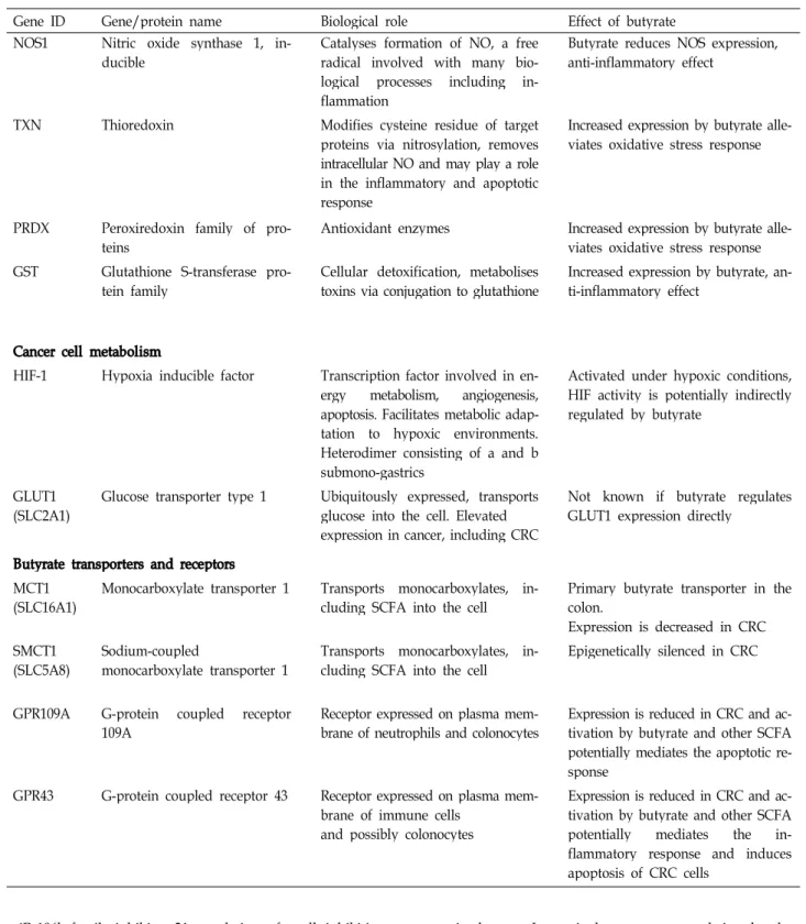
Table 3. Summary of the genes and proteins involved in the anti-tumorigenic effects of butyrate [31]
Gene ID Gene/protein name Biological role Effect of butyrate
Apoptosis, cell cycle, stress response
HDAC Histone deacetylase Family of proteins that regulate gene
transcription Butyrate inhibits HDAC activity
CDKN1A Cyclin-dependent kinase in-
hibitor 1A, p21 Cell cycle regulator Expression is induced by butyrate to cause cell cycle arrest
CDKN1B Cyclin-dependent kinase in-
hibitor 1B, p27 Cell cycle regulator Expression is induced by butyrate to cause cell cycle arrest
CDK Cyclin-dependent kinases Cell cycle regulator Expression is induced by butyrate to cause cell cycle arrest
CCND1 Cyclin D1 Cell cycle regulator Butyrate reduces expression
GADD Growth arrest and DNA-in-
ducible gene family Cell stress response Expression is induced by butyrate MAPK Mitogen-activated protein kin-
ase family,includes p38, JNK, ERK
Signalling cascade that mediates the
cell stress response Expression is induced by butyrate
Bcl-2 B-cell CLL/lymphoma 2 protein
family Regulates apoptosis and cell
survival.
Consists of Bcl2 (anti-apoptosis), Bax, Bak and Bad (pro-apoptotic)
Involved in butyrate-induced apopto- sis
HSP27 Heat shock protein 27 Cell stress response, apoptosis Expression is induced by butyrate
TNFR Tumour necrosis factor receptor
family Activation by TNF mediates the
apoptotic response via the extrinsic pathway
Butyrate sensitises the cell to the apoptotic response occurring via TNFR activation
CYCS Cytochrome c Apoptosis Butyrate induces cytochrome c re-
lease into the cytoplasm during apop- tosis
TP53 Tumour protein p53 Tumour suppressor and cell cycle
regulator Activation by butyrate is one known
mechanism involved in butyrate-in- duced apoptosis
MYC c-myc Transcription factor involved in cell
cycle progression and apoptosis Butyrate reduces expression Inflammation and the immune response
NF-kb Nuclear factor kappa beta fam-
ily Transcription factor involved in
many cellular processes including inflammation, immune response and apoptosis
Activity of NF-kB is influenced by bu- tyrate to mediate expression of cyto- kines and chemokines involved in in- flammation, e.g. interleukins, MCP1, GM-CSF, VEGF
COX2 (PGH2) Cyclo-oxygenase-2 (prostaglandin
G/H synthase 2) Involved in inflammation by media-
ting prostaglandin synthesis Reduced expression by butyrate in vi- tro, anti-inflammatory effect
TNF Tumour necrosis factor family
of proteins Family of pro-inflammatory cyto-
kines, binding to TNFR activates extrinsic apoptotic pathway
Butyrate sensitises cells to TNFR acti- vation to promote apoptosis
IL8 Interleukin 8 Inflammatory response Butyrate reduces IL8 expression,
anti-inflammatory effect
Journal of Life Science 2012, Vol. 22. No. 11 1579
Table 3. Continued
Gene ID Gene/protein name Biological role Effect of butyrate
NOS1 Nitric oxide synthase 1, in-
ducible Catalyses formation of NO, a free
radical involved with many bio- logical processes including in- flammation
Butyrate reduces NOS expression, anti-inflammatory effect
TXN Thioredoxin Modifies cysteine residue of target
proteins via nitrosylation, removes intracellular NO and may play a role in the inflammatory and apoptotic response
Increased expression by butyrate alle- viates oxidative stress response
PRDX Peroxiredoxin family of pro-
teins Antioxidant enzymes Increased expression by butyrate alle-
viates oxidative stress response GST Glutathione S-transferase pro-
tein family Cellular detoxification, metabolises
toxins via conjugation to glutathione Increased expression by butyrate, an- ti-inflammatory effect
Cancer cell metabolism
HIF-1 Hypoxia inducible factor Transcription factor involved in en- ergy metabolism, angiogenesis, apoptosis. Facilitates metabolic adap- tation to hypoxic environments.
Heterodimer consisting of a and b submono-gastrics
Activated under hypoxic conditions, HIF activity is potentially indirectly regulated by butyrate
GLUT1
(SLC2A1) Glucose transporter type 1 Ubiquitously expressed, transports glucose into the cell. Elevated expression in cancer, including CRC
Not known if butyrate regulates GLUT1 expression directly
Butyrate transporters and receptors MCT1
(SLC16A1) Monocarboxylate transporter 1 Transports monocarboxylates, in-
cluding SCFA into the cell Primary butyrate transporter in the colon.
Expression is decreased in CRC SMCT1
(SLC5A8) Sodium-coupled
monocarboxylate transporter 1 Transports monocarboxylates, in-
cluding SCFA into the cell Epigenetically silenced in CRC
GPR109A G-protein coupled receptor
109A Receptor expressed on plasma mem-
brane of neutrophils and colonocytes Expression is reduced in CRC and ac- tivation by butyrate and other SCFA potentially mediates the apoptotic re- sponse
GPR43 G-protein coupled receptor 43 Receptor expressed on plasma mem- brane of immune cells
and possibly colonocytes
Expression is reduced in CRC and ac- tivation by butyrate and other SCFA potentially mediates the in- flammatory response and induces apoptosis of CRC cells
miR-106b family inhibits p21 translation, after all, inhibiting the expression of miR-106b family will increase p21 translation. Consequently, butyrate generated from dietary fiber inhibits not only inflammation responses but also the generation of cancers (Fig. 5) [39]. Recent cancer research pays increasing attention to cancers generated due to epi-
genetic changes. In particular, recent research is related to
HDACi (histone deacetylase inhibitors) and pays increas-
ingly more attention to not only cancer cures but also che-
moprevention (cancer generation prevented through the use
of foods and/or drugs). Genetic defects can be restored in
epigenetics. HDACi change the acetylation of chromatin and
Fig. 5. Butyrate regulates p21 expression via HDAC inhibition and decreased expression of the miR-106b family [39]. Butyrate inhibits HDAC, allowing increased histone acetylation, decreased higher order chromatin folding, and increased transcription of p21. Butyrate also decreases the expression of miR-106b, and several other miRNAs with the same seed sequence region.
The miR-106b family inhibits p21 translation, and therefore decreased expression of the miR-106b family leads to increased p21 translation.
other proteins, resultantly alter the expression of genes, in- duce apoptosis, arrest cell cycle, and inhibit the angiogenesis and transition of cancer cells [57,68]. However, action modes of HDACi at the level of their molecules [51,92] cannot be satisfactorily explained to cancer patients. SCFA (butyrate) activates HDACi even at a low concentration (mM) and in- active genes in cancer cells. Examples are p21, which is a cell cycle inhibitor, and BAK (Bcl-2 homologous antago- nist/killer), which induces apoptosis. Butyrate activates these genes in common cells as well [16]. In Table 4, accord- ing to the results of most studies conducted on various fatty acids, butyrate induces apoptosis [26].
Regulation of Inflammation by butyrate
Anti-inflammation actions of SCFA include reducing the expression of IL-8 [29] and inhibiting the synthesis of NO in intestinal epithelial cells [77]. Butyrate inhibits the activ- ities of NF-kB, which has an effect on the generation of can- cers caused by inflammation. An example is ulcerative col- itis, which is an inflammatory bowel disease [34]. Butyrate activates GPR109A, GPR41, and GPR43, which are receptors of medium chain fatty acids [31]. Many studies have found physiological and pathological conditions of intestinal mi- croorganisms and demonstrated that microorganisms and their products play an important role in the gastrointestinal
tract, adipose tissue, immune system, and nervous system
[20,59,87]. When the distribution of intestinal micro-
organisms is changed, the concentration of substances that
are generated and secreted by microorganisms is changed,
which contributes to conditions in which illnesses may break
out, such as IBD, colon cancer, obesity, and type 1&2 dia-
betes[18, 59,87,88]. The SCFA concentration in the gastro-
intestinal tract and blood is effective to prevent such diseases
as IBD, cancer and diabetes [20,34,59,86]. Of SCFA’s, buty-
rate coordinates the proliferation and differentiation of cells
and is involved in immunity and inflammation reactions
[89]. In vitro, SCFA induces the chemotaxsis of neutrophil
and activates GPR109A, GPR41, and GPR43, which are re-
ceptors [16,43,53,58,76,88]. Anti-inflammation effects of buty-
rate include reducing the activities of NF-Kb, inhibiting the
generation of interferon γ, increasing PPARγ (peroxisome
proliferator-activated receptor-γ; multiplication and tran-
scription factor for beta cells of the pancreas), inhibiting
HDAC, and many others [35]. Butyrate is evaluated as a cure
for IBD; in research conducted by Hallert., et al (2003), 20
g of dietary fiber was administered to 22 ulcerative colitis
(UC) patients each day for 4 weeks, and it was found that
the concentration of butyrate was significantly increased in
their feces and the pain of their abdomen was significantly
improved [34]. Butyrate facilitates the expression and epi-
genetic remodeling of pluripotency-associated genes so as
Journal of Life Science 2012, Vol. 22. No. 11 1581
Table 4. Summary of fatty acid induction of apoptosis in CRC cell lines [26]
Author Fatty acid CRC cell
line Treatment
duration (hr) Apoptosis detection
method Apoptosis infuced
Hinnebusch, et al. 2002
5 mM Acetic acid
HT-29 24 PI staining via FCMa
NS
5 mM Propionic acid NS
5 mM Na Butg ≈6.5%b*
5 mMValeric acid NS
5 mMCaproate acid NS
Litvak, et al. 1998 5 mM Na But Caco-2 24-72 Annexin-V/PI via FCM 13.08±1.409%bc*
Hofmanova, et al. 2009 3 mM Na But HT-29
24-72 DAPI staining ≈12%bc*
HCT-116 ≈30%bc*
FHC ≈10%bc*
Pajak, et al. 2009
2 mM Na But+TNFα
COLO 205
24 67.09±6.15bd*
2 mM Na But 4 Microscopic examination ≈3%bc
2 mM Na But+TNFα 4 Annexin-V/PI via FCM ≈13%dc
5 mM Na But 4 ≈12%bc
Hýžďalová, et al. 2008 5 mM Na But HT-29 72 DAPI staining Detected*
FHC DetectedNS
Ruemmele, et al. 2003 20 mM Na But Caco-2 72 Annexin-V/PI via FCM 90%bc*
Roy, et al. 2009 2.5-10 mM Na But Caco-2 48 Apoptosis-PI staining-FCM 40-65.7%bc*
Apoptosis via ELISA ≈150-180%bc*
Lazarova, et al. 2004 5 mM Na But
COLO 201
24 Annexin-V/PI via FCM
6.7±1.2de
DLD-1 3.5±0.5de
HCT116 2.5±0.2de
HT29 1.5±0.2de
LoVo 2.4±0.1de
LS174T 3.1±0.7de
RKO 1.3±0.2de
SW48 1.7±0.2de
SW480 2.31±0.3de
SW620 1.7±0.1de
Buda, et al. 2003 4 mM Na But SW620
48 Annexin-VPE/7-AAD 9.0 ±0.5bd*
LS174T 9.3±0.6bd*
SW1222 2.4±0.2bdNS
Lévy, et al. 2003 5-7.5 mM Na But Caco-2 72-96 DNA fragmentation Detected
Domokos, et al. 2010 10 mM Na But HT29R 48 h PI staining via FCM 84.6%d*
HT29-21 8.90%
Narayanan, et al. 2001 5 μg DHAh Caco-2 24-48 h DNA fragmentation Detected
Kato, et al. 2007 125 μM DHA COLO 205 72 h Mitochondrial membrane 13.3%d* Compared to
analysis Linoleic acid
Schonberg, et al. 2006 70 μM DHA SW480 96 TUNEL assay NS
SW620 NS
Nano, et al. 2003 20 or 100 μM Caco-2 48 DNA fragmentation LA*, AA*, EPA*, DHA*
C14-C24 FA’s (100 μM)
Engelbrecht, et al. 2008 10 μM OAjAAk DHA Caco-2 48 Caspase 3 activity DHA*, PMANS, OANS
Toit-Kohn, et al. 2009 10 μM DHA Caco-2 72 Caspase 3 activity Detected
Kim, et al. 2002 5 μM CLAl (t10c12) Caco-2 72 DNA Fragmentation Detected
Annexin-VPE/PI ≈33%b*
Via FCM
*Significance assumed at p <0.05, not significant (NS),
aflow cytometry,
bdata expressed as mean % compared to control,
cmean,
d
![Table 1. Relative area of digestive intestines where fermentation takes place in each animal (%) [12]](https://thumb-ap.123doks.com/thumbv2/123dokinfo/5018387.550400/2.892.89.800.1051.1146/table-relative-digestive-intestines-fermentation-takes-place-animal.webp)
![Fig. 1. Key physiologic and microbiological features of the gut [21]. Relative concentrations of bacteria and the pH at various locations within the adult gut are also noted](https://thumb-ap.123doks.com/thumbv2/123dokinfo/5018387.550400/3.892.115.778.170.573/physiologic-microbiological-features-relative-concentrations-bacteria-various-locations.webp)
![Fig. 2. Polysaccharide-degrading bacteria in the ruminant and human gastrointestinal tracts [27]](https://thumb-ap.123doks.com/thumbv2/123dokinfo/5018387.550400/4.892.204.691.637.1024/fig-polysaccharide-degrading-bacteria-ruminant-human-gastrointestinal-tracts.webp)
![Fig. 3. A simplified schematic illustrating the relationships between primary degraders of insoluble plant fiber and other members of gut microbial commmono-gastricies [27]](https://thumb-ap.123doks.com/thumbv2/123dokinfo/5018387.550400/5.892.178.719.166.512/simplified-schematic-illustrating-relationships-degraders-insoluble-microbial-gastricies.webp)
![Table 2. Genome sequences concerned with plant polysaccharide breakdown in four species of gut bacteria [27] Bacteroides Thetaiotaomicron 5482 BifidobacteriumLongum NCC 2705 RuminococcusFlavefaciens FD1 FibrobacterSuccinogenes S85](https://thumb-ap.123doks.com/thumbv2/123dokinfo/5018387.550400/6.892.89.806.184.490/sequences-concerned-polysaccharide-bacteroides-thetaiotaomicron-bifidobacteriumlongum-ruminococcusflavefaciens-fibrobactersuccinogenes.webp)
![Fig. 4. Schematic representation of the proposed role of GPR43 as a functional tumor suppressor in colon cancer [80]](https://thumb-ap.123doks.com/thumbv2/123dokinfo/5018387.550400/7.892.231.664.166.446/schematic-representation-proposed-functional-tumor-suppressor-colon-cancer.webp)
![Table 3. Summary of the genes and proteins involved in the anti-tumorigenic effects of butyrate [31]](https://thumb-ap.123doks.com/thumbv2/123dokinfo/5018387.550400/8.892.101.807.187.1145/table-summary-genes-proteins-involved-tumorigenic-effects-butyrate.webp)
![Fig. 5. Butyrate regulates p21 expression via HDAC inhibition and decreased expression of the miR-106b family [39]](https://thumb-ap.123doks.com/thumbv2/123dokinfo/5018387.550400/10.892.243.655.171.462/butyrate-regulates-expression-hdac-inhibition-decreased-expression-family.webp)
![Table 4. Summary of fatty acid induction of apoptosis in CRC cell lines [26]](https://thumb-ap.123doks.com/thumbv2/123dokinfo/5018387.550400/11.892.93.811.184.1034/table-summary-fatty-acid-induction-apoptosis-crc-lines.webp)
