• 검색 결과가 없습니다.

Correlations between the Muscle Thickness of the Transverse Abdominis and the Multifidus Muscle with Spinal Alignment in College Students

N/A
N/A
Protected

Academic year: 2021

Share "Correlations between the Muscle Thickness of the Transverse Abdominis and the Multifidus Muscle with Spinal Alignment in College Students"

Copied!
6
0
0

로드 중.... (전체 텍스트 보기)

전체 글

(1)

Original Article Open Access

대학생의 배가로근과 뭇갈래근 두께와 척추정렬간의 상관관계

임재헌

서남대학교 물리치료학과

Correlations between the Muscle Thickness of the Transverse Abdominis and the Multifidus Muscle with Spinal Alignment in College Students

Jae-Heon Lim

Department of Physical Therapy, Seonam university

Received: November 15, 2014 / Revised: November 30, 2014 / Accepted: December 5, 2014

ⓒ 2014 Journal of Korea Proprioceptive Neuromuscular Facilitation Association

This is an Open Access article distributed under the terms of the Creative Commons Attribution Non-Commercial License (http://creativecommons.org/licenses/by-nc/3.0) which permits unrestricted non-commercial use, distribution, and reproduction in any medium, provided the original work is properly cited.

| Abstract |

Purpose: The transverse abdominis and themultifidus muscle are located in the core. They surround one’s trunk and help in body stabilization. Specifically, they control spine articulation to maintain posture and balance. Therefore, weakened deep muscle in the trunk may cause spinal malalignment. This study aims to compare the correlation between the thickness of the transverse abdominis and the multifidus muscle and the spine alignment among college students in their 20s.

Methods: This study measured the thickness of the transverse abdominis and the multifidus muscle of 42 healthy college students in their 20s using ultrasonic waves. The thickness of the muscle was measured for the length of the cross-section except for fascia. The thickness of the left and right muscles was measured, and the mean value was calculated. As the thickness of the transverse abdominis can increase because of pressure during exhalation, it was measured at the last moment of exhalation. Spinal alignment was measured by the kyphosis angle, lordosis angle, pelvic tilt, trunk inclination, lateral deviation, trunk imbalance, and surface rotation using Formetric III, which is a three-dimensional imaging equipment. They were measured for three times, and the mean values were calculated. The general characteristics of the subjects were analyzed using descriptive statistics. The correlations between each factor were analyzed using Pearson’s correlation analysis.

Results: The transverse abdominis showed asignificant correlation with trunk inclination (p<.05). The multifidus muscle showed a significant positive correlation with pelvic tilt and a negative correlation with surface rotation (p<.05).

Conclusion: The thickness of transverse abdominis and the multifidus muscle appears to influence spinal alignment.

Specifically, the multifidus muscle, which plays an important role on the sagittal plane, influences surface rotation, thus making it an important muscle for scoliosis patients. Therefore, a strengthening training program for the transverse abdominis and the multifidus muscle is necessary according to specific purposes among adults with spinal malalignment.

Key Words: Transverse abdominis, Multifidus, Spinal alignment

†Corresponding Author : Jae-Heon Lim ([email protected])

(2)

Ⅰ. 서 론

현재 대학생 및 성인들은 고등학교 부터 가방의 무 거운 하중을 척추에 지속적으로 전달되고 일정부분 진행된 측만 또는 전만의 척추만곡을 가지고 있다 . 척 추의 불균형의 원인은 근육 및 골격의 불균형 , 대사장 애나 유전적 소인이 있는 것으로 밝혀졌으나 그 중 1차적인 것이 근육의 불균형으로 나쁜 자세에서 기인 한다(O’Sullivan et al, 2002). 자세란 신체 모든 관절들의 위치의 합성으로 근육의 기능과 절대적인 관계가 있으 며 자세에 따라 근의 일이나 각 관절에 걸리는 부하의 크기도 달라진다. 올바른 척추의 정렬은 어떤 운동을 행하거나 체위를 유지함에 있어서 최소한의 근육 작용 을 동원하게 되고 , 그 과정에서 몸통 안정화근육인 심 부근육들이 동원된다(Hodges & Richardson, 1996).

신체 몸통의 안정화 역할을 하며 척추를 잡아주는 배가로근과 뭇갈래근은 가장 심부에 위치해 있으며, 배가로근은 몸통을 테두리를 감싸주며, 뭇갈래근은 후방으로 몸통을 고정시켜주는 역할을 한다 . 특히 척 추의 중립에 도움을 주며 신체의 안정화에 도움을 준 다(Ikezoe et al, 2012). 뭇갈래근은 척추의 고정화에 참여하여 척추를 중립하는데 도움을 주며, 척추의 비 틀림을 예방하여 척추정렬에 가장 크게 기여하는 근 육 중에 하나이다 . 무엇보다 척추 내 수핵을 보호하는 역할을 하여 척추의 동적 안정성과 기능적인 움직임 을 할 때 중요한 역할을 담당한다(Hides et al, 1994).

척추정렬의 안정성은 배가로근과 배속빗근이 협력 하여 작용되는 심부 근육인 뭇갈래근에 영향을 받는 다 . 이들의 회전은 척추분절의 회전축에 가까우며, 근 육의 길이가 짧기 때문에 각 척추분절을 조절하며 배 가로근은 가슴허리근막을 잡아 당기면서 복부 내압을 상승시키는 능력을 가지고 있기 때문에 척추분절의 안정성을 위한 주요 역할을 한다(Mannion et al, 2008).

배가로근의 이런 작용으로 견고한 원통을 만들게 되 고 척추의 심부근육들과 협력하여 요추와 골반에 대 한 중요한 분절 안정성을 제공한다. 뭇갈래근은 척추 를 후방으로 고정시켜 줌으로서 몸통을 굽힘할 때 척

추의 중립화 도움을 주며 , 자세유지 및 평행성에 중요 한 역할을 한다(Urquhart et al, 2005).

요통환자에 있어서 배부 근육의 위축을 관찰됨을 보고하는 등 척추 주위근은 척추의 기능이나 안정성 에 중요한 역할을 하고 있는 것으로 인식되고 있고 기능장애의 지표로써 그 크기를 재려는 시도들을 하 고 있다(Hides et al, 1995). 척추 주위근의 크기를 측정 하기 위한 방법으로 자기공명영상, 컴퓨터 단층촬영 과 초음파가 있는데 자기공명영상은 연부조직에 대한 해상도가 뛰어나고 해부학적인 위치를 잡는데 매우 우수하지만 비용이 많이 들고 촬영이 번거롭기 때문 에 실제 임상에서 사용하기 어렵다(Tracy et al, 1989).

초음파는 이용이 편리하고 비용이 저렴하여 근육의 두께를 측정하는데 도움이 된다. 자기공명영상으로 측정한 근육의 크기가 초음파로 측정한 크기와 상관 성이 있다고 제시한 바 있다(Hides et al, 1995).

배가로근과 뭇갈래근은 시상면에서 운동에 중요한 역할을 하며 주변 구조물에 의해 초음파로 식별이 용 이하여 특히 많이 이용되는 근육이다 . 척추 주위근의 크기를 측정하게 되면 근육의 위축이나 비대칭을 관 찰함으로 증상의 정도를 예측할 수 있으며 운동치료 의 효과를 검증할 수도 있다(Sipila & Suominen, 1993).

기존의 연구들이 만성요통환자나 노인들을 대상으로 이들 근육의 두께변화와 통증과 근력의 변화를 알아 본 연구들이 주를 이루었다.

하지만 아직까지 정상성인을 대상으로 척추의 심 부근인 배가로근과 뭇갈래근의 근 두께 변화가 척추 정렬과 어떤 관련성이 있는지 포괄적으로 알아본 연 구는 미흡하였다. 그래서 본 연구는 배가로근과 뭇갈 래근의 변화와 척추정렬과의 상관관계를 알아보아 다 양한 질환에서 심부근육의 근 두께와 척추정렬에 대 한 기초자료를 제공하는데 그 의의가 있다.

Ⅱ. 연구 방법

1. 연구 대상

이 연구의 대상자는 20대 남녀 대학생 43명을 대상

(3)

으로 연구의 목적에 대하여 자세한 설명을 들은 뒤, 실험에 참여하고자 하는 사람에 한하여 참가 동의서 에 서명을 한 후에 진행하였다 . 본 연구에 참가한 참가 자들의 일반적인 특성으로는 평균연령 24세, 평균 키 는 168.2㎝, 평균 몸무게는 61.9㎏이었다. 총 대상자 중 남성은 27명(63%), 여성은 37(37%)였다. 척추나 관 절에 염증이 있거나 척추앞전위증(spondylolisthesis), 허리부위에 통증이 있거나 수술한 자는 이 연구에서 제외하였다.

2. 측정방법 및 도구

1) 배가로근과 뭇갈래근의 두께 측정

배가로근과 뭇갈래근의 두께 측정은 초음파 영상 장치(MyLabOne, ESAOTE, Italy)를 이용하여 측정하 였다. 근육의 두께는 각 근육의 가로 단면에서 근막을 제외한 두께를 길이(㎜)로 측정하였다. 주파수 변조범 위는 6∼9MHZ 이고, gain의 범위는 20∼80이다. 초음 파 변환기는 배가로근을 측정할 때는 13MHz∼6MHz 선형탐촉자(linear transducer, SL3323)를 사용하였고, 뭇갈래근는 10MHz∼6MHz의 원형탐촉자(convex transducer, SC3123)를 이용하였다.

바로 누운 자세에서 배가로근 (transverse abdominis, TrA)의 두께를 측정하였고, 뭇갈래근(multifidus, MF) 은 엎드린 자세에서 측정하였다. 측정 위치는 TrA는 겨드랑이 선에서 외측을 따라 아래로 그은 선과 배꼽 이 만나는 점에서 앞쪽 2.5㎝ (Ota et al, 2012), MF는 허리뼈 4, 5번 가시돌기 사이의 외측 2㎝ 부분인 뼈 부착 부위에서 좌우 두께를 측정하였다(Kiesel et al, 2007). 근두께는 좌우 측정 후 좌우의 평균을 구하였 다. 배가로근의 근 두께는 날숨시에 증가될 수 있기 때문에 날숨 마지막 지점에서 측정되었다(Ainscough‐

Potts et al, 2006).

2) 척추정렬 측정

척추의 정렬상태를 분석하기 위해 3차원 영상장비 (Formetric Ⅲ, DIERS, Germany)를 이용하였다.

Formetric Ⅲ는 자동으로 인체의 특정부위를 감지하여 환자의 등 뒷면을 영상화 할 수 있어서 , 수동으로 해부 학적 표식을 부착해야 하는 번거러움이 없다 . 또한 할로겐 램프를 사용하여 X-ray처럼 방사선에 노출되 지 않으며 빠르게 측정할 수 있고, 장비의 흰 불빛 시스템이 등에 투사되어 등 뒤의 표면 윤곽으로 척추 의 변형을 알아낼 수 있다 . 소프트웨어는 DICAM basic 을 사용하였으며 , 공간의 길이는 3.0∼3.5 m, 폭은 1.5 m이며, 시간은 0.04∼60초로 측정하였다. 이 장비는 방사선 측정과 비교했을 때 , 높은 정확성과 신뢰도가 입증되었다.

측정변수는 몸통 뒤굽음각 (kyphosis angle), 몸통 앞 굽음각(lordosis angle), 골반기울기각(pelvic tilt), 몸통 전후기울기(trunk inclination), 몸통좌우기울기(lateral deviation), 몸통불균형(trunk imbalance), 가시돌기회전 각도(surface rotation)을 측정하였다.

대상자는 양팔을 편안하게 늘어뜨린 상태로 전방 을 주시하도록 하였으며 , 등이 보이도록 상의를 탈의 하고 위뒤엉덩뼈가시(posterior superior iliac spine, PSIS)가 보이기 위해 엉덩부위가 완전히 노출되도록 한 후에 촬영하였다 . 전방을 보고 선 자세에서 고개를 약간 10∼15도 정도 숙여 7번 목뼈가 보일 수 있도록 하였으며, 머리카락이 길어 목뼈가 보이지 않는 참가 자의 경우, 머리카락을 묶어 올려서 목뼈부위가 보일 수 있도록 하였다. “6초 동안 사진을 찍으니 움직이지 마세요”라는 지시와 함께 환자는 평상시 자세로 측정 하였다. 촬영에 영향을 줄 수 있는 목걸이와 시계 등은 착용하지 않도록 하였다 .

3. 자료분석

모든 자료들은 SPSS 12.0 통계프로그램을 이용하여

분석하였다. 전체 대상자들의 일반적 특성은 기술통

계를 이용하였으며, 각 자료의 상관성을 알아보기 위

해 피어슨상관분석을 이용하였다 . 통계적 유의성을

위한 유의수준은 0.05로 설정하였다.

(4)

Ⅲ. 연구결과

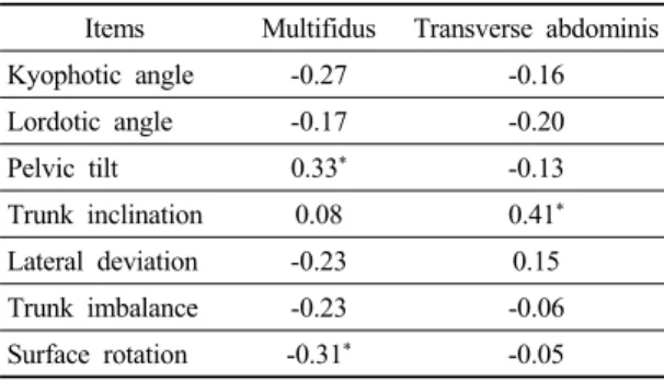
본 연구는 20대 대학생의 몸통 심부근인 배가로근 과 뭇갈래근 두께와 척추정렬 변화의 상관관계를 위 해 진행되었다 . 이를 위해 모든 대상자에게 초음파를 이용한 근두께 측정 그리고 척추 정렬과 골반의 변위 를 사용하여 상관관계를 통해 비교하였다(Table 1).

배가로근은 몸통전후기울기(trunk inclination)와 유 의한 양의 상관관계를 나타내었다(p<0.05). 뭇갈래근 은 골반기울기각(pelvic tilt)과 유의한 양의 상관관계 를 나타내었으며, 가시돌기회전각도(surface rotation) 과 유의한 음의 상관관계를 나타내었다(p<0.05).

Items Multifidus Transverse abdominis Kyophotic angle -0.27 -0.16 Lordotic angle -0.17 -0.20

Pelvic tilt 0.33

*

-0.13

Trunk inclination 0.08 0.41

*

Lateral deviation -0.23 0.15 Trunk imbalance -0.23 -0.06 Surface rotation -0.31

*

-0.05

*

p<0.05

Table 1. Pearson’s correlation coefficients analysis between multifidus, transverse abdominis and spinal alignment items.

Ⅳ. 고 찰

우리 인체의 골격은 척추를 중심으로 하여 대칭적 으로 배열하고 있고, 총체적인 모양을 유지하도록 이 루어져 있다. 무릎 주위의 넙다리네갈래근이 무릎관 절의 안정성에 기여하듯이 척추에서는 척추 주위근 특히 뭇갈래근과 배가로근이 중요하다고 알려져 있으 며, 만성 요통 환자들이 정상인에 비해 이들 근육의 약증이 발생되었다고 보고하였다(Cooper et al, 1992).

요통환자들에게 통증과 배가로근과 뭇갈래근의 크기 가 서로 관련성이 있다는 보고가 있다. 척추의 안정화

근육이 약화된다면 척추정렬이 비대칭적으로 되어 불 균형이 될 수 있다는 가정하에 본 연구를 진행하였다 . 척추 안정화 근육의 근두께는 초음파를 이용하여 측 정하였으며 , 본 연구에서 제시한 안정화 국소 근육인 배가로근과 뭇갈래근 초음파 측정의 신뢰도는 측정자 내 신뢰도 (급간내 상관계수)는 0.96∼0.99, 측정자간 신뢰도는 0.96∼0.99로 나타나 높은 상관관계를 나타 내어 신뢰도가 입증되었다(Koppenhaver et al, 2009).

몸통안정화 근육과 척추정렬과의 상관관계를 알아 본 연구 결과 배가로근은 몸통 전후 기울기와 유의한 양의 상관관계를 나타내었다. 몸통 전후 기울기는 목 뼈 7번과 양쪽 PSIS 사이의 선과 수직 전두면의 선 사이의 거리 또는 각도를 표시한 값으로 몸통이 앞으 로 기울어지는 정도를 각도로 나타낸 것이다 . 이는 배가로근이 두꺼워질수록 몸통의 전후 기울기가 점점 커진다는 것을 의미하며 기울기가 커지면 몸이 앞으 로 많이 기울어졌다는 것을 나타낸다 . 정상 여성들을 대상으로 척추안정화 운동과 몸통근육의 상관관계를 알아본 연구에서는 배가로근이 몸통 안정화와 상관성 이 있는 근육으로 보고하였다 (Lee, 2014). 하지만, 배가 로근 두께는 자세가 바뀜에 따라서 차이가 날 수 있으 며(Reeve & Dilley, 2009), 본 연구의 배가로근 두께는 좌우 배가로근의 대칭정도의 차이로 자세불안정성에 기여한 것으로 예측할 수 있다.

가시돌기회전각도(surface rotation)는 목뼈 7번과 허 리뼈 5번 사이 척추의 가시돌기들의 회전을 각도(°)로 나타낸 것이며 , 골반 기울기각(pelvic tilt)은 두 골반의 높이를 비교한 것으로써 다리길이의 차이를 의미한 다 . 본 연구의 결과 뭇갈래근은 골반기울기각과 양의 상관관계를 나타내어 뭇갈래근의 두께가 두꺼울수록 골반의 기울기 각이 증가한 것으로 나타났다 . 또한 척추분절 회전과는 유의한 음의 상관관계를 나타내어 뭇갈래근의 두께가 두꺼울수록 척추분절 회전의 각도 가 작아지는 것으로 나타났다.

뭇갈래근의 근 두께가 두꺼울수록 골반기울기를

나타내는 다리길이 차이는 더 커지는 것으로 나타났

다 . 골반은 척추와 하지를 연결하며 보행 능력에 가장

(5)

효과적으로 영향을 미치는 조절점이므로 하지 근육의 구조적인 특성이 변화할 때 많은 변화가 나타나 생역 학적 보상작용으로 척추에 영향을 미치게 된다(Lee et al, 2004). 결국 골반이 평행하지 않아서 다리에 차이 가 나타나는 요인은 척추 뿐 아니라 엉치엉덩관절의 변위차가 발생되는 것이 더 큰 요인될 수 있었기 때문 에 이와 같은 결과가 나온 것으로 판단된다.

척추분절 회전은 가시돌기가 회전하는 각도를 의 미하며 그 각도가 작아지는 것은 가시돌기의 회전이 적게 일어난 것을 의미하므로 뭇갈래근의 근육의 두 께가 척추의 뒤틀림을 예방하는 근육으로 예측해 볼 수 있다. 시상면에서 몸의 정렬을 바로 잡는 근육은 뭇갈래근으로 알려져 있으며, 근골격계에 문제가 있 어 몸통안정화 근육에 이상이 있는 경우 의도된 움직 임을 수행하기 어렵다. 허리에 통증을 호소하는 사람 을 대상으로 초음파 영상을 통해 본 결과 , 허리통증을 경험한 사람이 뭇갈래근과 배가로근의 두께가 차이가 나타났으며, 상지와 하지의 빠른 움직임을 할 때, 허리 통증을 호소하는 사람이 뭇갈래근과 배가로근의 근활 성이 지연된다고 하였다(Vasseljen & Fladmark, 2010).

만성요통환자를 대상으로 척추관절가동화를 1주 시 행한 후 통증이 감소하였는데, 관련 요인으로 뭇갈래 근의 두께 향상이라는 것을 밝혀내어 요통환자의 통 증의 원인이 뭇갈래근이라고 주장은 본 연구의 결과 를 뒷받침한다(Koppenhaver et al, 2011). 척추정렬에 영향을 줄 수 있는 많은 요인들 중에서 근두께에 제한 되었으므로 하지의 근육이나 배가로근이나 뭇갈래근 이외의 다른 근육과의 상관관계 연구들이 필요할 것 으로 생각된다.

Ⅴ. 결 론

본 연구는 20대 젊은 성인을 대상으로 척추의 심부 근인 배가로근과 뭇갈래근 두께를 초음파를 이용하여 측정하였고 , 3차원 영상장비를 이용해 척추의 정렬상 태를 알아보고 각각의 상관성을 제시하였다. 따라서 본 연구에서 제시한 배가로근과 뭇갈래근 두께와 자 세정렬과의 상관관계에 대한 평가는 오랫동안 앉아

있는 성인이나 올바른 자세를 갖지 못하는 성인들이 약화가 생길 수 있는 근육이 배가로근과 뭇갈래근이 라 할 수 있으므로 다양한 질환에서 이 근육들의 두께 를 향상시킬 수 있는 중재가 필요하다. 특히 시상면에 서 중요한 역할을 하는 뭇갈래근은 가시돌기의 회전 에 영향을 주므로 측만증 환자에게 강조해야 할 근육 이 될 것이다 . 따라서 척추부정렬이 있는 성인의 경우 특정 목적에 따라 배가로근과 뭇갈래근을 강화하는 운동프로그램이 필요할 것이다.

참고문헌

Ainscough‐Potts AM, Morrissey MC, Critchley D. The response of the transverse abdominis and internal oblique muscles to different postures. Manual Therapy.

2006;11(1):54-60.

Cooper RG, St Clair Forbes W, Jayson MI. Radiographic demonstration of paraspinal muscle wasting in patients with chronic low back pain. British Journal of Rheumatology. 1992;31(6):389-394.

Hides JA, Richardson CA, Jull GA. Magnetic resonance imaging and ultrasonography of the lumbar multifidus muscle.

Comparison of two different modalities. Spine (Phila Pa 1976). 1995;20(1):54-58.

Hides JA, Stokes MJ, Saide M, et al. Evidence of lumbar multifidus muscle wasting ipsilateral to symptoms in patients with acute/subacute low back pain. Spine (Phila Pa 1976). 1994;19(2):165-172.

Hodges PW, Richardson CA. Inefficient muscular stabilization of the lumbar spine associated with low back pain.

A motor control evaluation of transversus abdominis.

Spine (Phila Pa 1976). 1996;21(22):2640-2650.

Ikezoe T, Mori N, Nakamura M, et al. Effects of age and

inactivity due to prolonged bed rest on atrophy of

trunk muscles. European Journal of Applied

Physiology. 2012;112(1):43-48.

(6)

Kiesel KB, Uhl TL, Underwood FB, et al. Measurement of lumbar multifidus muscle contraction with rehabilitative ultrasound imaging. Manual Therapy.

2007;12(2):161-166.

Koppenhaver SL, Fritz JM, Hebert JJ, et al. Association between changes in abdominal and lumbar multifidus muscle thickness and clinical improvement after spinal manipulation. Journal of Orthopaedic and Sports Physical Therapy. 2011;41(6):389-399.

Koppenhaver SL, Hebert JJ, Fritz JM, et al. Reliability of rehabilitative ultrasound imaging of the transversus abdominis and lumbar multifidus muscles. Archives of Physical Medicine and Rehabilitation.

2009;90(1):87-94.

Lee HJ, Na YM, Lim KB. Biomechanical characteristics of lower extremities and pelvis in korean soccer players who complain of chronic low back pain. Korean Society of Sports Medicine. 2004;22(1):59-66.

Lee JS. Correlation Between Trunk stabilization and the abdominal muscles in healthy Females. Journal of Korea Entertainment Industry Association. 2014;

8(3):1-8.

Mannion AF, Pulkovski N, Toma V, et al. Abdominal muscle size and symmetry at rest and during abdominal hollowing exercises in healthy control subjects.

Journal of Anatomy. 2008;213(2):173-182.

O’Sullivan PB, Grahamslaw KM, Kendell M, et al. The effect of different standing and sitting postures on trunk muscle activity in a pain‐free population. Spine (Phila Pa 1976). 2002;27(11):1238-1244.

Ota M, Ikezoe T, Kaneoka K, et al. Age‐related changes in the thickness of the deep and superficial abdominal muscles in women. Archives of Gerontology and Geriatrics. 2012;55(2):e26-30.

Reeve A, Dilley A. Effect of posture on the thickness of transversus abdominis in pain-free subjects. Manual Therapy. 2009;14(6):679-684.

Sipila S, Suominen H. Muscle ultrasonography and computed tomography in elderly trained and untrained women.

Muscle and Nerve. 1993;16(3):294-300.

Tracy MF, Gibson MJ, Szypryt EP, et al. The geometry of the muscles of the lumbar spine determined by magnetic resonance imaging. Spine (Phila Pa 1976).

1989;14(2):186-193.

Urquhart DM, Hodges PW, Allen TJ, et al. Abdominal muscle recruitment during a range of voluntary exercises.

Manual Therapy. 2005;10(2):144-153.

Vasseljen O, Fladmark AM. Abdominal muscle contraction thickness and function after specific and general exercises: a randomized controlled trial in chronic low back pain patients. Manual Therapy. 2010;

15(5):482-489.

수치

Table 1. Pearson’s correlation coefficients analysis between multifidus, transverse abdominis and spinal alignment items.

참조

관련 문서

This study was to do a comparative analysis on kinematic differences and differences in muscle activity between the skilled and the unskilled in windsurfing

A study on the relationship between body mass index and the food habits of college students. Keilins respiratory chain concept and its

Greco-Roman wrestlers of high school, college, and the professionals players showed significant differences depending on their performance when measuring the maximum muscle

The purpose of this study was to investigate the lower extremity muscle strength and student health of male middle school students. Twenty middle school

The third was combination of levator palpebrae, superior rectus, inferior oblique muscle weakness(n=5) and the fourth was the third group plus medial rectus

The improvement factors of the performance showed significant increase in muscle strength, muscle endurance, cardiovascular endurance, power, agility and

Physical fitness changes on college students who played ultimate frisbee showed increases in muscular strength, muscle endurance(, cardiopulmonary endurance,

The aim of this study was not only to compare the differences of ego-resilience between students with high experience and students with low experience in