Abstract
Ⅰ. 서 론
혈관형성(angiogenesis)은 종양의 성장, 침투 및 전이에 꼭 필요 한 과정이다
1,2). 혈관화가 잘 이루어진 종양의 경우 국소적이나 원격전이에 의해 팽창하는 반면, 혈관화가 되지 않을 경우 지름 1~2cm 이상 성장할 수 없다고 알려져 있다
3). 따라서 혈관형성억 제제를 이용한 종양의 치료는 전도 유망한 치료방법이라고 할 수가 있다.
여러 가지의 혈관형성억제제, 예를 들면, fumagillin 유사체인 TNP470, 상어연골에서 유래된 억제제인 squalamine, v3 integrin에 대한 항체인 Vitaxin, vascular endothelial growth factor(VEGF)에 대 한 단클론 항체와 thalidomide 등이 현재 임상적으로 사용되고 있 다
4-8).
Thalidomide(α-N-phthalimidoglutarimide)는 진토(nausea)의 효력 을 갖는 진정제로서 사용되었었고, 토끼의 각막을 이용한 신생 혈관분석(corneal neovascularization assay)에서 강력한 혈관형성 억제의 효력을 갖는다고 입증되었으며
9), VEGF에 의한 혈관형성 을 억제해서 종양의 성장을 감소시킨다고 보고되었다
10). 여러 종 류의 고형성 종양(solid tumor)을 치료하기 위해서 thalidomide를 이용한 임상적인 시도가 보고되고 있다
11-13). 하지만 구강암을 치 료하기 위한 혈관형성억제제의 효력은 충분히 연구되지 않았다.
VEGF는 일차적으로 혈관내피세포(endothelial cell)에 특이성
누드마우스에 이종이식된 구강편평상피세포암종에 대한 thalidomide의 항암효과와 혈관형성억제에 관한 연구
김수곤∙명 훈∙김명진
서울대학교 치과대학 구강악안면외과학교실
김 명 진
100-744, 서울특별시 종로구 연건동 28-2 서울대학교 치과대학 구강악안면외과학교실 Kim Myung Jin
Dept. of OMFS, College of Dentistry, Seoul National University 28-2, Yeungun-Dong, Chongro-Ku, Seoul 100-744, Korea Tel : 82-2-760-2632 Fax : 82-2-766-4948
E-mail: [email protected]
ANTI-TUMOR AND ANTI-ANGIOGENIC EFFECT OF THALIDOMIDE ON ORAL SQUAMOUS CELL CARCINOMA XENOGRAFTS IN NUDE MICE
Su-Gon Kim, Hoon Myoung, Myung-Jin Kim
Department of Oral and Maxillofacial Surgery, College of Dentistry, Seoul National University
Angiogenesis is an essential process for the growth, invasion and metastasis of cancer. However, it is uncertain that antiangiogenic effects can be a major treatment strategy of oral cancer. The aim of this study was to investigate whether thalidomide, which is known to be a potent inhibitor of angiogenesis, have inhibitory effect on the growth and antiangiogenic effects of oral squamous cell carcino- ma(OSCC) xenografted in nude mice and whether antiangiogenesis of thalidomide can be included as a major treatment strategy of oral cancer.
After human oral squamous cell carcinoma strain KB was subcutaneously implanted in 20 nude mice, the volume of tumor was mea- sured every three days. When the tumor mass reached 75~100mm3, thalidomide(200mg/kg/d) was administered into 10 experimental nude mice and the same volume of distilled water was administered into 10 control nude mice and the tumor volume was measured every three days. The excised tumor masses on the 30th day after administration were frozen and processed for immunohistochemistry using vascular endothelial growth factor(VEGF) and CD31. We evaluated microvessel density and VEGF expression.
The results were as follows ;
1. Thalidomide retarded the growth of human OSCC as compared with the control group, but it was not statistically significant.
2. A statistically significant lower microvessel density was observed in the thalidomide-treated group than in the control group(p<0.01) and thalidomide significantly reduced VEGF expression (p<0.01).
Thalidomide exhibited significantly antiangiogenic effect, but did not inhibit the growth of human OSCC effectively. Antiangiogenic therapy of thalidomide alone is not likely to be effective in the treatment of human OSCC, but might be regarded as adjuvant chemotherapeutic strategy.
Key words: Antiangiogenesis, Thalidomide, Oral squamous cell carcinoma, CD31, VEGF
(specificity)을 갖는다. VEGF는 혈관 내피세포에 직접 작용해서 혈관내피세포를 증식시키고, 투과도를 증가시켜 병적인 상황과 생리적인 상황에서 혈관형성을 유도한다
14). 따라서 혈관형성은 VEGF의 발현에 의해 반영될 수가 있는 것이다. VEGF는 고등급 의 구강편평상피세포 이형성증과 침투성의 구강편평상피세포 암종에서 두드러지게 발현된다고 보고되고 있는데, 그것은 구강 편평상피세포암종의 성장이 부분적으로는 혈관형성에 의존한 다는 것을 제시한다
15). 따라서 혈관형성억제제는 구강편평상피 세포암종을 치료하거나, 최소한 암종의 성장을 억제한다고 추정 할 수가 있다. 본 연구에서는 강력한 혈관형성억제제라고 알려 져 있는 thalidomide가 누드마우스(BALB/c/nu)에 이종이식된 구 강편평상피세포암종의 성장을 억제하는지와 thalidomide의 혈관 형성억제 작용이 구강편평상피세포암종에도 나타나는지를 평 가하고, thalidomide에 의한 구강암의 치료가 향후 전략적인 치료 방법에 포함될 수 있는지를 평가하는데 그 목적이 있다.
Ⅱ. 실험재료 및 방법
1. 누드마우스 및 구강편평상피세포암종 세포접종체 (cell inoculum) 준비
누드마우스는 BALB/c/nu(Korea Research Institute of Bioscience and Biotechnology, Korea)를 기본종으로, 20마리 모두 생후 6~8 주, 체중이 약 20g의 암컷만을 사용하였다. 멸균 소독된 쥐장에 서 멸균된 물과 사료를 공급하였고, 층판기류(lamina flow)를 지 속적으로 유지한 크린벤취 속에 두어 청정한 환경을 유지하였으 며, 모든 술식 또한 크린벤취 속에서 시행하였다. 누드마우스의 주야간 생물학적 주기를 위하여 실내등을 12시간은 조영하고, 12 시간은 점멸하였다
16).
KB세포주(ATCC, VA, USA)를 기본세포주로 하여, 10% fetal bovine serum(Gibco, NY, USA), 100U/ml 페니실린과 100g/ml 스트 렙토마이신(Gibco, NY, USA) 항생제를 함유한 Dulbeco’s Modified Eagle Medium(Gibco, NY, USA) 10ml를 2일에 한 번씩 교 환하면서 지름 80mm의 세포배양용 플라스크(Nunc, Denmark)에 서 37℃, 습도 90%, 5%의 CO
2및 95%의 공기의 조건을 유지하는 세포배양기에서 배양 및 계대배양하여 증식하였다. 증식된 세포 는 계대배양시와 같이 Trypsin(0.1%)-EDTA 3ml를 넣고 배양기에 서 5분간 세포를 부양시켰다. 부유액을 원심분리(1200rpm, 3분) 하여 세포를 농축하고 hemocytometer로 세포수를 산정하여 세포 접종체를 준비하였다.
2. 종양유도, 종양크기 측정 및 thalidomide 투여
준비된 세포접종체를 이용하여, 누드마우스 한 마리당 0.2cc 의 배양액에 포함된 약 5×10
6개의 세포를 1cc 주사기 및 25게이 지의 주사바늘을 이용하여 누드마우스 등(flank)에 피하주사 접 종하였다. 종양의 길이와 너비를 caliper로 3일에 한 번씩 측정하 여 Gutman 등이 제안한 방법의 공식
17)으로 종양의 부피를 산출
하였다.
Volume(mm
3) = Length×Width
2×1/2 (Length : 장경, Width : 단경)
상대종양부피(RTV)를 다음과 같은 공식, 즉 RTV = Vi/Vo으로 산출하였는데, 여기서 Vi는 정해진 시간에 측정한 종양의 부피이 고, Vo는 투약을 개시한 시점에서의 종양의 부피로서 각각의 실 험동물은 약물의 독성을 평가하기 위해서 3일에 1회씩 체중을 측정하였다.
누드마우스에 유발된 종양의 부피가 75~100mm
3의 크기로 착 상(implantation) 되는데 경과한 시간은 7일이었으며, 7일째부터 임의로 두 군으로 나누었고, 각 군당 10마리씩 배분하여 치료를 시작하였다
18). 실험군은 증류수에 thalidomide(Calbiochem, San Diego, CA, USA)를 현탁시켜 200mg/kg/d을, 대조군은 동량의 증 류수를 하루에 1회씩 희생할 때까지 29게이지 주사바늘을 이용 하여 복강내 주사하는 방법으로 투여하였다.
3. 면역조직화학검사
누드마우스는 투약한지 30일째에 경추탈구의 방법으로 희생 하였고, 면역조직화학검사를 위해 종양조직을 절개한 후 동결표 본으로 만들었다.
4μm 두께의 동결조직절편을 준비하여 streptavidin-biotin-peroxi- dase(DAKO LSAB Kit, Peroxidase K680)방법으로 면역조직화학검 사를 시행하였다. 내인성 peroxidase의 활성을 억제하기 위해 동 결조직표본을 3% 과산화수소수 용액으로 5분간 처리한 후, 증류 수와 PBS로 세척하였다. 미세혈관 밀도를 구하기 위해 CD31 (platelet/endothelial cell adhesion molecule;PECAM-1)
19)JC70 단클론 항체를 1 : 20으로 희석한 뒤 30분간 상온에서 부란한 후 strepta- vidin-biotin-peroxidase complex를 결합(conjugation)하였다.
VEGF의 발현을 알아보기 위해 동결조직절편을 3% normal swine serum으로 한 시간 부란시켜 비특이적 IgG 결합을 방지하 였다. VEGF 다클론 항체(A-20, Santa Cruz Biotechnology, CA, USA) 를 1 : 400으로 희석하여 사용하였고, 4℃에서 밤새 반응시켰다.
Streptavidin-biotin-peroxidase complex를 결합시킨 후 발색은 3,3′- diaminobenzidine hydrochloride(DAB)으로 하였고, 대조염색은 Mayer’s hematoxylin을 이용하였다. 혈류 공급이 풍부하다고 이 미 알려져 있는 유방암종의 조직을 양성대조로 삼았으며, 음성 대조는 일차항체를 정상 swine serum으로 대체한 것으로 하였다.
4. 미세혈관 밀도 측정, VEGF의 발현도 측정 및 통계 분석
미세혈관 밀도는 Arapandoni-Dadioti 등이 기술한 방법으로 평
가하였다
20). 종양에서 멀리 떨어진 부위와 괴사부위의 미세혈관
은 제외하고 종양주위에서 단위면적당 가장 많은 수의 미세혈관
을 가진 부위를 저배율(×100)에서 세 군데를 찾은 뒤 고배율(×
250)에서 모세혈관의 수를 세어 그 평균값으로 하였다. VEGF 발 현강도는 Takahasi 등이 제시한 0에서 3+까지의 단위로 등급 (grade)을 매겼다
21); 0 : 발현이 전혀 안됨, 3+ : 가장 강한 발현, 그 리고 VEGF의 발현강도에 따라 구강편평상피세포암종을 두 그룹 으로 나누었는데, 발현이 전혀 안 되었거나, 약하게 되었을 경우, 즉 그룹 0과 1+에 해당하는 경우는 VEGF-poor로 간주하였고, 발 현의 강도가 중등도거나 강하게 되었을 경우, 즉 그룹 2+와 3+에 해당하는 경우 VEGF-rich로 간주하였다
22). 미세혈관 밀도 측정과 VEGF 발현도 측정은 두 명의 검사자가 2회씩 double blinded technique으로 측정하였다.
체중의 변화, 상대종양부피 및 미세혈관 밀도의 차이는 Mann- Whitney non-parametric analysis을 이용하여 분석하였고, VEGF 발 현강도의 차이는 Chi-square test를 이용하였으며, 유의수준은 p<0.05로 정하였다.
Ⅲ. 결 과
1. 체중의 변화
세포접종체를 피하주사한 날로부터 체중의 변화를 그래프로 나타내었을 때 대조군과 실험군 모두 치료가 진행되면서 체중의 감소와 증가가 불규칙하게 반복되는 양상을 나타내었고, 두 군 사이에 통계적 유의성은 없었다(Fig. 1).
2. 상대종양부피
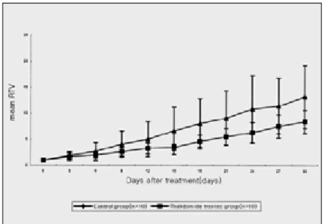
상대종양부피는 대조군에서 계속해서 증가하는 양상을 보여 치료한 지 6일째 2.68±1.73, 18일째 7.98±4.79, 그리고 30일째에 는 13.209±6.03을 나타내었다. Thalidomide로 치료한 실험군에서 도 10마리 모두 실험이 진행되는 동안 계속해서 증가하는 양상
Fig. 1. Body weight change of nude mice. Fig. 2. Mean relative tumor volume(RTV) after administration of thalidomide.
Fig. 3. CD31 immunohistochemistry findings in the control group(x200). Brown stained cytoplasmas of endothelial cells were shown.
Fig. 4. CD31 immunohistochemistry findings in the thalido- mide-treated group(x200). Microvessels were rarely or sparsely shown in the connective tissue layer around OSCC cells.
을 보여 치료한 지 30일째에 8.37±0.85로 나타났는데, 그것은 종 양부피가 실험하는 동안 평균 8.37배로 증가한 것을 의미한다.
Thalidomide는 대조군에 비해 종양의 성장을 지연시켰으나, 통계 적으로 유의성은 없었다(Fig. 2).
3. 미세혈관 밀도와 VEGF의 발현도
CD31 항체에 의해 혈관내피세포의 세포질이 갈색으로 염색되 었는데, 종양세포 주위 결합조직에서 대조군의 미세혈관이 실험 군에 비해 더 많이 관찰되었다(Fig. 3, Fig. 4). 미세혈관 밀도는 thalidomide로 치료한 실험군이 대조군보다 최소한 2배 이상 더 낮았으며, 통계적으로 유의성이 있었다(p<0.01, Fig. 5).
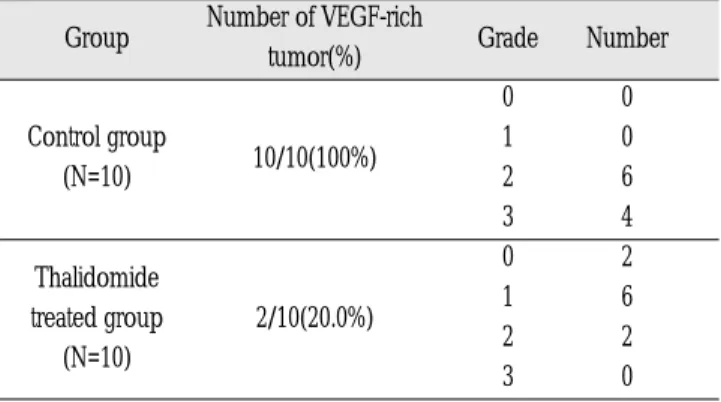
VEGF는 종양세포의 세포질에 갈색으로 염색되었는데, 대조군 에서 thalidomide로 치료한 실험군에 비해 훨씬 많이 관찰되었다 (Fig. 6, Fig. 7). 대조군은 10마리 모두 중등도 이상의 발현강도를 보여 VEGF-rich가 100%로 나타났으며, thalidomide로 치료한 실험 군에서는 10마리 중 2마리에서 중등도의 발현강도를 보여 VEGF-
rich가 20%로 나타났다(Table 1). Thalidomide는 VEGF의 발현을 통계적으로 유의성 있게 감소시켰다(p<0.01).
Ⅳ. 총괄 및 고안
고형성 종양(solid tumor)의 성장, 침투 및 전이는 혈관형성 (angiogenesis)을 통해서 이루어진다는 것은 잘 알려져 있다
1,2). 종 양세포는 혈관형성인자(angiogenic factors)들을 분비하여 혈관형 성을 유도한다
1). 이러한 혈관형성인자에는 aFGF(acidic fibroblast growth factor), bFGF(basic fibroblast growth factor), TGF-α(trans- forming growth factor-α), TGF-β(transforming growth factor-β), VEGF(vascular endothelial growth factor), interleukin-8, TNF(tumor necrosis factor)와 PD-ECGF(platelet-derived endothelial cell growth factor) 등이 있다
23). 이 중에서 VEGF와 PD-ECGF는 in vivo상에서 혈관형성력을 가지고 있다고 알려져 있고
23), 특히 VEGF는 혈관 내피세포의 분열촉진인자(mitogen)로서 최근에 종양의 혈관형성 을 핵심적으로 조절한다고 인정받고 있다
2). VEGF는 특이적으로
Fig. 7. VEGF immunohistochemistry findings in the thalido- mide-treated group(×200). Brown stained cytoplasms in OSCC cells were shown very rarely or sparsely.
Fig. 5. Comparison of microvessel density in both thalido- mide-treated group and control group.
Fig. 6. VEGF immunohistochemistry findings in the control group(×200). Brown stained cytoplasms of OSCC cells were shown very much.
Table 1. Correlations of VEGF expression in each drug treated group
Group Number of VEGF-rich
Grade Number tumor(%)
0 0
Control group
10/10(100%) 1 0
(N=10) 2 6
3 4
Thalidomide 0 2
treated group 2/10(20.0%) 1 6
(N=10) 2 2
3 0
혈관내피세포의 분열을 유도하는 cytokine으로서 혈관투과도 (vascular permeability)를 증가시키고, 혈관내피세포의 분화, 이동 및 생존을 촉진시켜 혈관형성을 유도함으로써 결국 종양세포의 성장을 증진시키는 작용을 한다
22,24). VEGF 발현 상실은 미세혈관 밀도와 혈관투과도를 떨어뜨리고 apoptosis를 증가시킨다
28). 따라 서 종양의 성장 잠재력을 반영하는 혈관형성은 VEGF의 발현으 로 평가할 수가 있다.
미세혈관 밀도가 방광암, 대장암, 위암과 흑색종에서 경부 임 파절 전이, 종양의 재발, 나쁜 예후와 상관관계가 있다고 보고되
었으며
26-29), 구강편평상피세포암종에 있어서도 종양의 성장, 종양
의 침습성, 경부 임파절 전이, 나쁜 예후와 미세혈관 밀도와의 관 계를 보여준 연구는 많이 있다
14,30,31).
혈관수의 측정은 Arapandoni-Dadioti 등이 기술한 방법으로 평 가하였는데, 종양에서 멀리 떨어진 부위와 괴사부위의 미세혈관 은 제외하였다. 미세혈관 계산시 근육층을 가지거나, 적혈구의 직경보다 8배 이상의 직경을 가진 큰 혈관은 제외하였으며, 혈관 내강의 유무와 상관없이 CD31 항체에 양성이고 인접세포군과 명백하게 분리되어 있는 내피세포 집락은 한 개의 혈관으로 인 정하였다. 그리고 염증에 의한 혈관증식을 제외하기 위해 종양 근처에서 혈관수가 많더라도 염증이 심한 부분은 혈관수 측정부 위에서 제외시켰다
20).
VEGF 발현을 평가하는데 있어서 본 연구에서 사용한 Takahasi 등
21)이 제시한 방법외에 세포막이나 세포질에 갈색으로 염색되 는 세포가 종양세포의 5% 이상이면 염색강도에 상관없이 양성 으로 판정하는 방법
32)과 종양세포의 30% 이상에서 인접한 정상 조직에 있는 근육층에 염색되는 강도보다 더 강하게 발현되는 경우 양성으로 판정하는 방법
33)등이 있지만, 더 객관적이고 양 적인 평가를 위해 중합효소 연쇄반응을 이용한 VEGF mRNA의 발현을 알아볼 필요가 있을 것 같다.
혈관형성이 고형성 종양의 성장, 침투 및 전이에 꼭 필요한 과 정이기 때문에, 종양의 치료를 위해 여러 가지 혈관형성억제제 가 현재 임상적으로 사용되어져 오고 있다
5-7,34). Thalidomide(α-N- phthalimidoglutarimide)는 1950년대에 강력한 항구토 작용을 갖는 안전한 진정제로서 소개되었으며, 임산부들에게 임신 1기의 오 심(입덧)을 진정시키기 위해 처방하였다
35). 1960년대 초반에 thal- idomide가 기형을 유발한다는 보고가 있은 후
36,37), 사용이 중단되 었지만, 최근에 bFGF와 VEGF가 유도하는 혈관형성억제를 포함 한 다양한 약리학적 효과로 새로운 관심을 끌고 있다
9). Thalidomide는 나성결절홍반(erythema nodosum leprosum)
38), 조 직을 이식받은 환자들의 graft-versus-host disease
39), HIV 감염자의 구강과 구인두에 발생한 아프타성 궤양
40)및 Crohn’s disease
41)를 치료하는데 효과적이라고 알려져 있다. D’Amato 등은 thalido- mide가 기형을 유발하는 것은 혈관형성억제 작용과 관련이 있을 수 있다고 제시하였다
42).
여러 종양의 세포주들을 실험동물에 이종이식한 후 혈관형성 억제제로서의 thalidomide을 평가한 실험적 연구가 다수 보고되 었지만, 구강편평상피세포암종 세포주에 대해 혈관형성억제제 로서의 thalidomide를 평가한 논문은 보고된 바가 없다. 누드마우
스에 이종이식된 전립선암(LNCaP)에서 낮은 미세혈관 밀도와 종 양성장억제 효과가 통계적으로 유의성 있게 나타났고
43), 토끼에 이종이식된 V2 암종에서는 thalidomide 단독 투여시에는 종양의 성장을 55% 억제하였고, 비스테로이드성 소염 진통제인 sulindac 과 병용 투여시에는 종양의 성장을 75% 억제하였으며, 낮은 미 세혈관 밀도를 보였다
44). 누드마우스에 이종이식한 식도암(ES63) 에서 낮은 미세혈관 밀도와 낮은 VEGF 발현도 및 종양성장 억제 효과가 유의성 있게 나타났지만, 다른 세포주의 식도암(ES80)에 서는 유의성이 없어 세포주에 따른 반응도의 차이를 보였다
45). 동계(syngenic)의 쥐에 이종이식된 흑색종(B16-F10)과 결장암(CT-
26)
에서는 종양성장 억제효과가 유의성 있게 나타나지 않았다
46). 본 연구에서는 구강편평상피세포암종에서 thalidomide가 미세혈 관 밀도와 VEGF의 발현을 감소시키는 등 혈관형성억제 작용을 유의성 있게 나타내었으나, 종양의 성장을 효과적으로 억제하지 는 못 하였다.
실험을 시작하기 전에 구강편평상피세포암종의 성장이 혈관 형성인자에 의한 혈관형성에 달려 있다면, 구강편평상피세포암 종의 성장은 thalidomide로 억제가 가능하리라는 가설을 세웠다.
하지만 thalidomide로 치료한 실험군에서 미세혈관 밀도와 VEGF 의 발현은 유의성 있게 감소하였는데도 불구하고 오히려 구강편 평상피세포암종의 성장은 비록 대조군에 비해서는 지연되었으 나, 실험이 진행중인 동안 계속해서 지속되었다는 사실은 다음 의 내용을 시사한다고 볼 수가 있다. 첫번째로 thalidomide는 혈 관형성을 완전히 억제하지는 못하였기 때문에, 억제되지 않고 남아 있는 아주 적은 양의 혈관형성도 구강편평상피세포암종의 성장을 유지하는데 충분하다고 생각된다. 두번째로 종양마다 주 된 혈관형성인자가 있기 때문에, 효과적인 치료결과를 얻기 위 해서는 종양의 혈관형성을 우세하게 조절하는 주된 혈관형성인 자를 평가해서 가장 적합한 혈관형성억제제를 선택해야 한다고 제안된 보고
45)로부터 VEGF는 구강편평상피세포암종의 주된 혈 관형성인자가 아닐 가능성이 있다. 세번째로 두경부 영역은 해 부학적으로 혈류 공급이 풍부하기 때문에 구강암의 성장은 다른 부위의 종양보다 혈관형성에 덜 의존적이고, 종양세포의 유전적 변이로 인해 종양세포는 무산소 환경에서도 성장을 할 수가 있 으며, 혈관형성은 종양의 성장을 조절하는 많은 인자 중 한 가지 에 지나지 않는다
47,48).
앞으로 thalidomide가 구강편평상피세포암종의 성장과 VEGF 에 의한 구강편평상피세포암종의 성장 반응도에 미치는 효과를 알아보기 위해서 여러 종류의 구강편평상피세포암종 세포주를 이용한 실험이 진행되어야 할 것으로 보인다.
Ⅴ. 결 론
구강편평상피세포암종인 KB 세포주를 기본주로 하여, 누드마 우스에 이종이식한 후 thalidomide의 in vivo 항암효과와 혈관형 성억제에 관한 연구를 통하여 다음과 같은 결론을 얻었다.
1. Thalidomide는 대조군에 비해 종양의 성장을 지연시켰으나,
통계적으로 유의성은 없었다.
2. 미세혈관 밀도는 thalidomide로 치료한 실험군이 대조군에 비해서 통계적으로 유의성 있게 감소하였으며(p<0.01), thal- idomide는 VEGF의 발현을 통계적으로 유의성 있게 감소시 켰다(p<0.01).
이상의 결과에서 thalidomide는 구강편평상피세포암종에서 혈 관형성억제 작용을 유의성 있게 나타내었으나, 성장을 효과적으 로 억제하지는 못 하였고, 구강편평상피세포암종을 치료하는데 있어서 thalidomide 단독으로는 한계를 보이며 보조화학요법의 한 방법으로 사용될 수 있을 것으로 사료된다.
참고문헌
1. Folkman J, Shing Y : Angiogenesis. J Biol Chem 267 : 10931-10934, 1992.
2. Folkman J : What is the evidence that tumors are angiogenesis dependent? J Natl Cancer Inst 82 : 4-6, 1990.
3. Folkman J, Cotran RS : Relationship of vascular proliferation to tumor growth. Int Rev Exp Pathol 16 : 207-248, 1976.
4. Kim KJ, Li B, Winer J, Armanini M, Gillett N, Phillips HS, Ferrara N : Inhibition of vascular endothelial growth factor-induced angiogene- sis suppresses tumour growth in vivo. Nature 362 : 841-844, 1993.
5. Barinaga M : Designing therapies that target tumor blood vessels.
Science 24(275) : 482-484, 1997.
6. Coleman KR, Braden GA, Willingham MC, Sane DC : Vitaxin, a humanized monoclonal antibody to the vitronectin receptor (alphavbeta3), reduces neointimal hyperplasia and total vessel area after balloon injury in hypercholesterolemic rabbits. Circ Res 84 : 1268-1276, 1999.
7. Matsumoto K, Ninomiya Y, Inoue M, Tomioka T : Intra-tumor injec- tion of an angiogenesis inhibitor, TNP-470, in rabbits bearing VX2 carcinoma of the tongue. Int J Oral Maxillofac Surg 28 : 118-124, 1999.
8. Schiller JH, Bittner G : Potentiation of platinum antitumor effects in human lung tumor xenografts by the angiogenesis inhibitor squalamine : effects on tumor neovascularization. Clin Cancer Res 5 : 4287-4294, 1999.
9. Kenyon BM, Browne F, D’Amato RJ : Effects of thalidomide and related metabolites in a mouse corneal model of neovascularization.
Exp Eye Res 64 : 971-978, 1997.
10. Kruse FE, Joussen AM, Rohrschneider K, Becker MD, Volcker HE : Thalidomide inhibits corneal angiogenesis induced by vascular endothelial growth factor. Graefes Arch Clin Exp Ophthalmol 236 : 461-466, 1998.
11. Figg WD, Bergan R, Brawley O : Randomized phase Ⅱ study of thalidomide in androgen independent prostate cancer (AIPC) [abstract]. Proceedings of American Society of Clinical Oncology 16 : 333a, 1997.
12. Fine HA, Loeffler JS, Kyritsis A : A phaseⅡtrial of the anti-angio- genic agent, thalidomide, in patients with recurrent high-grade gliomas [abstract]. Proceedings of American Society of Clinical Oncology 16 : 385a, 1997.
13. Nguyen M, Tran C, Barsky S : Thalidomide and chemotherapy com- binations: preliminary results of preclinical and clinical studies. Int J Oncol 10 : 965-969, 1997.
14. Moriyama M, Kumagai S, Kawashiri S, Kojima K, Kakihara K, Yamamoto E : Immunohistochemical study of tumour angiogenesis in oral squamous cell carcinoma. Oral Oncol 33 : 369-374, 1997.
15. Denhart BC, Guidi AJ, Tognazzi K, Dvorak HF, Brown LF : Vascular permeability factor/vascular endothelial growth factor and its recep- tors in oral and laryngeal squamous cell carcinoma and dysplasia.
Lab Invest 77 : 659-664, 1997.
16. 정기영, 김명진 : 누드마우스에 이종이식된 구강편평상피세포암종
에 대한 Paclitaxel(Taxol )의 항암효과에 관한 연구. 서울대학교 대
학원 치의학석사학위≥ Æ 1999년 2월.
17. Gutman M, Singh RK, Yoon S, Xie K, Bucana CD, Fidler IJ : Leukocyte-induced angiogenesis and subcutaneous growth of B16 melanoma. Cancer Biother 9 : 163-170, 1994.
18. Klauber N, Parangi S, Flynn E, Hamel E, D’Amato RJ : Inhibition of angiogenesis and breast cancer in mice by the microtubule inhibitors 2-methoxyestradiol and taxol. Cancer Res 57 : 81-86, 1997.
19. Parums DV, Cordell K, Micklem K, Heryet AR, Gatter KC, Mason DY : JC70, a new monoclonal antibody that detects vasecular endotheli- um associated antigen on routinely processed tussue sections, J Clini Pathol 43 : 752-757, 1990.
20. Arapandoni-Dadioti P, Giatromanolaki A, Trihia H, Harris AL, Koukourakis MI : Angiogenesis in ductal breast carcinoma.
Comparison of microvessel density between primary tumor and lymph node metastasis. Cancer Lett 137 : 145-150, 1999.
21. Takahashi Y, Kitadai Y, Bucana CD, Cleary KR, Ellis LM : Expression of vascular endothelial growth factor and its receptor, KDR, corre- lates with vascularity, metastasis, and proliferation of human colon cancer. Cancer Res 55 : 3964-3968, 1995.
22. Obermair A, Kohlberger P, Bancher-Todesca D, Tempfer C, Sliutz G, Leodolter S, Reinthaller A, Kainz C, Breitenecker G, Gitsch G : Influence of microvessel density and vascular permeability factor/vascular endothelial growth factor expression on prognosis in vulvar cancer. Gynecol Oncol 63 : 204-209, 1996.
23. Tokumo K, Kodama J, Seki N, Nakanishi Y, Miyagi Y, Kamimura S, Yoshinouchi M, Okuda H, Kudo T : Different angiogenic pathways in human cervical cancers. Gynecol Oncol 68 : 38-44, 1998.
24. Winlaw DS : Angiogenesis in the pathobiology and treatment of vascular and malignant diseases. Ann Thorac Surg 64 : 1204-1211, 1997.
25. Grunstein J, Roberts WG, Mathieu-Costello O, Hanahan D, Johnson RS : Tumor-derived expression of vascular endothelial growth factor is a critical factor in tumor expansion and vascular function. Cancer Res 59 : 1592-1598, 1999.
26. Dickinson AJ, Fox SB, Persad J : Quantification of angiogenesis as an independent predictor of prognosis in invasive bladder carcino- mas. Br J Urol 74 : 762-766, 1994.
27. Jaeger TM, Weidner N, Chew K : Tumor angiogenesis correlates with lymph node metastasis in invasive bladder cancer. J Urol 154 : 69-71, 1995.
28. Takahashi Y, Kitadai Y, Bucana CD : Expression of vascular endothelial cell growth factor and its receptor, KDR, correlates with vascularity, metastasis, and proliferation of human colon cancer.
Cancer Res 55 : 3964-3968, 1995.
29. Tanigawa N, Amaya H, Matsumura M : Extent of tumor vasculariza- tion correlates with the prognosis and hematogenous metastasis in gastric carcinomas. Cancer Res 56 : 2671-2676, 1996.
30. Shitani S, Rafael A, Yoshimura Y : Immunohistochemical detection of microvessels in oral squamous cell carcinoma. Jpn J Oral Maxillofac Surg 40 : 645-650, 1994.
31. Gasparini G, Weinder W, Maluta S : Intratumoral microvessel densi- ty and p53 protein : correlation with metastasis in head and neck squamous cell carcinoma. Int J Cancer 55 : 739-744, 1993.
32. Maeda T, Matsumura S, Hiranuma H : Expression of vascular endothelial growth factor in human oral squamous cell carcinoma : its association with tumor progression and p53 gene status. J Clin Pathol 51 : 771-775, 1998.
33. Koide N, Nishio A, Kono T, Yazawa K, Igarashi J, Watanabe H, Nimura Y, Hanazaki K, Adachi W, Amano J : Histochemical study of vascular endothelial growth factor in squamous cell carcinoma of the esophagus. Hepatogastroenterol 46(26) : 952-958, 1999.
34. Ueda N, Kamata N, Hayashi E, Yokoyama K, Hoteiya T, Nagayama M : Effects of an anti-angiogenic agent, TNP-470, on the growth of oral squamous cell carcinomas. Oral Oncol 35 : 554-560, 1999.
35. Klausner JD, Freedman VH, Kaplan G, : Short analytical review -
thalidomide as an anti-TNFαinhibitor : implications for clinical use.
Clin Immunol Immunopathol 81 : 219-223, 1996.
36. McBride WG : Thalidomide and congenital abnormalities. Lancet 2 : 1358, 1961.
37. Lenz W : Thalidomide and congenital abnormalities. Lancet 1 : 45, 1962.
38. Sheskin J : Thalidomide in the treatment of lepra reactions. Clin Pharmacol Ther 6 : 303-306, 1965.
39. Vogelsang GB, Farmer ER, Hess AD, Altamonte V, Beschorner WE, Jabs DA : Thalidomide for the treatment of chronic graft-versus-host disease. J Engl J Med 326 : 1055-1058, 1992.
40. Jacobson JM, Greenspan JS, Spritzler J, Ketter N, Fahey JL, Jackson JB : Thalidomide for the treatment of oral ophthous ulcers in patients with human immunodeficiency virus infection. J Engl J Med 336 : 1487-1493, 1997.
41. Wettstein AR, Meagher AP : Thalidomide in Crohn’s disease. Lancet 350 : 1445-1446, 1997.
42. D’Amato RJ, Loughnan MS, Flynn E, Folkman J : Thalidomide is an inhibitor of angiogenesis. Proc Natl Acad Sci USA 91: 4082-4085, 1994.
43. Guinan P, Shaw M, Mirochnik Y, Slobodskoy L, Ray V, Rubenstein M : Paclitaxel is more effective than thalidomide in inhibiting LNCaP
tumor growth in a prostate cancer model. Meth Find Exp Clin Pharmacol 20(9) : 739-742, 1998.
44. Verheul HM, Panigrahy D, Yuan J, D’Amato RJ : Combination oral antiangiogenic therapy with thalidomide and sulindac inhibits tumour growth in rabbits. Br J Cancer 79 : 114-118, 1999.
45. Kotoh T, Dhar DK, Masunaga R, Tabara H, Tachibana M, Kubota H, Kohno H, Nagasue N : Antiangiogenic therapy of human esophageal cancers with thalidomide in nude mice. Surgery 125 : 536-544, 1999.
46. Gutman M, Szold A, Ravid A, Lazauskas T, Merimsky O, Klausner JM : Failure of thalidomide to inhibit tumor growth and angiogene- sis in vivo. Anticancer Res 16 : 3673-3677, 1996.
47. Gleich LL, Biddinger PW, Pavelic ZP, Gluckman JL : Tumor angio- genesis in T1 oral cavity squamous cell carcinoma: role in predict- ing tumor aggressiveness. Head Neck 18 : 343-346, 1996.
48. Gleich LL, Zimmerman N, Wang YO, Gluckman JL : Angiogenic inhibition for the treatment of head and neck cancer. Anticancer Res 18 : 2607-2609, 1998.
49. 송승일, 김명진 : 7,12-Dimethylbenza(a)nthracene으로 유도된 햄스 터 협낭 구강암 발암과정에서 genistein의 혈관형성 억제에 관한 연 구. 서울대학교 대학원 치의학석사학위논문 2000년 2월.