I. 서 론
하악골체부에서의 하악관의 위치와 그 주변 구조물과의 관계 는 발치,
치근단수술, 낭종 및 종양에 대한 처치 그리고 악교정 수
술 등의 구강외과수술과 임플란트의 식립,근관치료 등 여러 치
과임상에 있어 중요성을 가지고 있다.하악관에 손상을 줄 수 있는 대표적인 외과적 시술로는 구치의 발거를 들 수 있다.1-3)전방의 대구치들 보다 제3대구치 발거에서 문제가 많이 나타나는데 이는 하악의 후방 구치부로 갈수록 치근 첨과 하악관과의 근접도가 증가하기 때문이다.4)
Stockdale
5)에 의하면 하악 영구치의 치근첨과 하악관사이의 거리는 치아가 맹 출하면서 계속 증가하다가 치아가 교합면에 이르면 치근첨과 하 악관이 어느 정도 안정적인 거리를 갖게 되고 제3대구치보다 제1대구치와 제2대구치에서 더 큰 간격을 갖게 된다. 제2대구치는
제3대구치에 비하여 하악관과 치근첨과의 거리가 크게 형성되지 만 제1대구치보다는 하악관과 근접된 거리를 유지하기 때문에 제2대구치의 간단한 발치 후에도 환자가 지각이상을 호소할 수 있다.6)하악관에 대한 손상은 하악지시상골절단술 시에도 일어날 수 있다.
하악지시상골절단술 시행 후 영구적인 지각이상이 높은 빈
도수로 나타나고 있다.7,8)하악관의 위치가 하악협측피질골판에 가까운 환자에서 두 개의 피질골판 분리 시 하악관이 노출되는 경우가 많으며,노출된 후 골 고정 시에 골판사이에 눌리거나 늘
어지면서 하지조신경이 손상될 수 있다.하악골에
implant 매식 시 나타나는 지각이상에 대한 연구들
을 보면,연구자의 기준이나 환자의 호소정도에 따라 차이가 나
지만 적게는1.7%에서 많게는 42.5%
의 지각이상을 나타낸다고한다.9,10)특히
LeSley, Ellies 등
10)의 연구를 보면 지각이상을 보이는 환자 중에서 이공 후방의 구치부에 식립한 경우에 있어 이 공의 전방에 식립한 경우보다
3배정도 지각이상을 보이는 경우
가 높았고,두 부위에 함께 식립한 경우에는 4배정도 높게 나타나
구치부의implant 식립시 방사선학적 위치결정이 더욱 중요하
다고 하였다.Dentascan
�을 이용한 하악구치부의 하악관 위치에 관한 임상적 연구
김준철∙이승훈∙이정근 아주대학교 의과대학 치과학교실
Abstract (J. Kor. Oral Maxillofac. Surg. 2002;28:341-347)
THE CLINICAL STUDY OF THE MANDIBULAR CANAL LOCATION IN MANDIBULAR MOLAR AREAS USING DENTASCAN
Jun-Cheol Kim, Seung-Hoon Rhee, Jeong-Keun Lee Dept. of Dentistry, School of Medicine, Ajou University
This study was designed to determine the location of the mandibular canal on lower molar areas. Thirty-three patients were examined with multi-planar reformatted CT scan(Dentascan
�). Three kinds of measurements were performed. The first was the distances between the upper border of the mandibular canal and the root apices of the first and second molars, the second was the distance between the cortical plate of the mandible and mandibu- lar canal, and the last was the location of the mandibular canal in the buccolingual plane.
The obtained results are as follows
1. The distance between the root apices of lower molars and the superior border of mandibular canal was largest at the mesial root of the first molar, and shortest at the distal root of the second molar(p<0.05).
2. The longest distance between the outer surface of the buccal cortical plate of the mandible and mandibular canal was measured from the distal root of the second molar, and this distance decrease gradually mesial- ly(p<0.05).
3. The distance between the mandibular base and inferior border of mandibular canal was longest at the dis- tal root of the second molar, and shortest at the mesial root of the first molar(p<0.05).
4. The location of mandibular canal was lingually positioned in relation to the axis of teeth and alveolar ridge in molar areas.
이 정 근
442-749, 경기도 수원시 팔달구 원천동 산5 아주대학교 의과대학 치과학교실
Jeong-Keun Lee
Dept. of Dentistry, School of medicine, Ajou University San5, Wonchon-Dong, Paldal-Gu, Suwon, 442-749, Korea Tel : 82-31-219-5333 Fax : 82-31-219-5329
E-mail: [email protected]
다평면 재구성 전산화단층촬영은 진단과 치료 시에 많은 변화 를 가져 왔다.
컴퓨터 프로그램으로 데이터를 재구성해 수직단면
상,횡단면상, 파노라마 상으로 나타내며 이런 영상의 단면은 조
직학적 절편과 매우 흡사하며1 m m오차 내에서 9 5 %의 정확도
를 보여준다.11)따라서 재구성된 상들은 중요한 내부구조물들을 파악하고 치조제의 해부학적 구조를 검사하는데 매우 효율적이 다.하악에 대한 여러 외과적 시술 시 하악관과 하악관내 내용물에 대한 합병증의 가능성은 항상 존재한다고 볼 수 있으며 하악관의 위치와 경로 그리고 그 주변조직과의 관계를 파악하는 것은 매우 중요하다
.
이 연구의 목적은Dentascan
�프로그램을 이용한CT scan에서 하악관과 하악 제1, 2대구치의 근첨과의 거리 및 하악
관과 하악 피질골과의 거리를 측정하여,이들 치아의 근첨에 대
한 하악관의 거리와 하악관의3차원적인 위치관계를 파악함으로
써 하악 제1, 2대구치의 상실 후 기능 회복을 위하여 임프란트 식 립 시 하치조 신경의 손상의 방지를 통한 수술 후 합병증을 감소 시키는데 있다.Ⅱ. 연구재료 및 방법 1. 연구대상
1999년부터 2001년까지 아주대학교 병원 치과에 내원하여 치
과용 전산화 단층촬영(Dentascan�)
을 시행한 환자들 중 다음 과 같은 기준으로 대상을 선정하였다.① 하악 제1, 2대구치가 완전히 맹출된
20세 이상의 성인이어
야 할 것② 교합평면의 심한 부조화가 없어야 할 것
③ 하악 제1, 2대구치 부위의 치근단 병소가 없어야 할 것
④ 기타 외상이나 병변에 의한 하악골의 외과적 수술병력이 없 어야 할 것
대상으로 선정된 증례는 총33증례였으며 그 중 남자는
19명 여
자는14명이었다. 촬영된 CT scan은 Dentascan
�프로그램을 이용하여1mm slide thickness, 확대율 1:1로 재구성 되었다.
2. 연구 방법
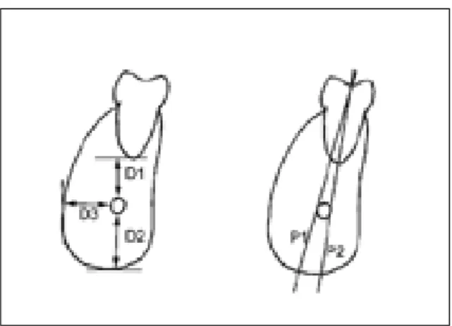
하악골체부 하악관의 수평 및 수직 위치 관계를 파악하기 위하 여 하악 제1대구치의 근심치근첨,
원심치근첨 및 하악제2대구치
의 근심치근첨,원심치근첨, 총 4곳을 선택하였다. 선택된 4곳에
서 하악관까지의 수직 거리(D1),
하악관에서부터 하방으로 하악 골체부 하연까지의 수직거리(D2) 그리고 하악관에서부터 하악 골체부 외연까지의 수평거리(D3)를 변수로 하여 각각에 대한 평 균과 표준편차를 구하였고 통계 처리 프로그램을 이용한 분산 분 석 및 사후 분석을 통하여 각 계측치간의 비교검정을 시행하였다(Table 8). 하악관의 수평적 위치 관계 파악에 있어서 하악골체
부의 장축(P1) 및 치아의 장축(P2)을 기준으로 하악관이 협설 방향으로 어느 부위에 존재하는가를 살펴보았다(Fig 1).계측 항목
1) 하악관의 수직적 위치관계
① 치근첨과 하악관 상연과의 거리
② 하악골의 하연과 하악관과의 거리
2) 하악관의 수평적 위치관계
① 하악골의 외연과 하악관과의 거리
② 치아의 장축에 대한 하악관의 협설측 위치
③ 하악골의 장축에 대한 하악관의 협설측 위치
치아의 장축은 다근치의 경우 치근 만곡이 나타나기 시작하는 근첨⅓을 제외한 근심부 치근을 기준으로 하였다
.
측정치의 계측은
0.5mm 단위로 시행되었으며 계통오차를 확인하기 위하여
각 값에 대하여 두 번의 계측을 시행하였다.
계통 오차는 쌍체비
교에 의하여 검정되었으며 두 계측치 간의 평균치의 차이는0.0025mm로 두 계측치 간에 유의차가 없었다. 무작위 오차는 0.0048, 신뢰도계수는 1을 나타내었다(Table 9). 따라서 계측
오차는 결과의 해석에 영향이 없을 것으로 사료된다.12)Ⅲ. 결 과 1. 하악관의 수직적 위치
4개의 치근 중에서 하악관과의 거리가 가장 큰 것은 제1대구치
근심치근으로 평균5.4mm이었으며,제1대구치 원심치근(
평균5.2mm),
제2대구치 근심치근(평균3.6mm) 그리고 제2대구치 원심치근(평균3.6mm) 순으로 나타났다(Table 1). 치근첨보다 하악관이 상방에 위치하는 증례는 하나도 없었으며 하악관이 치 근첨과 맞닿아 있는 경우가2증례 있었고 모두 하악 제2대구치
Fig. 1. schematic drawing of a tomographic section of
mandibular segment. D1:The distance between the
upper border of the mandibular canal and the root
apex of molar. D2:The distance between the
cortical plate of the mandible and mandibular canal
D3:The distance between the mandibular base and
inferior border of mandibular canal. P1: The Long
axis of the alveolar ridge and mandible. P2: The
long axis of tooth
치근에서 나타났다
.
하악관과 치근에2 m m
이하로 근접한 증례 는 하악 제1대구치부에서는 나타나지 않았으나 제2대구치에서 는12.1%
로 나타났다.또 치근에 3 m m이하로 근접하고 있는 경
우도 하악제1대구치 근심치근이12.1%로 가장 낮게 나타났고
하악제2대구치 근심치근에33.3%로 가장 높게 나타났다(Table 7). 하악관과 하악하연과의 거리는 제2대구치 원심치근 부위에
서 가장 크게 나타났고 제2대구치 근심치근,제 1대구치 원심치
근,제1대구치 근심치근 순으로 나타났다(Table 3).
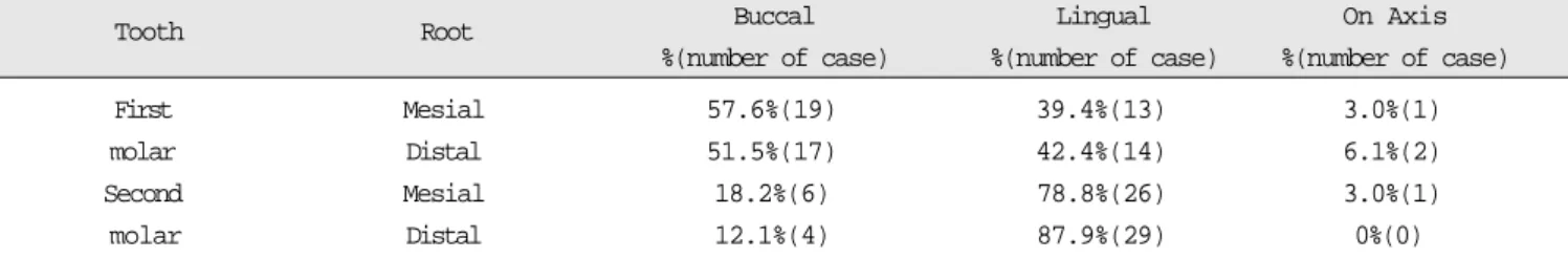
2. 하악관의 수평적 위치
하악골체부의 장축에 대해서 하악관이 협측에 위치한 경우가 제1대구치 근심치근에서
57.6%, 제1대구치 원심치근에서 51.5%로 나타났으나,
치아의 장축에 대한 협설측 관계를 보면 제1대구치 근심 원심치근 모두 협측에 위치한 경우가84.8%로
더 높게 나타났다.제2대구치 치근부위에서는 하악골체부의 장
축에 대한 위치에서 하악관이 협측에 위치한 경우가 근심치근에 서18.2%, 원심치근에서 12.1%를 보였고, 치아장축에 대해서는
근심치근이
45.5%,
원심치근이30.3%
를 보였다(Table 4, 5).
하악관과 하악골 협측 피질골판 사이의 거리는 하악 제1대구치 근심치근부위에서 가장 적게 나타났으며 제2대구치 근심치근 부 위에서 가장 크게 나타났다(Table 2). 하악관과 협측 피질 골판 외면까지의 거리가
4 m m이하인 경우가 하악 제1대구치 근심치
근에서30.3%로 나타났으며 제2대구치 원심치근에서는 3 %에
불과하였다(Table 6).분산 분석 및 사후 분석 시행 결과 치근첨과 하악관 상연과의
Table 2. Average distance(mm) between the buccal corti- cal plate of mandible and mandibular canal
First Mesial 5.5 33 1.9
molar Distal 5.7 33 1.8
Second Mesial 6.2 33 1.7
molar Distal 7.1 33 1.7
Tooth Root Mean N Std. Deviation
Table 3. Average distance(mm) between the lower bor- der of mandible and mandibular canal
First Mesial 6.9 33 2.9
molar Distal 7.2 33 2.4
Second Mesial 8.4 33 2.5
molar Distal 8.9 33 2.3
Tooth Root Mean N Std. Deviation
Table 1. Average distance(mm) between the root apex of the lower molar and mandibular canal
First Mesial 5.4 33 2.1
molar Distal 5.2 33 1.9
Second Mesial 3.6 33 1.4
molar Distal 3.6 33 1.5
Tooth Root Mean N Std. Deviation
Table 4. Position of the mandibular canal to tooth long axis in the buccolingual aspect
First Mesial 84.8%(28) 6.1%(2) 9.1%(3)
molar Distal 84.8%(28) 9.1%(3) 6.1%(2)
Second Mesial 45.5%(15) 48.5%(16) 6.1%(2)
molar Distal 30.3%(10) 69.7%(23) 0%(0)
Tooth Root Buccal Lingual On Axis
%(number of case) %(number of case) %(number of case)
Table 5. Position of the mandibular canal to mandibular body axis in the buccolingual aspect on cross-sectional view
First Mesial 57.6%(19) 39.4%(13) 3.0%(1)
molar Distal 51.5%(17) 42.4%(14) 6.1%(2)
Second Mesial 18.2%(6) 78.8%(26) 3.0%(1)
molar Distal 12.1%(4) 87.9%(29) 0%(0)
Tooth Root Buccal Lingual On Axis
%(number of case) %(number of case) %(number of case)
Table 7. The Number and percentage of cases that the distance between the mandibular canal and molar tooth apex are under 2mm
First Mesial 87.9%(29) 0%(0) 12.1%(4) molar Distal 78.8%(26) 0%(0) 21.2%(7) Second Mesial 54.5%(18) 12.1%(4) 33.3%(11)
molar Distal 57.6%(19) 12.1%(4) 30.3%(10) Tooth Root > 2 m m < 2 m m < 3 m m Table 6. The Number and percentage of cases that the
distance between the mandibular canal and outer surface of buccal cortical plate are under 4mm
First Mesial 69.7%(23) 30.3%(10) molar Distal 78.8%(26) 21.2%(7) Second Mesial 87.9%(29) 12.1%(4) molar Distal 97.0%(32) 3.0%(1)
Tooth Root > 4 m m < 4 m m
Table 8. ANOVA and post hoc test.
D1:The distance between the upper border of the mandibular canal and the root apex of molar. D2:The distance between the cortical plate of the mandible and mandibular canal. D3:The distance between the mandibular base and inferior border of mandibular canal. MR6: The point corresponding to mesial root apex of mandibular first molars. DR6: The point corre- sponding to distal root apex of mandibular first molars. MR7: The point corresponding to mesial root apex of mandibular second molars. DR7: The point corresponding to distal root apex of mandibular second molars.
D1 Dunnett T3 M R 6 D R 6 .1818 .4243 .999
MR7 1.7727* .4243 .001
D R 7 1.8030* .4243 .001
D R 6 M R 6 -.1818 .4243 .999
M R 7 1.5909* .4243 .001
D R 7 1.6212* .4243 .001
M R 7 M R 6 -1.7727* .4243 .001
D R 6 -1.5909* .4243 .001
D R 7 3.0303E-02 .4243 1.000
D R 7 M R 6 -1.8030* .4243 .001
D R 6 -.6212* .4243 .001
M R 7 -3.0303E-02 .4243 1.000
D 2 Dunnett T3 M R 6 D R 6 -.2424 .6332 .999
M R 7 -1.5000 .6332 .162
D R 7 -2.0152* .6332 .018
D R 6 M R 6 .2424 .6332 .999
M R 7 1.2576 .6332 .236
D R 7 -1.7727* .6332 .022
M R 7 M R 6 1.5000 .6332 .162
D R 6 -1.2576 .6332 .236
D R 7 -.5152 .6332 .948
D R 7 M R 6 2.0152* .6332 .018
D R 6 1.7727* .6332 .022
M R 7 .5152 .6332 .948
D 3 Dunnett T3 M R 6 D R 6 .2273 .4367 .997
M R 7 -.7576 .4367 .430
D R 7 -1.6818* .4367 .002
D R 6 M R 6 .2273 .4367 .997
M R 7 -.5303 .4367 .765
D R 7 -1.4545* .4367 .007
M R 7 M R 6 .7576 .4367 .430
D R 6 .5303 .4367 .765
D R 7 -.9242 .4367 .161
D R 7 M R 6 1.6818* .4367 .002
D R 6 1.4545* .4367 .007
M R 7 .9242 .4367 .161
Dependent Variable (I) ROOT (J) ROOT Mean Difference(I-J) Std. Error
거리(D1)는 제1대구치 근,
원심치근 부위 모두 제2대구치 근, 원
심치근 부위에서보다 유의성 있게 길게 나타났고(p<0.05), 하악 골 하연과 하악관과의 거리(D2)는 제2대구치 원심치근 부위보 다 제1
대구치의 근,
원심 치근 부위 모두 유의성 있게 거리가 짧은 것으로 나타났다(p<0.05). 하악골의 외연과 하악관과의 거리
(D3)도 제2대구치 원심치근 부위보다 제1대구치의 근, 원심 치
근 부위 모두 유의성 있게 거리가 짧은 것으로 나타났다(p<0.05).
Table 9. Random error estimation
Se; estimate of the random error of the measurements, Tt; the coefficient of reliability.
1st 2nd Difference 1st 2nd Difference 1st 2nd Difference
Mean 4.4508 4.4545 0.0037 7.8523 7.8485 0.0038 6.1212 6.1212 0.0000
Sd 1.8990 1.9057 0.0067 2.6849 2.6794 0.0055 1.8781 1.8699 0.0082
Variance 3.6062 3.6317 0.0000 7.2087 7.1792 0.0000 3.5273 3.4965 0.0001
Se 0.0047 0.0039 0.0058
Tt 1.0000 1.0000 1.0000
D 1 D 2 D 3
Fig. 2. Distance of the inferior alveolar canal from the mesial roots of the mandibular first molars
Fig. 3. Distance of the inferior alveolar canal from the distal roots of the mandibular first molars
Fig. 4. Distance of the inferior alveolar canal from the mesial roots of the mandibular second molars
Fig. 5. Distance of the inferior alveolar canal from the
distal roots of the mandibular second molars
Ⅳ. 총괄 및 고찰
하악관은 하악지 부위에서는 하악공으로부터 전하방으로 주행 하여 하악골체부위에서는 수평으로 주행하다가 치아의 치조와 로 향하게 된다.
하악관은 하치조신경과 혈관을 포함하며 치아의
치근으로 들어간다.
제2
소구치 치근 하방에서 하악관은 이공(mental foramen)과 절치관(incisive canal)으로 나뉘게
된다.하악관은 방사선학적으로 매우 다양한 변이를 보이는데,
대부분에 경우에 있어 하악관은 방사선투과성 선(radiolucent line)과 방사선불투과성 그림자(radiopague shadow)로 보이
게 된다.13)어떤 경우에서 하악관은2개의 희미한 흰 선(white line)사이에 골밀도의 변화가 없는 구조물로도 보이기도 한다.
일반적으로
2개의 선중 위의 선이 밀도가 높게 나타난다.
14)하악 관은 흔히 방사선 사진 상에서 구치부 치근의 하방에서 보이게 되지만 구치부 치근의 상방에 나타나거나 치근첨과 중첩되어 보 이게 되기도 한다.
6)하지만 일반적인 치과방사선 사진 상에서 치 아와 하악관과의 관계가 정확하게 나타나지는 않는다.구내방사
선사진과 단층사진을 비교한 연구에서 구내사진에서 치근과 하 악관이 연결돼 보이는 경우라도 단층사진상에서는 그렇게 보이 지 않는 경우가 있다고 하였다.15)H o w e와 Poynton은
16)3대구
치 치근과 하악관이 연결되어 보이는 환자들에게서 발치를 시행 하였을 때3명중 1명 꼴로만 술후 감각이상을 호소한다고 하였
다.일반적으로 전산화단층촬영은 김진 등17)의 연구에서와 같이 다발성 골절이나 종괴의 크기가 커서 제거술 및 재건술을 함께 고려하는 경우
,
악안면 기형 환자의 술전 평가 및 치료계획 수립 시와 같은 악안면 영역의 특수 상황의 진단 및 치료계획 수립 시 요구된다.이와 더불어 전산화단층촬영을 하였을 경우 수직단면
상에서 하악관의 위치나 모양을 쉽게 관찰 할 수 있다.
17)그러나 특별한 경우 하악관의 모양이나 위치가 쉽게 그 주위 조직과 구 별되지 않을 수도 있다. Abrahams 등18)은 이런 경우 하악관의위치를 찾아내는데 몇 가지 방법을 제시하였다
.
첫째로cortical niche sign을 이용하는 방법인데, 하악관이 하악골의 피질골과
근접되어 주행할 경우 피질골의 설측 내면(inner corticalmargin)을 따라서 움푹 들어간 부위(niche)를 형성하게 된다
는 것이다.두 번째는 여러 사진을 종합하여 판독하는 삼각계측
법(triangulation)으로 수직단면상을 판독할 때 횡단면상과 파노라마 상을 함께 참조하는 것이고,마지막으로 연속된 여러
부위의 수직단면상을 이용하는 방법인데 보이지 않는 부위는 하 악관이 잘 보이는 앞뒤의 사진을 이용하여 위치를 추정하는 것이 다.본 연구에서는 하악관의 경계가 명확하게 보이지 않는 경우
는 연구에서 제외하였다.
Fox는 500명의 환자를 대상으로 제2대구치에서 하악관의 위
치를 조사하였다.6)제2대구치 치근과2 m m이내의 근접도를 보
인high canal의 경우가 연구대상의 18.4%이었고 하악관과
치근이 직접 연결된 경우가2 %이었으며 방사선 사진 상 치근과
하악관이 중첩되어 보이나 연결되지 않는 경우는3.2%라고 하
였다. Heasman의 연구에서도 제2대구치 치근과2 m m이내의
근접도를 보인high canal
이15.6%
에서 나타난다고 하였다.
19) 하지만 남아프리카 환자를 대상으로 한Nortj의 연구에서는 high canal이 46.7%에 이르고 하악관과 하악골 하연의 사이
가 가까운Low canal
이48.9%
라고 하였으며 연령이 증가함에 따라 치근과의 근접도가 낮아지고 이는 치아의 맹출과 관련이 있 을 것이라고 하였다.20)154명의 한국인 마른 턱뼈를 대상으로 한
김희진 등의 연구21)에서 하악관이 치근과 가까이 지나가는“높 은”형태가10례로 6.5%의 발현율을 보인 것과 비교하여 본 연
구에서는 제2대구치 근심치근에서2 m m이내의 거리를 보이는
증례가12.1%, 3mm이내의 거리를 보이는 증례가 33.3%로 가
장 크게 나타났다.
하악관과 치근과의 거리에서 제1
대구치 치근 과의 거리는 제2대구치 치근과의 거리에 비해 비교적 큰 값을 보 였고 제1대구치 근심치근이 가장 큰 값을 보였다.Bjo
¨rk
은 하악관의 모양과 위치가 하악골의 성장동안 변하지 않는다고 하였고 대부분의 골성장은 하악골의 바깥쪽에서 나타 나며 하악관 자체의 변화는 거의 없다고 하였다.22)Nortj 등은
Fig. 6. Distance of the inferior alveolar canal from the lateral cortex of the mandible.
Fig. 7. Distance of the inferior alveolar canal from the
inferior cortex of the mandible.
하악관과 하악구치 치근과의 수직적인 관계가 안정되어 있다고 하였으며 하악골 내에서 하악관의 주행경로가 하악지에서 전방 으로 갈수록 하방주행하기 때문에 제3대구치에서 제일 가깝고 제2대구치와 제1대구치로 갈수록 증가한다고 하였는데14)본 연 구 결과 유사한 양상을 보이고 있었다.
발치나 치근단 수술 등의 술식을 시행할 때 뿐 아니라 하악지시 상골절단술을 시행할 때도 하악관이 손상될 가능성은 많다.
하악
골의 하악지 부분과 제1, 2대구치 부위의 피질골을 양측으로 분 리하기 때문에 하악관이 협측 피질골판에 가깝게 주행할 수록 수 술시 하악관의 손상이 일어나기 쉽다.23)T a ma
′s
는 하악관과 협측 피질골판 내측면과의 거리에 따라4
개의 군으로 분류하였는데 반 이상이 하악관과 협측피질골판 내측면과의 거리가3 - 4 m m인
군에 포함되었고1 m m이하의 군에도 2 0 %
정도가 포함된다고 하였다.24)본 연구에서는 하악골의 협측피질골판 외측면과의 거 리를 측정하였는데 각 치근별 평균거리는Table 2에 있다. 임프
란트 치료에 있어 지름3.75mm의 standard fixture의 식립이
가능하기 위하여 요구되는 최소한의 하악골 협설폭을4 m m로
볼 때
4 m m이하의 경우가 제1대구치 근심치근부위에서
30.3%(10증례)
로 가장 크게,제2대구치 부위에서 3%(1증례)
로 가장 적게 나타났는데 이는 임프란트 식립 시 제2대구치에서 보다 제1
대구치에서 협설폭의 확보가 더욱 어려울 수 있음을 암 시한다.Ⅴ. 결 론
1999년부터 2001년까지 아주대학교병원 치과에 내원하여 Dentascan을 촬영한 33명의 환자에서 하악관과 하악 제1, 2대
구치의 근첨 및 하악골 외연과의 거리를 측정하여,하악관의 이
들 구조물에 대한 거리와 수직적 및 수평적인 위치관계를 조사하 여 다음과 같은 결론을 얻었다.1.
하악 제1, 2
대구치의4
개의 치근 중에서 하악관과의 거리가 가장 큰 것은 제1대구치 근심치근이었으며 제2대구치 원심치 근이 가장 적게 나타났다(p<0.05).2.
하악관과 하악골 협측피질골판사이의 거리는 하악 제1대구 치 근심치근부위에서 가장 적게 나타나고 제2대구치 원심치 근부위에서 가장 크게 나타났다(p<0.05).3.
하악관과 하악하연과의 거리는 제2대구치 원심치근 부위에 서 가장 크게 나타났고 제1대구치 근심치근부위에서 가장 적 게 나타났다(p<0.05).4.
하악관의 협설측 관계를 보면 근심에서 원심으로 이동할수록 설측으로 위치하는 경향이 나타났으며,
치아의 장축에 대한 협설측 위치보다 하악골의 장축에 대한 협설측 위치에서 설측 에 많이 위치하였다.위의 결과를 바탕으로 하악구치부의 임프란트 치료를 계획할 때,
다른 조건이 모두 동일한 경우 하치조 신경의 손상을 방지하
기 위해서는 후방으로 갈수록 짧은 길이의 임프란트를 선택하며 협측으로 가까이 식립하는 것이 중요하다고 사료된다.참고문헌