Autoimmune epilepsy is a newly emerging area of epilepsy. The concept of “autoimmune” as an etiology has recently been revisited thanks to advances in autoimmune encephalitis and precision medicine with immunotherapies. Autoimmune epilepsy presents with specific clini- cal manifestations, and various diagnostic approaches including cerebrospinal fluid analysis, neuroimaging, and autoantibody tests are essential for its differential diagnosis. The diagnosis is often indeterminate despite performing a thorough evaluation, and therefore empirical im- munotherapy may be applied according to the judgment of the clinician. Autoimmune epilep- sy often manifests as new-onset refractory status epilepticus (NORSE). A patient classified as NORSE should receive empirical immunotherapy as soon as possible. On the other hand, a more- cautious, stepwise approach is recommended for autoimmune epilepsy that presents with epi- sodic events. The type of autoimmune epilepsy is also an important factor to consider when choosing from among various immunotherapy options. Clinicians should additionally take the characteristics of antiepileptic drugs into account when using them as an adjuvant therapy. This expert opinion discusses the diagnostic and treatment approaches for autoimmune epilepsy from a practical point of view.
Key Words autoimmune epilepsy, autoimmune encephalitis, NORSE,
autoimmune epilepsy treatment, autoimmune epilepsy immunotherapy, refractory status epilepticus.
Clinical Approach to Autoimmune Epilepsy
INTRODUCTION
Autoimmune epilepsy is a group of epilepsies mediated by all kinds of autoimmune reac- tions.1 Most cases of autoimmune epilepsy consist of autoimmune limbic encephalitis, with inflammation of the limbic area causing seizure, memory loss, unconsciousness, and psy- chiatric symptoms. In a broad spectrum, autoimmune epilepsy also includes febrile illness- related refractory seizures (FIRES), new-onset refractory status epilepticus (NORSE), cere- bral autoinflammatory diseases, and epilepsy induced by systemic autoimmune disorders.
This expert opinion aims to provide clinical approaches to diagnosing and treating autoim- mune epilepsy.
CLINICAL CLUES OF AUTOIMMUNE EPILEPSY
Prodromal symptoms
Fever, headache, dizziness, insomnia, or upper respiratory infection may present as prodro- mal symptoms. Short-term fever generally manifests after immunization against self-anti- gens. A viral infection should be suspected if the fever persists for more than 3 days. How- ever, since viral encephalitis can be followed by autoimmune encephalitis,2,3 clinicians need to consider both possibilities depending on the other symptoms and diagnostic test results of the patient.
Yoonhyuk Janga, Dong Wook Kimb Kwang Ik Yangc, Jung-Ick Byund Jong-Geun Seoe, Young Joo Nof Kyung Wook Kangg
Daeyoung Kimh, Keun Tae Kimi Yong Won Choi, Soon-Tae Leea on behalf of the Drug Committee of Korean Epilepsy Society
a Department of Neurology, Seoul National University Hospital, Seoul, Korea
b Department of Neurology, Konkuk University School of Medicine, Seoul, Korea
c Department of Neurology, Soonchunhyang University
College of Medicine, Cheonan Hospital, Cheonan, Korea
d Department of Neurology, Kyunghee University Hospital at Gangdong, Seoul, Korea
e Department of Neurology, School of Medicine,
Kyungpook National University, Daegu, Korea
f Department of Neurology,
Samsung Noble County, Yongin, Korea
g Department of Neurology,
Chonnam National University Hospital, Chonnam National University School of Medicine, Gwangju, Korea
h Department of Neurology,
Chungnam National University Hospital, Chungnam National University School of Medicine, Daejeon, Korea
i Department of Neurology, Keimyung University
Dongsan Medical Center, Daegu, Korea
Received January 29, 2020 Revised March 2, 2020 Accepted March 4, 2020 Correspondence Soon-Tae Lee, MD, PhD Department of Neurology, Seoul National University Hospital, 101 Daehak-ro, Jongno-gu, Seoul 03080, Korea Tel +82-2-2072-4757 Fax +82-2-3672-7553 E-mail [email protected] Yong Won Cho, MD, PhD Department of Neurology, Keimyung University Dongsan Medical Center, 1095 Dalgubeol-daero, Dalseo-gu, Daegu 42601, Korea Tel +82-53-258-7832 Fax +82-53-258-4380 E-mail [email protected]
cc This is an Open Access article distributed under the terms of the Creative Commons Attribution Non-Com- mercial License (https://creativecommons.org/licenses/by-nc/4.0) which permits unrestricted non-commercial use, distribution, and reproduction in any medium, provided the original work is properly cited.
JCN
Open AccessREVIEW
pISSN 1738-6586 / eISSN 2005-5013 / J Clin Neurol 2020;16(4):519-529 / https://doi.org/10.3988/jcn.2020.16.4.519
Autoimmune Epilepsy
JCN
Clinical course
The clinical course of autoimmune epilepsy has an acute-to- subacute progression. The clinical symptoms aggravate rap- idly from onset to nadir during the acute amplification period of the self-reactive lymphocytes. While a viral infection gen- erally stabilizes within 2 weeks by the production of antibod- ies, autoimmune epilepsy, if not treated by immunotherapy, often progresses to status epilepticus refractory to conven- tional antiepileptic drugs (AEDs). Some cases of autoim- mune epilepsy show spontaneous remission, but in the pres- ence of triggers, it can recur even several years later. NORSE can be the most-severe form of autoimmune epilepsy, and it is defined as a clinical presentation—not a specific diagno- sis—in a patient with the new onset of refractory status epi- lepticus without a clear acute or active structural, toxic, or metabolic cause.4
Comorbid symptoms of autoimmune encephalitis
Autoimmune epilepsy is accompanied by altered mental sta- tus, psychiatric symptoms, or memory deficits.5 The pres- ence of comorbid symptoms often indicates a diagnosis of autoimmune encephalitis, with N-methyl-D-aspartate re- ceptor (NMDAR) encephalitis characterized by psychosis, dyskinesia, hypoventilation, and autonomic dysfunction. Fa- ciobrachial dystonic seizure (FBDS) and hyponatremia are unique features in leucine-rich glioma-inactivated protein 1 (LGI1)-antibody encephalitis.6 Along with refractory sta- tus epilepticus, epilepsia partialis continua frequently pres- ents in γ-aminobutyric acid (GABA)-A encephalitis.7 Pa- tients with glutamic acid decarboxylase (GAD) encephalitis may develop limbic encephalitis (characterized by seizures and memory decline) or other neurologic syndromes such as cerebellitis and stiff-person syndrome. Nonneurologically, the GAD antibody is associated with type I diabetes mellitus.8Tumor
Autoimmune epilepsy can be associated with a tumor as a paraneoplastic syndrome, but association with an incidental tumor is also common. Ovarian teratoma, thymoma, small- cell lung cancer, and neuroendocrine tumors are especially prone to inducing paraneoplastic syndromes, with approxi- mately 40% of NMDAR encephalitis patients having an ovar- ian teratoma.9,10 Additionally, contactin-associated protein-like 2-antibody encephalitis is associated with thymoma in about 5% of patients.10 Small-cell lung cancer is detected in 70% of patients with GABA-B encephalitis, while lung cancer and thy- moma are found in approximately 70% of patients with α-amino- 3-hydroxy-5-methyl-4-isoxazolepropionic acid (AMPA) en- cephalitis. On the other hand, no tumor has been found to be associated with GAD. These observations indicate that clini-
cians should consider applying cancer screening to patients with autoimmune epilepsy.
NORSE
A multicenter study found the NORSE etiology in about half of the patients (47%), with autoimmune etiologies (37%, comprising 19% nonparaneoplastic and 18% paraneoplastic) being more common than infection (8%).11 These results in- dicate that an autoimmune pathogenesis is much more likely than a viral infection in NORSE. Therefore, after performing a thorough evaluation of infection, it is possible to consider NORSE as potentially autoimmune epilepsy requiring active immunotherapy.
DIAGNOSTIC TESTS
Cerebrospinal fluid analysis
Autoimmune encephalitis generally presents with an abnor- mal cerebrospinal fluid (CSF) profile, including lymphocytic pleocytosis and elevated protein levels.12,13 However, an infec- tious cause must always be excluded. Atypical bacteria such as listeria, tuberculosis, or borrelia can mimic the symptoms of autoimmune epilepsy. The immunoglobulin G (IgG) in- dex and oligoclonal band in the CSF are helpful to confirm the presence of intrathecal antibody synthesis. While the CSF protein in the acute stage is sensitive to active ongoing inflam- mation, high levels of CSF protein in the chronic stage can in- dicate remaining inflammation.
Neuroimaging
The findings of brain magnetic resonance imaging (MRI) in patients with certain types of autoimmune encephalitis can vary from normal to T2-weighted hyperintensities in the mesial temporal lobes or multifocal brain lesions. NORSE patients exhibit progressive medial temporal atrophy even af- ter the NORSE stops,14 which might be due to the initial in- jury or an ongoing inflammatory process. The hippocampal atrophy that occurs in NMDAR encephalitis is potentially reversible.15 On the other hand, the hippocampus of pa- tients with LGI1-antibody encephalitis becomes atrophied if immunotherapy is delayed.16 Multifocal T2-weighted hyper- intensities appear in the cortex and subcortex regions of the temporal and frontal lobes in GABA-A encephalitis.718F-flu- orodeoxyglucose positron-emission tomography (18F-FDG PET) reveals remarkable occipital hypometabolism in NM- DAR encephalitis as well as prominent hypermetabolism in the hippocampus and basal ganglia in LGI1-antibody enceph- alitis.17 Arterial spin labeling perfusion MRI can detect epi- leptic foci.18 However, 18F-FDG PET and arterial spin label- ing alone cannot reliably distinguish the different causes of
Jang Y et al.
JCN
encephalitis. Finally, clinicians should be aware that a certain proportion of patients with autoimmune encephalitis with no detectable antibody have less-severe or even normal brain MRI or 18F-FDG PET findings despite suffering from relative- ly severe symptoms.
Electroencephalography
There is no specific electroencephalography (EEG) sign for distinguishing the different types of autoimmune epilepsy. Ex- ceptionally, the extreme delta brush has been suggested to be a specific EEG sign of NMDAR encephalitis, and is found in 30% of patients.19 It is noteworthy that FBDS (the pathogno- monic feature of LGI1-antibody encephalitis) is diagnosed based on the phenomenology alone rather than using EEG.
FBDS manifests as a brief (<3 seconds) dystonic movement of the arm that also includes the ipsilateral face or the leg.6,20 Since EEG reveals nonspecific abnormalities, clinicians should rely on the clinical history and neurologic examinations. Never- theless, EEG still plays a significant role in detecting seizures and differentiating them from behavioral symptoms or altered mentality in autoimmune epilepsy.
Autoantibody tests
Autoantibody detection provides a confirmatory diagnosis of autoimmune encephalitis, and thus is recommended in all suspected patients. Clinicians should be aware that no defi- nite autoantibody will be present in 40–50% of cases.21,22 Au- toantibody screening is applied to both the serum and CSF.
In NMDAR encephalitis, the disease severity is correlated with the antibody titer in the CSF but not that in the serum. Al- though the presence of a systemic antibody (thyroid peroxi- dase antibody and antinuclear antibodies) is not pathogenic, this can indicate the presence of an autoimmune response.
Brain biopsy
A brain biopsy can be the final option for patients who do not respond to empirical immunotherapy. A specimen must be carefully obtained from focal lesions indicated by brain MRI showing contrast enhancement, T2-weighted fluid-at- tenuated inversion recovery hyperintensity, or high-intensity diffusion-restricted area in diffusion-weighted imaging. An- alyzing the biopsy specimen will make it possible to more accurately exclude other etiologies such as lymphoma, pri- mary angiitis of the central nervous system (CNS), demye- linating disease, or other malignancy. Moreover, the compo- sition of infiltrative lymphocytes (e.g., CD3, CD4, CD8, CD20, and CD68) in the specimen provides information about the main immunity type responsible for autoimmune epilepsy, suggesting the immunotherapy target; for example, cyclophos- phamide can be administered to a patient with a high-T-cell
infiltrative lesion, and a patient who had a CD68+ microgli- al lesion responded well to anakinra.23 Nevertheless, a brain biopsy is invasive, and so cases should be selected carefully.
DIFFERENTIAL DIAGNOSIS AND DIAGNOSTIC APPROACH
The two important steps to addressing autoimmune epilep- sy are 1) excluding other etiologies and 2) applying empiri- cal treatment during the diagnosis. From the viewpoint of the
“autoimmune” etiology, the detailed criteria are well described in the guideline for autoimmune encephalitis, and the defini- tion can be applied to autoimmune epilepsy with various de- grees of certainty.5 The initial approach starts with a detailed history-taking and neurologic examination (see the sections entitled “Prodromal symptoms” and “Clinical course”). The initial diagnostic workups include blood laboratory, EEG, brain MRI, and CSF studies, which include tests for CNS in- fection and autoantibodies. All of these tests should also con- sider other causes of epilepsy, such as CNS infection and vas- cular, neoplastic, metabolic, degenerative, and genetic diseases.
Accordingly, a wide variety of diagnostic tests is required for the differential diagnosis (Fig. 1).
The differential diagnosis in cases of viral encephalitis should screen for herpes simplex virus (HSV) 1, HSV2, varicella- zoster virus, Epstein-Barr virus, enterovirus 71, cytomegalo- virus, mumps, Japanese/West Nile encephalitis, and measles.
Moreover, atypical bacteria such as listeria, Lyme disease (bor- relia), tuberculosis, rickettsia, and neurosyphilis can mimic the clinical manifestations of autoimmune epilepsy. Rare infec- tious agents including Creutzfeldt-Jakob disease, JC virus, HIV, fungi, amoebas, and parasites should also be excluded.
In addition to infection, the following vascular causes man- ifesting as atypical symptoms are often confused with auto- immune epilepsy: vasculitis, embolic infarction, cerebral ve- nous sinus thrombosis, posterior reversible encephalopathy syndrome, strategic infarction, hemiplegic migraine, and du- ral arteriovenous fistula. Brain tumors such as glioma, lep- tomeningeal seeding, lymphoma, germ-cell tumor and he- mophagocytic lymphohistiocytosis, as well as metabolic diseases including metabolic encephalitis, hyperglycemia/
hypoglycemia, porphyria, and vitamin deficiencies are oth- er major conditions that show symptoms similar to autoim- mune epilepsy and hence need to be considered in the differ- ential diagnosis. Furthermore, degenerative diseases such as Alzheimer’s disease, and developmental diseases such as mi- tochondrial diseases, schizophrenia, cortical dysplasia, and autism can mimic autoimmune epilepsy. Finally, the side effects of some drugs such as 5-fluorouracil, ifosfamide, and metro- nidazole can also produce the clinical symptoms of autoim-
Autoimmune Epilepsy
JCN
mune epilepsy.
Because the results of tests for viruses and autoantibodies can take several days or even weeks to obtain, empirical im- munotherapy such as IVIg can be started if initial labs still support autoimmune epilepsy. In these cases, systemic tu- mors should be screened for and removed when present.
When the final results of autoantibody and virus tests are available, the patients can be reclassified into 1) definite auto- immune epilepsy with the responsible autoantibody, 2) auto- immune epilepsy without definite antibody, and 3) non-au- toimmune epilepsy, where immunotherapy has no effect.5
TREATMENT
OF AUTOIMMUNE EPILEPSY
The treatment of autoimmune epilepsy can be categorized into two axes: 1) disease-modifying treatment by immuno- therapy and 2) the administration of appropriate AEDs. For precise immunotherapy, careful history-taking, neurologic
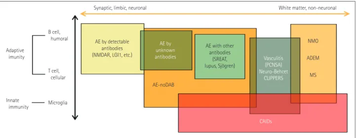
examinations, and pathomechanism analysis of autoimmune epilepsy are mandatory. The pathomechanism of autoim- mune epilepsy can be categorized as shown in Fig. 2. Howev- er, the disease spectrum illustrated in Fig. 2 is hypothetical, and the pathomechanism remains largely unknown.
Therapeutics: immunotherapy
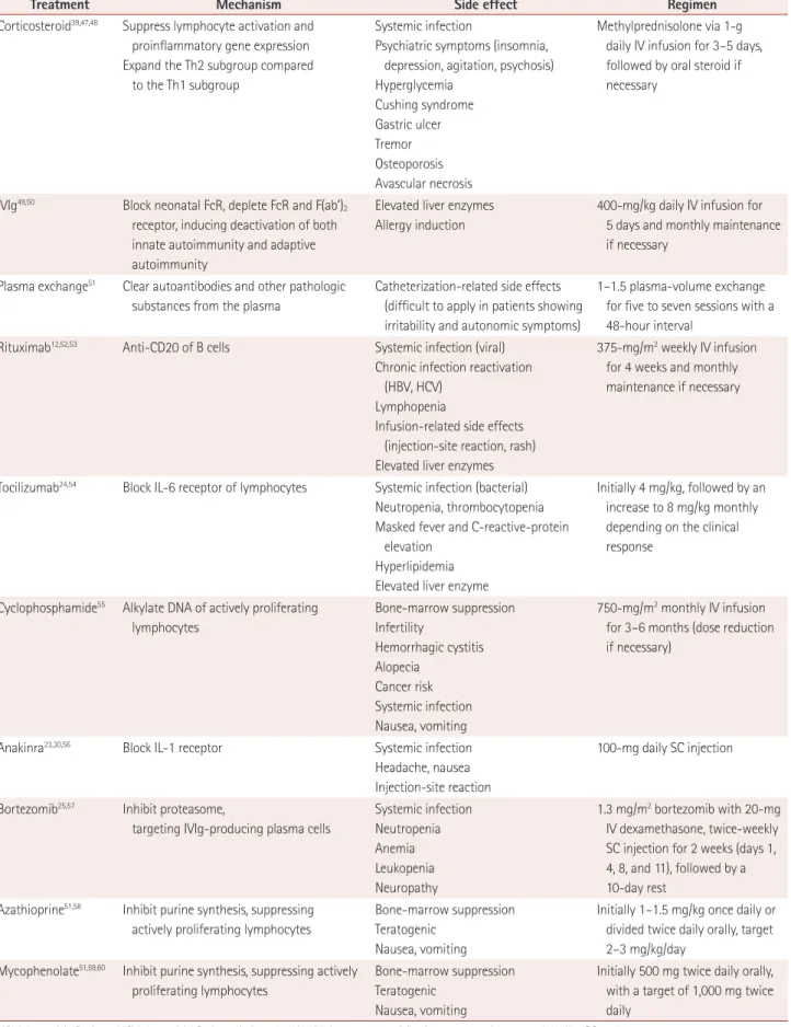
(Table 1)Current evidence for immunotherapy in autoimmune encephalitis
High-dose steroids and intravenous immunoglobulin (IVIg) have been the initial immunotherapies for autoimmune en- cephalitis, affecting a broad spectrum of autoimmune respons- es including humoral and cellular immune reactions (Table 1).
However, more than half of the patients do not respond to the initial therapy, in which cases rituximab has been effective as the next treatment of choice. Rituximab improved the prog- nosis in patients with autoimmune encephalitis regardless of the autoantibody status and whether they responded to the
Suspicious Autoimmune epilepsy
History-taking Neurologic exam
Results of autoantibody and virus tests
Empirical immunotherapy Tumor screening
Possible autoimmune epilepsy Non-autoimmune epilepsy
Non-autoimmune epilepsy Definite autoimmune epilepsy Autoimmune epilepsy without definite antibody
Blood laboratory test EEG Brain MRI CSF study Autoantibody test
Defferential diagnosis - vascular, infectious, neoplastic, metabolic, degenerative, genetic
Fig. 1. Diagnostic approach to autoimmune epilepsy. The initial approaches start with detailed history-taking and neurologic examinations. To ex- clude other etiologies of autoimmune epilepsy, various diagnostic workups including blood laboratory, EEG, brain MRI, and CSF studies are per- formed. Empirical immunotherapy can be applied during the diagnostic tests. The final diagnosis is made based on the results of the tests and the response to immunotherapy. CSF: cerebrospinal fluid, EEG: electroencephalography, MRI: magnetic resonance imaging.
Jang Y et al.
JCN
Fig. 2. Pathomechanism of autoimmune epilepsy. The pathomechanism of autoimmune epilepsy can be categorized into two axes: the targets of autoimmunity and the types of autoimmunity. For the end of one axis, AE with the detectable antibody is representative of synaptic, limbic, and neuronal damage. NMO, ADEM, and MS are white-matter diseases at the other end of the axis. AE-noDAB and AE with other antibodies, vasculitis, neuro-Bechet disease, and CLIPPERS are on the spectrum along this axis. Most of the diseases are caused by a combination of adaptive autoimmu- nity and innate autoimmunity. However, CAIDs are mainly caused by an innate autoimmune reaction. ADEM: acute disseminated encephalomyelitis, AE: autoimmune encephalitis, CAIDs: cerebral autoinflammatory diseases, CLIPPERS: chronic lymphocytic inflammation with pontine perivascular enhancement responsive to steroids, LGI1: leucine-rich glioma-inactivated protein 1, MS: multiple sclerosis, NMDAR: N-methyl-D-aspartate recep- tor, NMO: neuromyelitis optica, noDAB: no detectable antibody, PCNSA: primary central nervous system angiitis, SREAT: steroid-responsive encepha- lopathy-associated thyroiditis.
initial treatment.12 Most (60%) of the nonresponders to the initial immunotherapy showed a favorable outcome after rituximab treatment. Tocilizumab is the next treatment op- tion, showing efficacy in 60% of the patients who did not respond to rituximab.24 However, approximately 10% of pa- tients with autoimmune encephalitis do not respond well after receiving combined treatment with a high-dose cortico- steroid, IVIg, rituximab, and tocilizumab. The optimal man- agement of this group of patients remains unclear, but other drugs that should be investigated include bortezomib,25 ty- rosine kinase inhibitors, and high-dose methotrexate.
Current evidence for immunotherapy in NORSE
Immunotherapy was first applied to NORSE a few years ago.
The first case series showed that plasma exchange (PLEX) therapy could stop refractory seizures in patients with NORSE.26 Combination therapy of high-dose steroid and IVIg was asso- ciated with good outcomes in patients with NORSE.27 High- dose steroid with IVIg or PLEX can be applied to patients with refractory status epilepticus that is strongly suspected to have an autoimmune origin.28 The available data indicate that a certain proportion of patients with FIRES and NORSE respond well to treatment with high-dose steroids (11% and 15%, re- spectively), IVIg (both 5%), and PLEX (2% and 6%).29 How- ever, an analysis of the NORSE etiology showed that while it was caused by infection in only 8% of patients, 52% were cryp- togenic and an autoantibody was found in 37%. Thus, immu-
notherapy is receiving increasing attention as an important treatment option in patients with NORSE.11
Consistent with the above-mentioned findings, a child with superrefractory status epilepticus secondary to FIRES exhibited the overproduction of proinflammatory cytokines such as interleukin (IL)-6 and IL-8 in the CSF, and her sei- zures were controlled by the IL-1 receptor blocker anakinra.30 There is evidence that the serum and CSF levels of IL-6 are higher in patients with NORSE.31 Additionally, the IL-6 re- ceptor blocker tocilizumab stopped NORSE in patients who did not respond to the initial immunotherapy;32 and one or two doses (4–8 mg/kg) of tocilizumab dramatically amelio- rated NORSE in six out of seven patients. The findings of that study imply that tocilizumab can be the primarily-suggested treatment of choice in certain NORSE patients.
Nevertheless, clinicians should be aware that the evidence supporting immunotherapy against NORSE is not yet strong enough for this to be established as a treatment guideline. The current evidence mostly comes from single-center studies or cases, and so further studies involving larger numbers of pa- tients are still required. However, considering the urgency of treating NORSE, we cautiously suggest that immunotherapy would be a recommendable option to clinicians who other- wise have no choice but AEDs in the practical setting.
AE by detectable antibodies (NMDAR, LGI1, etc.)
AE by unknown antibodies
AE with other antibodies
(SREAT, lupus, Sjögren)
CAIDs Vasculitis
(PCNSA) Neuro-Behcet
CLIPPERS AE-noDAB
Synaptic, limbic, neuronal White matter, non-neuronal
B cell, humoral
T cell, cellular
Microglia Adaptive
imunity
Innate immunity
NMO ADEM MS
Autoimmune Epilepsy
JCN
Table 1. Immunotherapy options for autoimmune epilepsy
Treatment Mechanism Side effect Regimen
Corticosteroid39,47,48 Suppress lymphocyte activation and proinflammatory gene expression Expand the Th2 subgroup compared
to the Th1 subgroup
Systemic infection
Psychiatric symptoms (insomnia, depression, agitation, psychosis) Hyperglycemia
Cushing syndrome Gastric ulcer Tremor Osteoporosis Avascular necrosis
Methylprednisolone via 1-g daily IV infusion for 3–5 days, followed by oral steroid if necessary
IVIg49,50 Block neonatal FcR, deplete FcR and F(ab’)2
receptor, inducing deactivation of both innate autoimmunity and adaptive autoimmunity
Elevated liver enzymes Allergy induction
400-mg/kg daily IV infusion for 5 days and monthly maintenance if necessary
Plasma exchange51 Clear autoantibodies and other pathologic substances from the plasma
Catheterization-related side effects (difficult to apply in patients showing irritability and autonomic symptoms)
1–1.5 plasma-volume exchange for five to seven sessions with a 48-hour interval
Rituximab12,52,53 Anti-CD20 of B cells Systemic infection (viral) Chronic infection reactivation
(HBV, HCV) Lymphopenia
Infusion-related side effects (injection-site reaction, rash) Elevated liver enzymes
375-mg/m2 weekly IV infusion for 4 weeks and monthly maintenance if necessary
Tocilizumab24,54 Block IL-6 receptor of lymphocytes Systemic infection (bacterial) Neutropenia, thrombocytopenia Masked fever and C-reactive-protein
elevation Hyperlipidemia Elevated liver enzyme
Initially 4 mg/kg, followed by an increase to 8 mg/kg monthly depending on the clinical response
Cyclophosphamide55 Alkylate DNA of actively proliferating lymphocytes
Bone-marrow suppression Infertility
Hemorrhagic cystitis Alopecia
Cancer risk Systemic infection Nausea, vomiting
750-mg/m2 monthly IV infusion for 3–6 months (dose reduction if necessary)
Anakinra23,30,56 Block IL-1 receptor Systemic infection Headache, nausea Injection-site reaction
100-mg daily SC injection
Bortezomib25,57 Inhibit proteasome,
targeting IVIg-producing plasma cells
Systemic infection Neutropenia Anemia Leukopenia Neuropathy
1.3 mg/m2 bortezomib with 20-mg IV dexamethasone, twice-weekly SC injection for 2 weeks (days 1, 4, 8, and 11), followed by a 10-day rest
Azathioprine51,58 Inhibit purine synthesis, suppressing actively proliferating lymphocytes
Bone-marrow suppression Teratogenic
Nausea, vomiting
Initially 1–1.5 mg/kg once daily or divided twice daily orally, target 2–3 mg/kg/day
Mycophenolate51,59,60 Inhibit purine synthesis, suppressing actively proliferating lymphocytes
Bone-marrow suppression Teratogenic
Nausea, vomiting
Initially 500 mg twice daily orally, with a target of 1,000 mg twice daily
HBV: hepatitis B virus, HCV: hepatitis C virus, IL: interleukin, IV: intravenous, IVIg: intravenous immunoglobulin, SC: subcutaneous.
Jang Y et al.
JCN
How rapidly should immunotherapy be applied in autoimmune epilepsy?
Since NORSE is an emergency condition, immunotherapy should be administered as soon as possible, preferably with- in hours or (at worst) days . The occurrence of an uncontrolled seizure during an ongoing differential diagnosis can itself be fatal, and so the conventional management of status epilepti- cus with AEDs or anesthetics is essential. As empirical im- munotherapy, IVIg can be the first treatment of choice be- cause it is both effective in autoimmune epilepsy and safe in viral encephalitis.33 Moreover, the coadministration of anti- virals should be considered until the autoimmune etiology is confirmed if there is suspicion of infection and a relatively rapid aggravation of symptoms. If a patient does not fully re- spond to the initial immunotherapy, clinicians should move to an alternative immunotherapy such as rituximab, tocili- zumab, anakinra, or cyclophosphamide. Rituximab, cyclophos- phamide, and tocilizumab have demonstrated efficacy in auto- immune encephalitis,9,12,24 while tocilizumab was additionally found to successful in treating NORSE.32
Moreover, the occurrence of only sporadic seizures in au- toimmune epilepsy will give clinicians more time to com- plete the differential diagnosis, and treatment and immuno- therapy can be applied over days and even weeks. The first step is to control seizures by the appropriate administration of AEDs. However, an AED is an adjuvant treatment in auto- immune epilepsy, and few cases of seizure are prevented by an AED alone.20,34-36 The next step is to apply a maximum di- agnostic effort. Empirical immunotherapy is often required since it takes days or even weeks for the results to be obtained in the extensive testing required for a differential diagnosis.
Therefore, clinicians should fully discuss the diagnosis and treatment approaches with the patient and their caregivers before deciding whether they will apply immunotherapeu- tics. IVIg and corticosteroids can be administered as the ini- tial step of immunotherapy. If an infectious origin is excluded and the patient does not respond well to the initial treatment, an alternative immunotherapy such as rituximab, tocili- zumab, or cyclophosphamide can be considered. No guide- line on when to try the next therapy has been established, and hence this currently depends on 1) the response of the pa- tient to the initial immunotherapy and 2) the expected prog- nosis when the next immunotherapeutics are delayed. It is suggested that rituximab or cyclophosphamide produces bet- ter outcomes in patients who have failed to respond to steroids or IVIg.9 However, further investigations are necessary for the upfront or combined initial combined use of monoclonal antibody immunotherapeutics.
Protocol for immunotherapies
High-dose corticosteroidCorticosteroids have historically been applied as the initial treatments for a wide range of inflammatory and autoim- mune diseases. The recommended regimen for autoimmune epilepsy is a 1,000-mg daily IV infusion for 3–5 days. In situ- ations where the etiology is unclear, such as autoimmunity vs. infection, an empirical steroid might aggravate the infec- tion. In addition, steroids can be ineffective against antibody- mediated immune diseases,37-39 and so their efficacy in each type of autoimmune encephalitis needs to be investigated sys- temically. Also, the psychologic side effects of corticosteroids such as delirium, depressive mood, anxiety, and sleep depriva- tion can also interfere the ability of the clinician to accurately judge the treatment response.
IVIg
IVIg is recommended as a 400-mg/kg daily IV infusion for 5 days. The half-life of IVIg is 21–28 days,40 and an additional half-dose infusion can be considered after 1 month. The re- sponse rate for a single administration of IVIg is unknown due to the lack of prospective studies. IVIg is relatively safe, with transient hepatic enzyme elevation being the most-com- mon side effect, and eosinophilia or allergy can be induced.
PLEX
The recommended regimen of PLEX in autoimmune epi- lepsy is 1–1.5 plasma-volume exchanges with 5% albumin replacement fluid. The required consecutive sessions can be performed with a 48-hour interval for five times and more according to the patient’s condition. However, the application of PLEX is restricted in patients with autoimmune epilepsy, since they often show irritability and autonomic symptoms that can result in catheterization-related side effects.
Rituximab
Rituximab is a monoclonal antibody binding to CD20 on the surface of B cells. Rituximab has been classified as a sec- ond-line therapy, but due to successful outcomes in autoim- mune encephalitis, administration of rituximab in combi- nation with steroid or IVIg has recently been considered.
Rituximab shortens the recovery time and reduces the relapse rate.9,12 While the common recommended regimen is a 375- mg/m2 weekly IV infusion for 4 weeks, the dose and inter- val can be adjusted according to the patient’s condition. Be- cause an infusion reaction is common, premedication is required before administering rituximab. The depletion of memory B cells in hepatitis B carriers means that they need to take a preventive dose of antiviral agents.
Autoimmune Epilepsy
JCN
Tocilizumab
Tocilizumab is a monoclonal antibody that blocks the cell- surface IL-6 receptors in a broad spectrum of lymphocytes.
IL-6 is the critical cytokine for inducing both cellular and humoral immune responses in autoimmunity. In particular, plasma cells need IL-6 for their survival, and so tocilizumab might reduce the bone-marrow burden of pathogenic plasma cells. The recommended regimen is to start with a 4-mg/kg in- jection followed by increasing the dosage to 8 mg/kg/month depending on the clinical response. Neutropenia is the most- important adverse effect. The mode of action of tocilizumab can reduce both fever and the elevation of C-reactive protein.
Cyclophosphamide
Cyclophosphamide is an alkylating agent against actively proliferating lymphocytes that reduces both the B- and T- cell burdens. It can therefore be a useful treatment option in patients with vasculitis, excessive T-cell infiltration lesion at biopsy, or autoimmune encephalitis with no detectable anti- body accompanying prominent T2-weighted changes in MRI.
Pulse infusion at 750 mg/m2 monthly for 3–6 months (with dose adjustment according to the clinical condition) is more advantageous than oral intake, given that autoimmune en- cephalitis shows an acute-to-subacute progression. Blood- count monitoring is necessary at 2–3 weeks after the infusion.
Infusion for up to six cycles is recommended, depending on the clinical responses of the patient. Because cyclophospha- mide affects all proliferating cells, infertility is one of the se- rious side effects. Leuprolide acetate, a synthetic gonadotropin- releasing hormone analog, should therefore be considered in all female patients of reproductive age in order to protect them against premature ovarian failure during cyclophos- phamide therapy.41
Early immunotherapy
Previous studies have shown that the earlier initiation of im- munotherapy will result in faster recovery and a better prog- nosis. The prognosis depends on the depth of autoimmunity and the presence of neuronal damage. In NMDAR encephali- tis, although the receptors are regenerated when the disease antibody is removed, delayed immunotherapy will prolong the disease duration and increase morbidity. The autoanti- body of LGI1-antibody encephalitis sometimes induces the loss of hippocampal neurons when treatment is delayed.42,43 Autoimmune epilepsy without definite antibody can result in neuronal damage, since the absence of an autoantibody implies the presence of a novel antibody of unknown patho- genesis or other types of immune reaction, such as innate immunity or T-cell activation, instead of the involvement of plasma cells. Therefore, early empirical immunotherapy should
be actively considered for these patients. However, since there is always a risk of prescribing empirical immunotherapy to patients without autoimmune encephalitis, the importance of a thorough clinical evaluation and the exclusion of alternative diagnoses cannot be overemphasized.
Duration of immunotherapy
There are insufficient data for establishing a consensus on how long to maintain immunotherapy. The timescale gener- ally depends on how the patient responds to immunothera- py: the clinician can consider applying short-term immu- notherapy and monitoring the response if a patient recovers quickly, while in other cases more-aggressive strategies might need to be considered. It is not yet known whether a monthly IVIg-boosting infusion is advantageous and whether the monthly maintenance of rituximab is beneficial for recov- ery, and so additional randomized trials are needed. Mainte- nance immunotherapy is necessary in some cases to prevent recurrence.
Choice of disease-modifying immunotherapeutics according to the pathomechanism
Synaptic antibody
The pathogenesis of autoimmune encephalitis caused by synaptic antibodies are mediated by B cells. Thus, therapeutics against B cells can be an efficient strategy for this disease, with IVIg, high-dose corticosteroids, and PLEX being the first treatment of choice for immunotherapy, and rituximab and tocilizumab being the next treatment choices. Cyclophospha- mide should be cautiously applied in young patients due to its adverse effect on reproductive organs. If remission is in- duced, the patient will usually not need maintenance immu- notherapy to prevent recurrences, especially if they have been treated with alternative immunotherapies.
Intracellular/paraneoplastic antibody
Intracellular antigens are exposed by cytolysis induced by cy- totoxic T cells and complement activation. Thus, the detec- tion of anti-intracellular molecule antibodies indicates that the cellular immune response is the main process in auto- immune encephalitis. The initial immunotherapy in this group of patients involves IVIg and high-dose corticosteroids, which target a broad spectrum of immune responses. Cyclo- phosphamide and rituximab can be treatment options for al- ternative immunotherapy. A maintenance immunosuppressant is required if complete remission is not induced. In addition to immunotherapy, eliminating the source autoantigens con- stitutes another component of the therapy for this group of patients. If autoimmune epilepsy develops in patients with
Jang Y et al.
JCN
malignancy, cancer treatment should be the primary inter- vention as long as the autoimmune epilepsy is managed ade- quately.44 Further research is required into harmonizing che- motherapy and immunotherapy in this situation.
Autoimmune encephalitis with no detectable antibody in T2-weighted hyperintense or contrast-enhanced lesions Other causes of the disease such as vasculitis, lymphoma, other demyelinating diseases, and infectious encephalitis need to be thoroughly evaluated in this group of autoimmune en- cephalitides. However, most cases are miscellaneous. The considered treatment options can include immunothera- peutics targeting a broad spectrum of immune responses, such as high-dose corticosteroids, IVIg, cyclophosphamide, and methotrexate.
Autoimmune encephalitis with no detectable antibody and normal MRI findings
The presence of a clinicoradiologic mismatch in which there is no or only a minimal MRI lesion but severe clinical dete- rioration is suggestive of antibody-mediated encephalitis.
Immunotherapy similar to that for autoimmune encephalitis with a synaptic antibody can be attempted.
NORSE
As mentioned above, the active management of status epilep- ticus according to the standard protocol is the very first treat- ment step for NORSE.45 The sequential application of im- munotherapies including IVIg, rituximab, tocilizumab, and anakinra should then be cautiously but promptly considered in an hourly and daily fashion (please see the section entitled
“How rapidly should immunotherapy be applied in autoim- mune epilepsy?”).
AEDs in autoimmune epilepsy
Which AED to apply first is decided by the clinician. As a symptomatic treatment, no significant difference has been found among AEDs in the management of seizures in auto- immune epilepsy. Fewer side effects, no drug–drug interac- tions, and rapid loading of the drug are major considerations when choosing AEDs for autoimmune epilepsy. The candi- date first-line treatments are next-generation AEDs such as levetiracetam, lacosamide, perampanel, zonisamide, and pre- gabalin, with levetiracetam being the most-used drug.34,36 Moreover, the specific pathogenesis in each type of autoim- mune epilepsy means that certain AEDs would be theoreti- cally preferred or avoided; for example, in NMDAR encepha- litis, NMDAR antagonists such as ketamine should be avoided.
The safety of perampanel, which is an AMPA antagonist, has not yet been studied in AMPA-receptor encephalitis. On the
other hand, GABA-promoting AEDs such as benzodiazepines and barbiturates can be considered in patients with GABA-A encephalitis. However, as discussed below, the side-effect profiles prevent some AEDs from being used in specific sit- uations. The duration of AED use also should be personalized in individual patients, and AEDs can be tapered off when the autoimmune encephalitis is in full remission.36
Drug interactions
As an adjuvant treatment, AEDs should be chosen carefully so as not to affect the efficacy of certain immunotherapeu- tics. Since cytochrome P (CYP) hepatic enzyme inducers re- duce the blood levels of immunotherapeutics, they must be avoided in patients who have taken high-dose corticosteroids, cyclophosphamide, and oral immunosuppressants. Moreover, the use of valproic acid, a CYP2C9 enzyme inhibitor that enhances the blood level of immunosuppressants, should also be restricted due to bone-marrow toxicity.
Psychiatric symptoms and memory decline
Psychiatric symptoms and memory decline are common symptoms in limbic encephalitis, and so AEDs with similar side effects should be avoided as the first choice in these pa- tients. Levetiracetam and perampanel can induce or aggra- vate psychiatric and behavior abnormalities. If levetiracetam or perampanel is used to control seizures in the acute stage, it will be necessary to switch to other AEDs for long-term main- tenance. Topiramate and valproic acid can aggravate cognitive decline in limbic encephalitis.
Rash
A rash can occur as an infusion-related symptom of IVIg and rituximab or as a side effect of other immunosuppressants.
Thus, oxcarbazepine and lamotrigine have the disadvantage that it is difficult to distinguish whether a rash is an immu- notherapy-related symptom or a side effect of the AEDs. In particular, the use of aromatic AEDs has a high risk of idio- syncratic cutaneous reactions in LGI1-antibody encephali- tis.46 In addition, hyponatremia (the side effect of aromatic AEDs) can mimic the symptoms of LGI1-antibody encepha- litis. We therefore recommend avoiding the use of aromatic AEDs in LGI1-antibody encephalitis.
Cytopenia
Cytopenia is one of the serious side effects of immunother- apies such as rituximab, tocilizumab, and cyclophospha- mide. The next immune treatment approach should be de- layed until the blood cell counts recover. Valproic acid and oxcarbazepine can also induce cytopenia, and so they must be carefully considered so as not to aggravate this symptom.
Autoimmune Epilepsy
JCN
CONCLUSION
Considerable progress has been made in the field of autoim- mune epilepsy since the discovery of autoimmune enceph- alitis. In particular, the introduction of various immuno- therapy options has made some cases of the disease curable.
However, the diagnosis still depends heavily on the individual judgment of the clinician, with autoantibody confirmation tests results being negative in more than half of the patients, and thus requiring this type of diagnosis. In this context, em- pirical immunotherapy is simultaneously a therapeutic meth- od and a part of the diagnostic approach for autoimmune epilepsy. Clinicians are well aware of the dilemma that al- though empirical immunotherapy is expensive and has po- tential critical side effects, it has to be applied as soon as pos- sible to a specific group of patients in order to improve the prognosis. Thus, this review may help in the development of practical treatment approaches for autoimmune epilepsy—
the clues for identifying autoimmune epilepsy suggested in this study provide a basic understanding of when to begin em- pirical immunotherapy. Moreover, a method has been provid- ed for selecting immunotherapy options according to the pathomechanisms of different types of autoimmune epilepsy.
Approximately 15% of patients still do not respond well to immunotherapy treatment, and so future research needs to focus on patients with refractory autoimmune epilepsy. The pathomechanism first has to be clarified in order to deter- mine why current immunotherapies are ineffective. Other therapeutic options including those that target the innate immunity are worth considering. The duration of immuno- therapy maintenance should also be addressed. This review is mainly based on evidence gathered from previous studies, but expert opinions are also presented to address certain un- known areas. Further studies are needed to provide support- ing data for these expert opinions.
Author Contributions
Conceptualization: all authors. Funding acquisition: Yong Won Cho, Soon- Tae Lee. Methodology: all authors. Project administration: Soon-Tae Lee, Yong Won Cho. Supervision: all authors. Visualization: Yoonhyuk Jang, Soon-Tae Lee. Writing—original draft: Yoonhyuk Jang, Soon-Tae Lee. Writ- ing—review & editing: Yoonhyuk Jang, Soon-Tae Lee.
ORCID iDs
Yoonhyuk Jang https://orcid.org/0000-0002-3346-3357 Dong Wook Kim https://orcid.org/0000-0003-4484-0602 Kwang Ik Yang https://orcid.org/0000-0001-6343-6520 Jung-Ick Byun https://orcid.org/0000-0002-6224-4575 Jong-Geun Seo https://orcid.org/0000-0002-3944-5731 Young Joo No https://orcid.org/0000-0002-0145-5707 Kyung Wook Kang https://orcid.org/0000-0001-9362-8670 Daeyoung Kim https://orcid.org/0000-0001-9056-0017 Keun Tae Kim https://orcid.org/0000-0002-7124-0736 Yong Won Cho https://orcid.org/0000-0002-6127-1045
Soon-Tae Lee https://orcid.org/0000-0003-4767-7564 Conflicts of Interest
The authors have no potential conflicts of interest to disclose.
Acknowledgements
Soon-Tae Lee have an advisory role and received research grants from GC Pharma outside of the current work.
REFERENCES
1. Scheffer IE, Berkovic S, Capovilla G, Connolly MB, French J, Guil- hoto L, et al. ILAE classification of the epilepsies: position paper of the ILAE Commission for Classification and Terminology. Epilepsia 2017;58:512‐521.
2. Prüss H, Finke C, Höltje M, Hofmann J, Klingbeil C, Probst C, et al.
N-methyl-D-aspartate receptor antibodies in herpes simplex enceph- alitis. Ann Neurol 2012;72:902‐911.
3. Leypoldt F, Titulaer MJ, Aguilar E, Walther J, Bönstrup M, Havemeis- ter S, et al. Herpes simplex virus-1 encephalitis can trigger anti-NMDA receptor encephalitis: case report. Neurology 2013;81:1637‐1639.
4. Hirsch LJ, Gaspard N, van Baalen A, Nabbout R, Demeret S, Lodden- kemper T, et al. Proposed consensus definitions for new-onset refrac- tory status epilepticus (NORSE), febrile infection-related epilepsy syn- drome (FIRES), and related conditions. Epilepsia 2018;59:739‐744.
5. Graus F, Titulaer MJ, Balu R, Benseler S, Bien CG, Cellucci T, et al. A clinical approach to diagnosis of autoimmune encephalitis. Lancet Neu- rol 2016;15:391‐404.
6. Irani SR, Michell AW, Lang B, Pettingill P, Waters P, Johnson MR, et al. Faciobrachial dystonic seizures precede Lgi1 antibody limbic en- cephalitis. Ann Neurol 2011;69:892‐900.
7. Spatola M, Petit-Pedrol M, Simabukuro MM, Armangue T, Castro FJ, Barcelo Artigues MI, et al. Investigations in GABAA receptor an- tibody-associated encephalitis. Neurology 2017;88:1012‐1020.
8. Lancaster E. The Diagnosis and treatment of autoimmune encepha- litis. J Clin Neurol 2016;12:1‐13.
9. Titulaer MJ, McCracken L, Gabilondo I, Armangué T, Glaser C, Iizu- ka T, et al. Treatment and prognostic factors for long-term outcome in patients with anti-NMDA receptor encephalitis: an observational cohort study. Lancet Neurol 2013;12:157‐165.
10. Dalmau J, Graus F. Antibody-mediated encephalitis. N Engl J Med 2018;378:840‐851.
11. Gaspard N, Foreman BP, Alvarez V, Cabrera Kang C, Probasco JC, Jongeling AC, et al. New-onset refractory status epilepticus: etiology, clinical features, and outcome. Neurology 2015;85:1604‐1613.
12. Lee WJ, Lee ST, Byun JI, Sunwoo JS, Kim TJ, Lim JA, et al. Rituximab treatment for autoimmune limbic encephalitis in an institutional co- hort. Neurology 2016;86:1683‐1691.
13. Blinder T, Lewerenz J. Cerebrospinal fluid findings in patients with autoimmune encephalitis-a systematic analysis. Front Neurol 2019;10:
14. Hocker S, Nagarajan E, Rabinstein AA, Hanson D, Britton JW. Pro-804.
gressive brain atrophy in super-refractory status epilepticus. JAMA Neurol 2016;73:1201‐1207.
15. Iizuka T, Yoshii S, Kan S, Hamada J, Dalmau J, Sakai F, et al. Revers- ible brain atrophy in anti-NMDA receptor encephalitis: a long-term observational study. J Neurol 2010;257:1686‐1691.
16. Finke C, Prüss H, Heine J, Reuter S, Kopp UA, Wegner F, et al. Eval- uation of cognitive deficits and structural hippocampal damage in encephalitis with leucine-rich, glioma-inactivated 1 antibodies. JAMA Neurol 2017;74:50‐59.
17. Jang Y, Lee ST, Bae JY, Kim TJ, Jun JS, Moon J, et al. LGI1 expression and human brain asymmetry: insights from patients with LGI1-anti- body encephalitis. J Neuroinflammation 2018;15:279.
Jang Y et al.
JCN
18. Yoo RE, Yun TJ, Yoon BW, Lee SK, Lee SY, Kang KM, et al. Identifica- tion of cerebral perfusion using arterial spin labeling in patients with seizures in acute settings. PLoS One 2017;12:e0173538.
19. Schmitt SE, Pargeon K, Frechette ES, Hirsch LJ, Dalmau J, Friedman D.
Extreme delta brush: a unique EEG pattern in adults with anti-NMDA receptor encephalitis. Neurology 2012;79:1094‐1100.
20. Irani SR, Stagg CJ, Schott JM, Rosenthal CR, Schneider SA, Pettingill P, et al. Faciobrachial dystonic seizures: the influence of immunother- apy on seizure control and prevention of cognitive impairment in a broadening phenotype. Brain 2013;136:3151‐3162.
21. Granerod J, Ambrose HE, Davies NW, Clewley JP, Walsh AL, Mor- gan D, et al. Causes of encephalitis and differences in their clinical presentations in England: a multicentre, population-based prospec- tive study. Lancet Infect Dis 2010;10:835‐844.
22. Dubey D, Pittock SJ, Kelly CR, McKeon A, Lopez-Chiriboga AS, Len- non VA, et al. Autoimmune encephalitis epidemiology and a com- parison to infectious encephalitis. Ann Neurol 2018;83:166‐177.
23. Jang Y, Woo KA, Lee ST, Park SH, Chu K, Lee SK. Cerebral autoin- flammatory disease treated with anakinra. Ann Clin Transl Neurol 2018;
5:1428‐1433.
24. Lee WJ, Lee ST, Moon J, Sunwoo JS, Byun JI, Lim JA, et al. Tocilizum- ab in autoimmune encephalitis refractory to rituximab: an institution- al cohort study. Neurotherapeutics 2016;13:824‐832.
25. Shin YW, Lee ST, Kim TJ, Jun JS, Chu K. Bortezomib treatment for severe refractory anti-NMDA receptor encephalitis. Ann Clin Transl Neurol 2018;5:598‐605.
26. Li J, Saldivar C, Maganti RK. Plasma exchange in cryptogenic new onset refractory status epilepticus. Seizure 2013;22:70‐73.
27. Gall CR, Jumma O, Mohanraj R. Five cases of new onset refractory status epilepticus (NORSE) syndrome: outcomes with early immuno- therapy. Seizure 2013;22:217‐220.
28. Lopinto-Khoury C, Sperling MR. Autoimmune status epilepticus. Curr Treat Options Neurol 2013;15:545‐556.
29. Gaspard N, Hirsch LJ, Sculier C, Loddenkemper T, van Baalen A, Lancrenon J, et al. New-onset refractory status epilepticus (NORSE) and febrile infection-related epilepsy syndrome (FIRES): state of the art and perspectives. Epilepsia 2018;59:745‐752.
30. Kenney-Jung DL, Vezzani A, Kahoud RJ, LaFrance-Corey RG, Ho ML, Muskardin TW, et al. Febrile infection-related epilepsy syndrome treated with anakinra. Ann Neurol 2016;80:939‐945.
31. Sakuma H, Tanuma N, Kuki I, Takahashi Y, Shiomi M, Hayashi M. In- trathecal overproduction of proinflammatory cytokines and chemo- kines in febrile infection-related refractory status epilepticus. J Neurol Neurosurg Psychiatry 2015;86:820‐822.
32. Jun JS, Lee ST, Kim R, Chu K, Lee SK. Tocilizumab treatment for new onset refractory status epilepticus. Ann Neurol 2018;84:940‐945.
33. Iro MA, Martin NG, Absoud M, Pollard AJ. Intravenous immuno- globulin for the treatment of childhood encephalitis. Cochrane Data- base Syst Rev 2017;10:CD011367.
34. Byun JI, Lee ST, Jung KH, Sunwoo JS, Moon J, Kim TJ, et al. Preva- lence of antineuronal antibodies in patients with encephalopathy of unknown etiology: data from a nationwide registry in Korea. J Neuro- immunol 2016;293:34‐38.
35. Liu X, Yan B, Wang R, Li C, Chen C, Zhou D, et al. Seizure outcomes in patients with anti-NMDAR encephalitis: a follow-up study. Epilep- sia 2017;58:2104‐2111.
36. de Bruijn MAAM, van Sonderen A, van Coevorden-Hameete MH, Bastiaansen AEM, Schreurs MWJ, Rouhl RPW, et al. Evaluation of seizure treatment in anti-LGI1, anti-NMDAR, and anti-GABABR en- cephalitis. Neurology 2019;92:e2185‐e2196.
37. Slade JD, Hepburn B. Prednisone-induced alterations of circulating human lymphocyte subsets. J Lab Clin Med 1983;101:479‐487.
38. Lahood N, Emerson SS, Kumar P, Sorensen RU. Antibody levels and response to pneumococcal vaccine in steroid-dependent asthma. Ann Allergy 1993;70:289‐294.
39. Sunwoo JS. Corticosteroid treatment in autoimmune encephalitis. J Neurocrit Care 2017;10:60-68.
40. Lünemann JD, Quast I, Dalakas MC. Efficacy of intravenous immu- noglobulin in neurological diseases. Neurotherapeutics 2016;13:34‐46.
41. Somers EC, Marder W, Christman GM, Ognenovski V, McCune WJ.
Use of a gonadotropin-releasing hormone analog for protection against premature ovarian failure during cyclophosphamide therapy in wom- en with severe lupus. Arthritis Rheum 2005;52:2761‐2767.
42. van Sonderen A, Thijs RD, Coenders EC, Jiskoot LC, Sanchez E, de Bruijn MA, et al. Anti-LGI1 encephalitis: clinical syndrome and long- term follow-up. Neurology 2016;87:1449‐1456.
43. Thompson J, Bi M, Murchison AG, Makuch M, Bien CG, Chu K, et al. The importance of early immunotherapy in patients with faciobra- chial dystonic seizures. Brain 2018;141:348‐356.
44. Gultekin SH, Rosenfeld MR, Voltz R, Eichen J, Posner JB, Dalmau J.
Paraneoplastic limbic encephalitis: neurological symptoms, immu- nological findings and tumour association in 50 patients. Brain 2000;
123:1481‐1494.
45. Brophy GM, Bell R, Claassen J, Alldredge B, Bleck TP, Glauser T, et al. Guidelines for the evaluation and management of status epilepti- cus. Neurocrit Care 2012;17:3‐23.
46. Shin YW, Ahn SJ, Moon J, Kim TJ, Jun JS, Byun JI, et al. Increased ad- verse events associated with antiepileptic drugs in anti-leucine-rich glioma-inactivated protein 1 encephalitis. Epilepsia 2018;59 Suppl 2:
108‐112.
47. Vandevyver S, Dejager L, Tuckermann J, Libert C. New insights into the anti-inflammatory mechanisms of glucocorticoids: an emerging role for glucocorticoid-receptor-mediated transactivation. Endocri- nology 2013;154:993‐1007.
48. Mahata B, Zhang X, Kolodziejczyk AA, Proserpio V, Haim-Vilm- ovsky L, Taylor AE, et al. Single-cell RNA sequencing reveals T helper cells synthesizing steroids de novo to contribute to immune homeo- stasis. Cell Rep 2014;7:1130‐1142.
49. Schwab I, Nimmerjahn F. Intravenous immunoglobulin therapy: how does IgG modulate the immune system? Nat Rev Immunol 2013;13:
176‐189.
50. Jang Y, Lee ST, Kim TJ, Jun JS, Moon J, Jung KH, et al. High albumin level is a predictor of favorable response to immunotherapy in auto- immune encephalitis. Sci Rep 2018;8:1012.
51. Shin YW, Lee ST, Park KI, Jung KH, Jung KY, Lee SK, et al. Treat- ment strategies for autoimmune encephalitis. Ther Adv Neurol Disord 2017;11:1756285617722347.
52. Johnson PW, Glennie MJ. Rituximab: mechanisms and applications.
Br J Cancer 2001;85:1619‐1623.
53. Martin ST, Cardwell SM, Nailor MD, Gabardi S. Hepatitis B reactiva- tion and rituximab: a new boxed warning and considerations for sol- id organ transplantation. Am J Transplant 2014;14:788‐796.
54. Shetty A, Hanson R, Korsten P, Shawagfeh M, Arami S, Volkov S, et al. Tocilizumab in the treatment of rheumatoid arthritis and beyond.
Drug Des Devel Ther 2014;8:349‐364.
55. Emadi A, Jones RJ, Brodsky RA. Cyclophosphamide and cancer:
golden anniversary. Nat Rev Clin Oncol 2009;6:638‐647.
56. Mertens M, Singh JA. Anakinra for rheumatoid arthritis: a systemat- ic review. J Rheumatol 2009;36:1118‐1125.
57. Meister S, Schubert U, Neubert K, Herrmann K, Burger R, Gramatz- ki M, et al. Extensive immunoglobulin production sensitizes myelo- ma cells for proteasome inhibition. Cancer Res 2007;67:1783‐1792.
58. Johnson PJ, McFarlane IG, Williams R. Azathioprine for long-term maintenance of remission in autoimmune hepatitis. N Engl J Med 1995;333:958‐963.
59. Iaccarino L, Rampudda M, Canova M, Della Libera S, Sarzi-Puttinic P, Doria A. Mycophenolate mofetil: what is its place in the treatment of autoimmune rheumatic diseases? Autoimmun Rev 2007;6:190‐195.
60. Ginzler EM, Aranow C. Mycophenolate mofetil in lupus nephritis.
Lupus 2005;14:59‐64.