• 검색 결과가 없습니다.

Implications of Emphysema and Lung Function for the Development of Pneumonia in Patients with Chronic Obstructive Pulmonary Disease

N/A
N/A
Protected

Academic year: 2021

Share "Implications of Emphysema and Lung Function for the Development of Pneumonia in Patients with Chronic Obstructive Pulmonary Disease"

Copied!
7
0
0

로드 중.... (전체 텍스트 보기)

전체 글

(1)

Implications of Emphysema and Lung Function for the Development of Pneumonia in Patients with Chronic Obstructive Pulmonary Disease

Yoonki Hong, M.D. 1 , Jae Seung Lee, M.D. 2 , Kwang Ha Yoo, M.D. 3 , Ji-Hyun Lee, M.D. 4 , Woo Jin Kim, M.D. 1 , Seong Yong Lim, M.D. 5 , Chin Kook Rhee, M.D. 6 , Sang-Do Lee, M.D. 2 and Yeon-Mok Oh, M.D. 2

1

Department of Internal Medicine, Kangwon National University Hospital, Kangwon National University College of Medicine, Chuncheon,

2

Department of Pulmonary and Critical Care Medicine and Clinical Research Center for Chronic Obstructive Airway Diseases, Asan Medical Center, University of Ulsan College of Medicine, Seoul,

3

Department of Internal Medicine, Konkuk University School of Medicine, Seoul,

4

Department of Internal Medicine, CHA Bundang Medical Center, CHA University, Seongnam,

5

Division of Pulmonary and Critical Care Medicine, Department of Medicine, Kangbuk Samsung Hospital, Sungkyunkwan University School of Medicine, Seoul,

6

Department of Internal Medicine, Seoul St. Mary’s Hospital, College of Medicine, The Catholic University of Korea, Seoul, Korea

Background: Chronic obstructive pulmonary disease (COPD) is sometimes complicated with pneumonia, but little is known about the risk factors that promote the development of pneumonia in COPD. These risk factors were evaluated in the present study.

Methods: The data of 324 patients with COPD from a prospective multi-center observational cohort with obstructive lung disease were evaluated retrospectively. To identify risk factors for the development of pneumonia in COPD, the clinical and radiological data at enrollment and the time to the first episode of pneumonia were analyzed by Cox proportional hazard analysis.

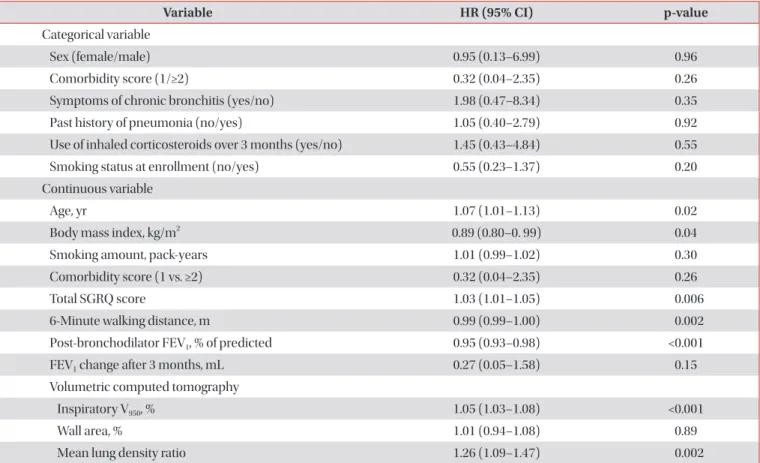
Results: The median follow-up time was 1,099 days and 28 patients (8.6%) developed pneumonia. The Cox analysis showed that post-bronchodilator forced expiratory volume in one second (FEV

1

, % of predicted) and the computed tomography (CT) emphysema extent (inspiratory V950) were independent risk factors for the development of pneumonia (post-bronchodilator FEV

1

: hazard ratio [HR], 0.97; 95% confidence interval [CI], 0.94–1.00; p=0.048 and inspiratory V950: HR, 1.04; 95% CI, 1.01–1.07; p=0.01).

Conclusion: Emphysema severity measured by CT and post-bronchodilator FEV

1

are important risk factors for the development of pneumonia in COPD.

Keywords: Pulmonary Disease, Chronic Obstructive; Emphysema; Pneumonia; Tomography, X-Ray Computed; Risk Factors

Copyright © 2016

The Korean Academy of Tuberculosis and Respiratory Diseases. All rights reserved.

Address for correspondence: Yeon-Mok Oh, M.D.

Department of Pulmonary and Critical Care Medicine and Clinical Research Center for Chronic Obstructive Airway Diseases, Asan Medical Center, University of Ulsan College of Medicine, 88 Olympic-ro 43-gil, Songpa-gu, Seoul 05505, Korea

Phone: 82-2-3010-3136, Fax: 82-2-3010-6968, E-mail: [email protected]

*The abstract was submitted and presented at the 17th Congress of the Asian Pacific Society of Respirology which was held on 14–16 Dec 2012 at the Hong Kong Convention and Exhibition Centre.

Received: Nov. 4, 2015, Revised: Dec. 7, 2015, Accepted: Dec. 28, 2015

cc

It is identical to the Creative Commons Attribution Non-Commercial License (http://creativecommons.org/licenses/by-nc/4.0/).

(2)

Introduction

Chronic obstructive pulmonary disease (COPD) is a com- plex syndrome with significant heterogeneity, having differ- ent clinical presentation, imaging, and response to therapy

1

. COPD is a major cause of chronic morbidity and mortality throughout the world and its incidence is expected to increase over the coming decades

2

. Pneumonia is also a major cause of hospitalization and a common cause of death

3

. Epidemio- logical studies show that COPD is the most common disease associated with pneumonia

4

.

COPD as a risk factor for the development of pneumonia was primarily identified by studies on population-based co- horts

5

or cohorts of patients with pneumonia

6,7

. It has been suggested that COPD severity is an important predictor of pneumonia hospitalization in the Unites States

5

. Elevations in baseline C-reactive protein levels and dyspnea index have also been suggested to be markers of pneumonia risk

8

. More- over, common comorbid conditions, such as advanced age, diabetes mellitus, and cardiovascular disease, in patients with COPD may also associate with an increased risk of pneumo- nia. Recently, concerns is raised on that inhaled corticoste- roids (ICS) use may associate with an increased incidence of pneumonia

9-11

. Despite these reports, it is still unknown which components of COPD associate with the development of pneumonia.

Given recent advances in computed tomography (CT) tech- nology, it may be a useful tool for evaluating the severity, ex- tension, and distribution of the disease components of COPD at both a qualitative and a quantitative level. Indeed, recent studies have reported that particular radiological characteris- tics of COPD are associated with various clinical, genetic, and physiological features, such as greater lung emphysema as- sociated with COPD exacerbations or Gly16 variant in ADRB2 gene associated with airway wall changes

12-14

. However, it is not clear about the role of CT for predicting the development of pneumonia in patients with COPD.

It would be of interest to know which types or components of COPD are associated with the development of pneumonia because being able to predict which patients with COPD are at greatest risk of developing pneumonia may improve the un- derstanding of the heterogeneity in COPD and facilitate better health care for these individuals. Therefore, the present ret- rospective study analyzed clinical features, exercise capacity, lung function, and CT measurements to identify risk factors for the development of pneumonia in patients with COPD on a prospective observational cohort.

Materials and Methods

1. Subjects

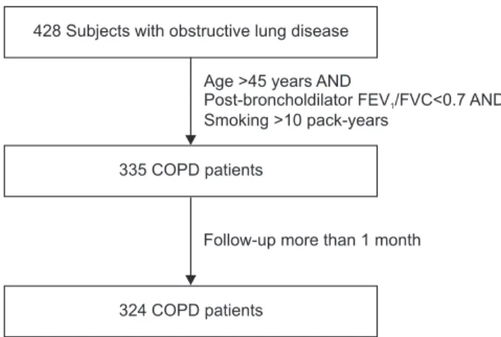
The data of 324 patients diagnosed with COPD were ana- lyzed retrospectively. These patients were selected from the Korean obstructive lung disease (Korean OLD, KOLD) Cohort (Figure 1), which consisted of 428 stable patients with OLD who had been prospectively recruited from the pulmonary clinics of 16 hospitals in South Korea between June 2005, and February 2012. The inclusion criteria for patients with OLD have been described previously

15

. The patients were diag- nosed with COPD if they were aged >45 years, had >10 pack- years of cigarette smoking, and had a post-bronchodilator forced expiratory volume in one second (FEV

1

)/forced vital capacity <0.7, but did not have bronchiectasis or sequelae of pulmonary tuberculosis. Patients who had exacerbation of COPD within the past 2 months were excluded. Patients who had been enrolled for less than 1 month were excluded be- cause adverse events were evaluated on a monthly basis.

At the enrollment visit, all patients were evaluated with medical interviews, physical examinations, spirometry, bron- chodilator reversibility tests, and lung volume, and 6-minute walk tests. Health-related quality of life was evaluated by calculating the total score of St. George’s Respiratory Ques- tionnaire (SGRQ). Comorbidity scores were calculated by using an updated Charlson comorbidity index

16

. Chronic bronchitis was defined by a questionnaire that identified the patients who had a chronic cough and phlegm production for 3 months per year for at least 2 consecutive years. In addition, volumetric CT was performed to evaluate airway wall thick- ness, emphysema severity, and mean lung density (MLD) ratio at full expiration and inspiration at the enrollment visit.

Our Institutional Review Board approved the analyses of

Figure 1. Selection of study subjects from the initial cohort with ob- structive lung disease. FEV

1

: forced expiratory volume in 1 second;

FVC: forced vital capacity; COPD: chronic obstructive pulmonary disease.

428 Subjects with obstructive lung disease

335 COPD patients

Age >45 years AND

Post-broncholdilator FEV /FVC<0.7 AND Smoking >10 pack-years

1

324 COPD patients

Follow-up more than 1 month

(3)

the clinical and imaging data (Institutional Review Board of Asan Medical Center, 2012-0484). Individual informed written consent was obtained from all patients.

2. Pulmonary function tests

The method for pulmonary function tests have been de- scribed previously

15

. Spirometry was performed by using a Vmax 22 (Sensor-Medics, Yorba Linda, CA, USA) or a PFDX (MedGraphics, St. Paul, MN, USA). To assess post-broncho- dilator FEV

1

increases, spirometry was performed before bronchodilation and 15 minutes after inhalation of salbuta- mol 400 g through a metered-dose inhaler with a spacer.

Bronchodilator reversibility was evaluated by measuring post- bronchodilator FEV

1

increase in liters. Lung volumes were measured by body plethysmography (V6200; Sensor-Medics or PFDX). Diffusing capacity for carbon monoxide (D

Lco

) was measured by the single-breath method using a Vmax229D (Sensor-Medics) or a Masterlab Body (JaegerAB, Würtsburg, Germany). All pulmonary function tests were performed as recommended by the American Thoracic Society (ATS)/Eu- ropean Respiratory Society (ERS).

3. Computed tomography

Volumetric CT scans were obtained by using a 16-multide- tector CT scanner (Somatom Sensation Instrument; Siemens, Erlangen, Germany; GE Lightspeed Ultra instrument; General Electric Healthcare, Milwaukee, WI, USA; Philips Brilliance instrument; Philips Medical Systems, Best, The Netherlands) as previously described

17

. The volume fraction (%) of the lung below −950 Hounsfield units (HU) at full inspiration was cal- culated automatically (Inspiratory V

950

) from the CT data. The ratio of MLD on expiration and inspiration was calculated.

The airway dimensions, wall area (WA), lumen area (LA), and wall area percent [WA%; i.e., WA/(WA+LA)×100] were mea- sured near the origin of two segmental bronchi (the right api- cal and left apico-posterior) that were selected by consensus by two radiologists. Emphysema index as Inspiratory V

950

, air trapping index as MLD ratio, and airway thickening as WA%

were significantly correlated with clinical parameters in pa- tients with COPD

17

.

4. Pneumonia definition

No definition of pneumonia existed in the KOLD study pro- tocol. Therefore, episodes of pneumonia were identified from the adverse events that were reported at unscheduled visits and at regular visits every 3 months. In this study, a pulmonary physician reviewed the clinical and radiological information of all respiratory adverse events, retrospectively. The event of pneumonia was selected among the record if the attend- ing physician had treated for pneumonia, and the reviewing

physician agreed with diagnosis of pneumonia on the basis of respiratory symptoms, laboratory finding, and pulmonary infiltration on chest radiography. Only episodes of commu- nity-acquired pneumonia were included in this study. Three episodes of healthcare-associated or nosocomial pneumonia were identified and all were excluded from the analysis in this study.

5. Statistical analysis

To investigate risk factors associated with the development of pneumonia, Cox proportional hazard analysis was used for univariate and multivariate analyses. Censoring occurred at the date of the first episode of pneumonia, the date of drop- out, and the date of death reported on death certificate or the date when the participant was last known to be alive. Selected potentially independent variables were age, sex, body mass index (BMI), total SGRQ score, Charlson comorbidity score, symptom of chronic bronchitis, 6-minute walking distance (6MWD), current smoking status, smoking pack-years, post- bronchodilator FEV

1

(% of predicted), bronchodilator revers- ibility (FEV

1

changes in liter), 3-month treatment response of FEV

1

(L), and CT measurements (V

950

, WA%, and MLD ratio).

Multicollinearity was assessed using variation index factor for analyzing multivariate factors. Kaplan-Meier plots were gen- erated to show the development of pneumonia over time in patient groups that were categorized according to the severity of emphysema. Additionally, the patients were classified into three groups on the basis of emphysema extent, as indicated by the CT measurement of inspiratory volume fraction (%) of the lung below –950 HU (V

950

). The patients in the 33th percentile or less, the 34th to 66th percentile, and the 67th per- centile or greater were catagorized as having mild, moderate, and severe emphysema, respectively.

The SAS version 9.2 (SAS Institute Inc., Cary, NC, USA), and SPSS version 15 (SPSS Inc., Chicago, IL, USA) were used for data analysis, with p-values <0.05 considered to be significant.

Results

The mean age of the patients with COPD was 66.7±7.5 (mean±standard deviation) years, the mean smoking amount was 47.5±26.9 pack-years, and the mean post-bronchodilator FEV

1

was 53.8±16% of predicted values. Men made up 97% of the patients. Of the 324 patients with COPD, 28 (8.6%) devel- oped pneumonia. The median follow-up time for all patients was 1,099 days (range, 40–2,428 days) and the median time to the development of pneumonia was 772 days (70–2,172 days) (Table 1).

The unadjusted Cox proportional hazard analyses showed

that age, BMI, SGRQ, 6MWD, post-bronchodilator FEV

1

, and

the CT measurements V

950

and MLD ratio associated with the

(4)

development of pneumonia in the patients with COPD (Table 2). Multivariate Cox proportional hazard analysis showed that only post-bronchodilator FEV

1

and the CT measurements of emphysema extent were independent risk factors (Table 3).

The subjects were classified into three groups on the basis of the magnitude of CT measurements of emphysema ex- tent. The patients in the 33th percentile or less were catego- rized as having mild emphysema (CT emphysema extent [mean±standard deviation], 7.4±4%); those in the 34th to 66th

percentile were categorized as having moderate emphysema (21.4±4.7%); and those in 67th percentile or greater were cat- egorized as having severe emphysema (42.6±7.6%). Figure 2 shows that the patients with severe emphysema were more likely to develop pneumonia over time (log-rank [Mantel-Cox]

test, p<0.001).

Discussion

This study showed that the extent of emphysema, as evalu- ated by CT imaging, and airflow obstruction of FEV

1

, as measured by spirometry, were independent risk factors for the subsequent development of pneumonia by patients with COPD during a median follow-up period of 3 years. It also sug- gested that the extent of emphysema associated more strongly with the subsequent development of pneumonia than other physiological measurements in patients with COPD. On this basis, the extent of emphysema, as evaluated by CT imaging, seems to be important for evaluating the condition of patients with COPD.

A population-based study reported that pneumonia risk associates with older age, male gender, comorbid conditions, smoking status, and the degree of lung function impairment

5

. In addition, COPD itself is the underlying disease that is most frequently associated with pneumonia

4

. Moreover, in the To- wards a Revolution in COPD Health (TORCH) study, two of the ICS-containing arms associated with increased incidences of pneumonia, with the combination arm exhibiting a dou- bling of the incidence from 4% to 8% over the 3-year period

9

, while some studies argue that ICS may be in fact protective of severe pneumonia and improve mortality

18

. The present study showed that age, male gender, smoking status, comorbid con- ditions, and the use of ICSs were statistically not independent risk factors for pneumonia in COPD, although it should be noted that the statistical power of the study may have been low: only 28 of the 324 patients developed pneumonia. More- over, the statistical significance may have been low for some variables because comorbid conditions occurred infrequently in our COPD cohort and because most patients had been treated with ICSs. Moreover, smoking status after enrollment could have changed because most patients were advised to stop smoking and the patients who entered the cohort were thought to have good compliance. However, with respect to the study’s focus upon which types or components of hetero- geneous COPD associate with the development of pneumo- nia, the clinical significance of the study may be increased if the effect of other factors, such as comorbid conditions, use of ICSs, and smoking status were mitigated.

Recently, it was reported that the airflow limitation grade and the presence of pulmonary emphysema were indepen- dently associated with the development of severe pneumonia in a Korean COPD population admitting a hospital

19

. It is Table 1. Baseline characteristics of the patients with COPD

(n=324)*

Characteristic Value

Median follow-up time, day 1,099 (40–2,428 )

Age, yr 66.7±7.5

Female/Male 10/314

Body mass index, kg/m

2

23±3.3

Smoking status at enrollment (yes/no) 107/324

Smoking amount, pack-years 47.5±26.9

MMRC scale (0/1/2/3/4) 41/111/100/56/16

Total SGRQ score 35.1±18.3

6-Minute walking distance, m 432±88

Comorbidity scores

(1/≥2) 32/292

Chronic bronchitis (yes/no) 41/283

Past history of pneumonia (yes/no) 52/269 Use of inhaled corticosteroids over 3 months

(yes/no) 225/50

Pulmonary function

Pre-bronchodilator FEV

1

, % of predicted 48.4±15.4 Post-bronchodilator FEV

1

, % of predicted 53.8±16

TLC, % of predicted 105.4±17.4

D

Lco

, % of predicted 72.1±25

Volumetric computed tomography

Inspiratory V

950

, % 23.7±15.6

Wall area, % 66.4±6.5

Mean lung density ratio 0.946±0.034 Wall area (%)=wall area/(wall area+lumen area)×100. Mean lung density ratio=mean lung density ratio of full expiration and inspiration.

*Plus-minus values are means±standard deviation.

Comorbidity scores were calculated by using the updated Charlson comorbidity index.

COPD: chronic obstructive pulmonary disease; MMRC scale:

Modified Medical Research Council dyspnea scale; SGRQ: St

George’s Respiratory Questionnaire; FEV

1

: forced expiratory

volume in 1 second; TLC: total lung capacity; D

Lco

: diffusing

capacity for carbon monoxide; V

950

: volume fraction (%) of the lung

below –950 Hounsfield unit.

(5)

consistent that the present study also showed an independent risk factors of the airflow limitation, FEV

1

, and an emphysema in development of pneumonia. Meanwhile, it is known that the presence of emphysema on CT scan does not indicate a severe COPD, emphysema is a phenotype of COPD. We analyzed emphysema extent quantitatively, and showed the extent of emphysema associated with the subsequent devel- opment of pneumonia.

CT has become an increasingly important tool for evaluat- ing smoking-related lung disease. Quantitative measurements of COPD by CT include emphysema severity, airway wall thickness, and air trapping. Increasing emphysema severity, as defined by CT, associates with poor health status

20

and increased mortality

21

. Recently, greater lung emphysema severity and airway wall thickness, as measured by CT, were found to associate with COPD exacerbations

13

. The present study showed that of the CT measurements, only emphy- sema extent associated with pneumonia. Patients with COPD present with anatomo-pathological changes in the bronchial mucosa that facilitate colonization of the lung by potentially pathological microorganisms, which may lead to episodes of pneumonia or COPD exacerbation. However, it is unclear how each phenotype, as defined by CT, relates to the develop- ment of pneumonia and the exacerbation of COPD. On the other hand, our findings did show that chronic bronchitis did not associate with the development of pneumonia, whereas it had been previously reported that the chronic bronchitis phe- notype in patients with COPD associated with a higher risk of Table 2. Cox proportional hazard analysis of risk factors for the development of pneumonia in patients with COPD (univariate analysis)

Variable HR (95% CI) p-value

Categorical variable

Sex (female/male) 0.95 (0.13–6.99) 0.96

Comorbidity score (1/≥2) 0.32 (0.04–2.35) 0.26

Symptoms of chronic bronchitis (yes/no) 1.98 (0.47–8.34) 0.35

Past history of pneumonia (no/yes) 1.05 (0.40–2.79) 0.92

Use of inhaled corticosteroids over 3 months (yes/no) 1.45 (0.43–4.84) 0.55

Smoking status at enrollment (no/yes) 0.55 (0.23–1.37) 0.20

Continuous variable

Age, yr 1.07 (1.01–1.13) 0.02

Body mass index, kg/m

2

0.89 (0.80–0. 99) 0.04

Smoking amount, pack-years 1.01 (0.99–1.02) 0.30

Comorbidity score (1 vs. ≥2) 0.32 (0.04–2.35) 0.26

Total SGRQ score 1.03 (1.01–1.05) 0.006

6-Minute walking distance, m 0.99 (0.99–1.00) 0.002

Post-bronchodilator FEV

1

, % of predicted 0.95 (0.93–0.98) <0.001

FEV

1

change after 3 months, mL 0.27 (0.05–1.58) 0.15

Volumetric computed tomography

Inspiratory V

950

, % 1.05 (1.03–1.08) <0.001

Wall area, % 1.01 (0.94–1.08) 0.89

Mean lung density ratio 1.26 (1.09–1.47) 0.002

Wall area (%)=wall area/(wall area+lumen area)×100. Mean lung density ratio=mean lung density ratio of full expiration and inspiration.

COPD: chronic obstructive pulmonary disease; HR: hazard ratio; CI: confidence interval; SGRQ: St George’s Respiratory Questionnaire; FEV

1

: forced expiratory volume in 1 second; V

950

: volume fraction (%) of the lung below –950 Hounsfield unit.

Table 3. Cox proportional hazard analysis of risk factors for the development of pneumonia in COPD (multivariate analysis)

Variable HR (95% CI) p-value Post-bronchodilator FEV

1

, % of predicted 0.97 (0.94–1.00) 0.048 Inspiratory V

950

, % 1.04 (1.01–1.07) 0.011 COPD: chronic obstructive pulmonary disease; HR: hazard ratio;

CI: confidence interval; FEV

1

: forced expiratory volume in 1 second;

V

950

: volume fraction (%) of the lung below –950 Hounsfield unit.

(6)

exacerbations

22

. It is also unclear how each clinical phenotype related to the development of pneumonia and the exacerba- tion of COPD. If the association between possible etiological factors, such as airway inflammation or emphysema, and COPD exacerbation or pneumonia is clarified, our under- standing of COPD heterogeneity and pathophysiology will im- prove. It seems apparent that the role of CT scan is advancing in patients with COPD. In practice, however, more evidences are required to perform CT scan on all patients, considering effectiveness and adverse effects of CT such as radiation expo- sure, availability and cost.

Our study showed that the development of pneumonia became more common over time in the group of patients with severe emphysema. In addition, the three emphysema groups differed significantly in terms of their initial treatment responses (3-month FEV

1

change after combined treatment with long-acting beta2 agonist and ICS): as the emphysema severity increased, the less increase in FEV

1

after treatment was observed. The mean FEV

1

increase was 178±237 mL in the mild emphysema group, 135±293 mL in the moderate emphysema group, and 62±185 mL in the severe emphysema group (Table 4). The poor treatment response of the patients with severe emphysema suggests that there should be caution regarding the use of ICSs in these patients, as these drugs may only serve to increase the pneumonia risk. Large randomized control trials examining both the risks and benefits of ICSs may be needed to help direct the proper use of ICSs in pa- tients with emphysema.

The present study also had other limitations. First, the retro- spective nature of this study may have hampered the accurate identification of episodes of pneumonia. This reflects the fact that a definition of pneumonia did not exist in the study proto- col. It is possible that unreported pneumonia existed. It is also possible that patients may have been misdiagnosed with a COPD exacerbation when it was actually pneumonia, or vice versa. However, every event in all patients was reviewed close- ly. Moreover, the incidence of pneumonia in patients who were treated with ICSs was similar to that found in other stud- ies

10,11

. Second, our study subjects were predominantly male smokers, probably due to the very low prevalence of female smokers in Korea. Further investigations are warranted for identifying gender differences of pneumonia risk in patients with COPD. Third, there was little information in the present study regarding the severity of the pneumonia episodes, the pathogenic microorganisms that were involved, the pneumo- coccal vaccination status of the patients, and the appropri- ateness of the therapy the patients received for the infection.

The characteristics of pneumonia that occur in patients with COPD warrant more research. Fourth, the influence on the pa- tient’s dose (i.e., medium vs. high) of ICS and the compliance to the medication regimen were not investigated. It remains a controversial topic and further research is required.

In conclusion, this study showed that emphysema severity measured by CT and post bronchodilator FEV

1

are important risk factors for the development of pneumonia in COPD.

Conflicts of Interest

No potential conflict of interest relevant to this article was reported.

Probability of pneumonia

0 100 200 300

40

30

20

10

400 Time to first pneumonia (wk)

0

Severe emphysema

Moderate emphysema

Mild emphysema

Figure 2. Kaplan-Meier plot showing the development of pneumo- nia over time in the patient groups with mild, moderate, and severe emphysema extent, as determined by computed tomography (CT). The patients were classified into three groups on the basis of emphysema extent, as indicated by the CT measurement of inspi- ratory volume fraction (%) of the lung below –950 Hounsfield unit (V

950

). The patients in the 33th percentile or less, the 34th to 66th percentile, and the 67th percentile or greater were categorized as having mild, moderate, and severe emphysema, respectively. Log- rank (Mantel-Cox) test, p<0.001.

Table 4. Initial 3-month treatment response according to the emphysema severity

Group Mean FEV

1

increase (mL)

Mild emphysema 178±237

Moderate emphysema 135±293

Severe emphysema 62±185

Initial 3-month treatment response was defined by 3-month FEV

1

change after combined treatment with long-acting 2 agonist and ICS.

The patients in the 33th percentile or less, the 34th to 66th percentile, and the 67th percentile or greater were categorized as having mild, moderate, and severe emphysema group, respectively.

FEV

1

: forced expiratory volume in 1 second; ICS: inhaled

corticosteroids.

(7)

Acknowledgements

This study was supported by a grant from the Korea Health- care Technology R&D Project, the Ministry for Health, Welfare and Family Affairs, Republic of Korea (A102065).

References

1. Han MK, Agusti A, Calverley PM, Celli BR, Criner G, Curtis JL, et al. Chronic obstructive pulmonary disease phenotypes:

the future of COPD. Am J Respir Crit Care Med 2010;182:598- 604.

2. Mannino DM, Buist AS. Global burden of COPD: risk factors, prevalence, and future trends. Lancet 2007;370:765-73.

3. Centers for Disease Control and Prevention (CDC). Pneu- monia and influenza death rates: United States, 1979-1994.

MMWR Morb Mortal Wkly Rep 1995;44:535-7.

4. Falguera M, Martin M, Ruiz-Gonzalez A, Pifarre R, Garcia M.

Community-acquired pneumonia as the initial manifestation of serious underlying diseases. Am J Med 2005;118:378-83.

5. Mannino DM, Davis KJ, Kiri VA. Chronic obstructive pulmo- nary disease and hospitalizations for pneumonia in a US cohort. Respir Med 2009;103:224-9.

6. Molinos L, Clemente MG, Miranda B, Alvarez C, del Busto B, Cocina BR, et al. Community-acquired pneumonia in pa- tients with and without chronic obstructive pulmonary dis- ease. J Infect 2009;58:417-24.

7. Pifarre R, Falguera M, Vicente-de-Vera C, Nogues A. Charac- teristics of community-acquired pneumonia in patients with chronic obstructive pulmonary disease. Respir Med 2007;101:

2139-44.

8. Calverley PM, Stockley RA, Seemungal TA, Hagan G, Wil- lits LR, Riley JH, et al. Reported pneumonia in patients with COPD: findings from the INSPIRE study. Chest 2011;139:505- 12.

9. Calverley PM, Anderson JA, Celli B, Ferguson GT, Jenkins C, Jones PW, et al. Salmeterol and fluticasone propionate and survival in chronic obstructive pulmonary disease. N Engl J Med 2007;356:775-89.

10. Drummond MB, Dasenbrook EC, Pitz MW, Murphy DJ, Fan E. Inhaled corticosteroids in patients with stable chronic ob- structive pulmonary disease: a systematic review and meta- analysis. JAMA 2008;300:2407-16.

11. Singh S, Amin AV, Loke YK. Long-term use of inhaled cortico-

steroids and the risk of pneumonia in chronic obstructive pul- monary disease: a meta-analysis. Arch Intern Med 2009;169:

219-29.

12. Han MK, Kazerooni EA, Lynch DA, Liu LX, Murray S, Curtis JL, et al. Chronic obstructive pulmonary disease exacerba- tions in the COPDGene study: associated radiologic pheno- types. Radiology 2011;261:274-82.

13. Kim WJ, Oh YM, Sung J, Lee YK, Seo JB, Kim N, et al. CT scan- ning-based phenotypes vary with ADRB2 polymorphisms in chronic obstructive pulmonary disease. Respir Med 2009;103:

98-103.

14. Fishman A, Martinez F, Naunheim K, Piantadosi S, Wise R, Ries A, et al. A randomized trial comparing lung-volume-re- duction surgery with medical therapy for severe emphysema.

N Engl J Med 2003;348:2059-73.

15. Kim WJ, Oh YM, Sung J, Kim TH, Huh JW, Jung H, et al. Lung function response to 12-week treatment with combined inhalation of long-acting beta2 agonist and glucocorticoid according to ADRB2 polymorphism in patients with chronic obstructive pulmonary disease. Lung 2008;186:381-6.

16. Quan H, Li B, Couris CM, Fushimi K, Graham P, Hider P, et al.

Updating and validating the Charlson comorbidity index and score for risk adjustment in hospital discharge abstracts using data from 6 countries. Am J Epidemiol 2011;173:676-82.

17. Lee YK, Oh YM, Lee JH, Kim EK, Lee JH, Kim N, et al. Quan- titative assessment of emphysema, air trapping, and airway thickening on computed tomography. Lung 2008;186:157-65.

18. Almirall J, Bolibar I, Serra-Prat M, Palomera E, Roig J, Hospital I, et al. Relationship between the use of inhaled steroids for chronic respiratory diseases and early outcomes in commu- nity-acquired pneumonia. PLoS One 2013;8:e73271.

19. Eom JS, Song WJ, Yoo H, Jeong BH, Lee HY, Koh WJ, et al.

Chronic obstructive pulmonary disease severity is associated with severe pneumonia. Ann Thorac Med 2015;10:105-11.

20. Han MK, Bartholmai B, Liu LX, Murray S, Curtis JL, Sciurba FC, et al. Clinical significance of radiologic characterizations in COPD. COPD 2009;6:459-67.

21. Martinez FJ, Foster G, Curtis JL, Criner G, Weinmann G, Fish- man A, et al. Predictors of mortality in patients with emphy- sema and severe airflow obstruction. Am J Respir Crit Care Med 2006;173:1326-34.

22. Kim V, Han MK, Vance GB, Make BJ, Newell JD, Hokanson JE,

et al. The chronic bronchitic phenotype of COPD: an analysis

of the COPDGene Study. Chest 2011;140:626-33.

수치

Figure 1. Selection of study subjects from the initial cohort with ob- ob-structive lung disease
Table 3. Cox proportional hazard analysis of risk factors  for the development of pneumonia in COPD (multivariate  analysis)
Table 4. Initial 3-month treatment response according to  the emphysema severity

참조

관련 문서

For the study of inhibitory synapse development, we analyzed the intensity, area, and density of Gephyrin or VGAT clusters in primary hippocamapal

Therefore, this study aims to identify the relations between gastroesophageal reflux disease and body mass index, and waist measurement as indexes of obesity.. For the

Injury is a burden on athletes, but the rehabilitation exercise of this study suggests that the improvement of knee muscle function and knee function

Wrestler anaerobic exercise on body composition and function by analyzing the impact of physical man wrestler necessary for the efficient exercise

The purpose of this study was to identify the frequency and related factors of advanced airway management for patients with cardiac arrest by the

In the current study, I investigated the association between vascular risk factors as chronic ischemia of the prostate and BPH by conducting experiments to elucidate

: Development of Microstructure and Alteration of Mechanical Properties.. 4.6 The homogeneous nucleation rate as a function of undercooling ∆T. ∆T N is the critical

The result of this study on the intervention of function words in children with language development delay shows that intervention of function words by grammatically