국내 호수에서 Microcystins의 생합성에 관여하는 mcyA 유전자의 염기서열 다양성 분석
오경희·한아원·조영철*
충북대학교 공과대학 환경공학과
Analysis of Sequence Diversity of mcyA Gene Involved in Microcystin Synthesis in Korean Reservoirs
Kyoung-Hee Oh, Ah-Won Han, and Young-Cheol Cho*
Department of Environmental Engineering, Chungbuk National University, Cheongju 361-763, Republic of Korea (Received April 9, 2010/Accepted May 5, 2010)
The sequence diversity of mcyA gene involved in synthesis of microcystins was analyzed in Microcystis spp. isolated from the Korean reservoirs and in the environmental samples taken from the Daechung, Chungju, Yongdam, Soyang, and Euam Reservoirs at the cyanobacterial blooming season. It was estimated that the sequences of mcyA gene in the isolated Microcystis spp. were much conserved when compared with those in GenBank database. A few kinds of clones were dominant in the investigated environmental samples, occupying 87 to 100% of total clones. No mcyA sequences originated from Anabaena spp. or Planktothrix spp. was found. These results indicated that microcystins are produced mainly by Microcystis spp. and the sequences of mcyA genes are much conserved in the investigated Korean reservoirs.
Keywords: Korean reservoirs, mcy gene, mcyA, Microcystis, microcystins
부영양화된 수생태계에서 일어나는 조류 대발생은 악취와 이취미를 유발하여 물의 이용도를 감소시키며, 간독소 또는 신 경독소를 생산하여 가축의 죽음과 인간의 질병을 일으키기도 한다(9). 조류가 생산하는 독소 중에서 가장 널리 알려진 것은 간독성을 가진 microcystins로, 주요 원인 종은 Microcystis, Anabaena, Planktothrix (Oscillatoria), Nostoc 속에 속하는 남 조세균이다(32). 국내 호소에서는 Microcystis 속 남조세균에 의한 조류대발생이 주로 일어난다(1).
1996년에 브라질에서 microcystins에 의해 오염된 물로 인 해 다수의 신장투석환자가 사망한 사건(19)을 계기로, 세계보 건기구(WHO)에서는 남조세균이 생산하는 독소 중에서 유일하 게 microcystins에 대해 1.0 μg microcystin-LR/L의 먹는 물 가이드라인을 정하였다(5). 아직까지 국내에서는 조류 독소에 의한 심각한 피해가 보고된 적은 없으나, 주로 식수원으로 사 용되는 호소에서 독소를 생산하는 남조세균이 대량 발생한다 는 것을 고려할 때 이에 대한 연구가 지속적으로 이루어질 필 요가 있다.
Microcystins는 총 7개의 아미노산으로 이루어진 circular
* For correspondence. E-mail: [email protected]; Tel: +82-43-261- 3577; Fax: +82-43-261-3577
peptides이며, 이를 구성하는 아미노산의 종류에 따라 -LR, -LA, -YR, -RR 등을 포함하여 약 70여 종이 알려져 있다(29, 32).
Microcystins의 종류에 따라 독성 강도가 다르며, microcystin- LR의 독성이 가장 강하다(32). 국내 12개 호소에서 micro- cystins의 농도를 조사한 결과, 20-1,500 μg/(g-freeze-dried bloom materials)의 범위였으며, microcystins의 종류는 -RR, -YR, -LR과 desmethyl-7-microcystin-LR이었다(18). 국내 대부 분의 호소에서는 microcystin-RR의 농도가 가장 높다(10, 18).
Microcystis sp.에서 microcystins를 합성하는데 관여하는 mcy gene cluster는 두 개의 operon (mcyABC와 mcyDEFGHIJ) 에 10개의 open reading frame을 암호화하는 약 55 kb의 유전 자로 구성되어 있다(4). Microcystin 생합성 유전자의 염기서열 은 분리된 Microcystis sp.의 독소 생산능을 평가하거나(7, 14), mcy gene의 진화학적 연관성(20), 환경 시료에서 유전자 염기 서열 다양성(23), 및 실시간 PCR을 이용한 독소생산 Micro- cystis의 계수(28) 등의 연구에 활용되었다. 국내에서는 주로 Microcystis의 독소 생산 여부를 확인하기 위한 분자생물학적 지표를 개발하는 연구가 이루어졌다(3, 12, 13).
mcy gene cluster 중 가장 많이 연구된 것은 mcyA 유전자이 다. mcyA 유전자로부터 설계된 분자생물학적 지표는 남조세균
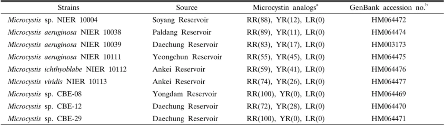
Table 1. Origin and characteristics of toxin for Microcystis strains used in this study
Strains Source Microcystin analogsa GenBank accession no.b
Microcystis sp. NIER 10004 Soyang Reservoir RR(88), YR(12), LR(0) HM064472
Microcystis aeruginosa NIER 10038 Paldang Reservoir RR(89), YR(11), LR(0) HM064474 Microcystis aeruginosa NIER 10039 Daechung Reservoir RR(83), YR(17), LR(0) HM003173 Microcystis aeruginosa NIER 10111 Yeongchun Reservoir RR(55), YR(45), LR(0) HM064475 Microcystis ichthyoblabe NIER 10112 Ankei Reservoir RR(59), YR(41), LR(0) HM064476 Microcystis viridis NIER 10113 Ankei Reservoir RR(74), YR(26), LR(0) HM064477
Microcystis sp. CBE-08 Yongdam Reservoir RR(100), YR(0), LR(0) HM064469
Microcystis sp. CBE-12 Daechung Reservoir RR(72), YR(28), LR(0) HM064470
Microcystis sp. CBE-29 Daechung Reservoir RR(100), YR(0), LR(0) HM064471
a Numbers in parentheses indicate the % portion of analogs in total of microcystins.
b GenBank accession no. of mcyA gene sequence
Table 2. Sampling locations and concentrations of chlorophyll a and microcystins
Reservoir Location Sampling date Chlorophyll a (μg/L) Microcystins (μg/L) Microcystin analogsa
Daechung Chuso-ri Oct. 27, 2007
Jul. 01, 2008 n.d.
33.7 n.d.
48.6 n.d.
RR(78), YR(20), LR(2.6)
Yongdam Yongpyungdaegyo Jun. 27, 2008 40.6 11.0 RR(100)
Chungju Hacheondaegyo Sep. 02, 2008 64.2 14.2 RR(100)
Soyang Shineui-ri Sep. 05, 2008 4.60 1.34 RR(100)
Euam Gongji-chun Sep. 17, 2008 41.0 6.85 RR(100)
a Numbers in parentheses indicate the % portion of analogs in total of microcystins.
n.d., not determined.
의 독소 생산 여부를 확인하거나(8, 21, 25), microcystins를 생 산하는 남조세균을 genus 수준에서 구분하기 위한 연구(7)에서 사용되었다. 국내에서도 mcyA 유전자는 독성 Microcystis를 검 출하기 위한 지표를 개발하기 위한 연구(3, 13)와 국내에서 분 리된 Microcystis 속 남조세균의 독소생산 유전자의 다양성을 규명하는 연구(14)에 사용되었다. 하지만, 아직까지 여러 환경 시료에서 mcyA 유전자의 다양성에 대해서는 많이 알려지지 않 았다. 따라서, 본 연구에서는 mcyA 유전자 염기서열을 이용하 여 개발된 분자생물학적 지표를 이용하여, 국내의 여러 대표적 인 호수를 대상으로 microcystins의 농도 및 종류와 mcyA 유 전자의 염기서열 다양성을 분석하였다.
재료 및 방법
남조세균의 배양
실험에 사용된 독소 생산 Microcystis의 종류를 Table 1에 나타내었다. Microcystis 중 NIER로 표시된 6종은 대청호, 팔 당호, 소양호, 영천호 및 안계호에서 분리된 것으로 국립환경 과학원(National Institute of Environmental Research, NIER) 으로부터 제공을 받았으며, 3종(CBE-08, CBE-12, CBE-29)은 대청호와 용담호에서 분리된 종이다(14). 남조세균의 배양 배 지는 CB 배지를 사용하였다(26). 남조세균을 배양하기 위하여 25±1°C를 유지한 배양실에서 150 rpm으로 교반하였으며, 14 시간의 명기(약 40 μE/m2/sec 강도의 빛)와 10시간의 암기를 교대로 반복하였다.
현장 시료의 채취 및 엽록소 a의 농도 분석
분석에 사용된 수시료는 대청호, 용담호, 충주호, 의암호, 소 양호에서 채취하였다(Table 2). 대청호의 경우 2007년 10월과 2008년 7월에 시료를 채취하였고, 나머지 호수에서는 2008년 6월과 9월에 걸쳐 시료를 채취하였다. 채취된 시료는 냉장 상 태를 유지하여 실험실로 옮긴 후, 분석하였다. 엽록소 a의 농 도를 측정하기 위해 적당량의 시료를 GF/C 여과지(Whatman, UK)로 여과한 후, 아세톤 용액(90%, v/v)으로 여과지의 색소 를 추출하였다. 분광광도계(Optizen 2120UV, Mecasys, Korea) 로 추출액의 흡광도를 측정한 결과를 이용하여 엽록소 a의 농 도를 계산하였다(2).
DNA 추출 및 정제
배양된 남조세균 및 수시료로부터 genomic DNA의 추출은 Rhochelle 등(24)의 방법을 변형시켜 사용하였다. 배양된 남조 세균의 경우, 배양액을 22,000×g에서 10분간 원심분리한 후, lysozyme이 포함된 완충용액(0.15 M NaCl, 0.1 M EDTA, 15 mg/ml lysozyme, pH 8.0)을 넣고 37°C에서 1시간 동안 처리 하였다. 처리된 용액에 SDS가 포함된 완충용액[0.1 M NaCl, 0.5 M Tris-HCl, 6%(w/v) SDS, pH 8.0]을 넣고 5 분간 초음 파세척기(150W, 대한과학, Korea)를 처리하여 남조세균을 파 쇄하였다. 남조세균의 파쇄 효율을 높이기 위해 급속 해냉동 (-70°C/ 60°C, 각각 15분) 처리를 3회 실시하였다. 용액 중 DNA는 phenol:chloroform법을 사용하여 정제하였다. 수시료 의 경우, GF/C 여과지로 시료를 여과한 후, lysozyme 완충용
액 및 SDS 완충용액, 초음파세척기, 및 3회의 급속해냉동법을 실시하였으며, 사용된 완충용액의 조성 및 물리적 처리 조건은 배양된 남조세균에서 DNA를 추출하는 조건과 동일하게 사용 하였다.
PCR 및 library 구축
mcyA 유전자를 증폭하기 위해 mcyA-Cd 1F (5′-AAA AGT GTT TTA TTA GCG GCT CAT-3′)와 mcyA-Cd 1R (5′
-AAA ATT AAA AGC CGT ATC AAA-3′)을 PCR primer 로 사용하였다(7). PCR 반응액은 5× Mg2+-free PCR buffer (Promega, USA), dNTP (200 μM), Mg2+ (1.5 mM), bovine serum albumin (500 μg/ml)에 GoTaq Flexi polymerase (20 mU/μl; Promega)를 중합효소로 사용하였다. 반응액 50 μl에 10 ng의 주형 DNA를 더하여 PCR을 수행하였다. PCR 조건 은 95°C에서 3분간 전처리한 후, 변성(95°C, 30초), 결합 (54°C, 40초), 중합(72°C, 40초) 단계를 30주기 수행하였으며, 72°C에서 10분간 후처리를 하였다. PCR을 위한 thermal cycler는 MJ Mini Gradient Thermal Cycler (Bio-Rad, USA) 를 사용하였다.
수시료에서 mcyA 유전자에 대한 library를 구축하기 위하여, PCR 산물을 PCR Purification kit (TaKaRa, Japan)로 정제한 후, T-blunt vector (Solgent, Korea)를 사용하여 클로닝을 하였 다. Ampicillin (50 μg/ml)과 kanamycin (50 μg/ml)이 포함된 LB 배지에서 1일 간 배양한 후, 시료 당 약 100개의 colony를 무작위로 선택하였다. 선택된 colony에 PCR 산물이 삽입된 것 을 확인하기 위하여, mcyA-Cd 1F/mcyA-Cd 1R을 primer로 사 용하여 다시 PCR을 수행하였다. 염기서열은 Solgent에 의뢰 하여 분석하였으며, DNA 분석기는 ABI PRISM 3730XL DNA analyzer (Applied Biosystems, USA)를 사용하였다. 국 내 호소에서 분리된 남조세균의 염기서열의 GenBank 분류번 호는 Table 1에 나타내었으며, 수시료에서 얻은 clone의 염기 서열은 GenBank 분류번호 HM003174-HM003178과 HM064483- HM064494에 수록되었다. 분석된 염기서열은 CLUSTALW2 (11)로 정렬한 후, 각 시료에서 동일한 clone의 출현 빈도를 계 산하였다. 유전학적 계통분석은 MEGA4 (27)를 사용하였으며, 계통도는 neighbor-joining tree algorithm을 사용하여 얻었다.
Microcystins의 추출 및 정량
수시료에 포함된 microcystins의 농도를 측정하기 위하여, 시료를 GF/C 여과지로 여과한 후, Maatouk 등(15)의 방법을 일부 변형시켜 추출 정제하였다. 여과지에 methanol 용액(75%, v/v)을 첨가한 후, 강한 vortexing을 3분간 수행하였다. 이를 원심분리(11,000×g, 5분)하고, 상등액을 주사기 필터(0.2 μm, PTFE; Dismic®-13; Advantec MFS, Inc., Japan)로 여과하였다.
여과액은 C18 column cartridge (Sep-Pak Vac 3cc; Waters, USA) 를 사용하여 정제하였으며, 용리액은 진공농축기(Modulspin31;
한일과학, Korea)를 사용하여 농축하였다.
Microcystins는 UV 흡광 검출기(UV 725S; 영린과학, Korea) 와 Xterra RP18 column (5 μm 입자크기, 15 cm×3.9 mm
I.D.; Waters)이 장착된 HPLC를 사용하여 정량하였으며, triflu- oroacetic acid (0.05%, v/v)가 포함된 acetonitrile/water (32%:
68%)를 이동상으로 사용하였다. 순수한 microcystins -LR, -YR, -RR (Wako Pure Chemical Ind., Japan)을 외부 표준물 질로 사용하여, microcystins의 peak를 판별하고 정량하였다.
결과 및 토의
엽록소 a 및 microcystins의 분석
대상 호소 중 소양호를 제외하고, 엽록소 a의 농도는 33.7- 64.2 mg/L의 범위로 매우 높게 나타나 조사 시점에 심한 조류 대발생이 일어났음을 확인할 수 있었다(Table 2). 2001년부 터 2008년까지 대청호 지역의 댐 수문자료(한국수자원공사, www.kwater.or.kr)를 조사한 결과, 2008년에 강한 강우가 없 었기 때문에 댐의 방류량이 적어 수체가 정체됨으로써 조류 대 발생이 극심했던 것으로 판단된다. 2001년에도 적은 강우량으 로 인해 극심한 조류 대발생이 관찰되었다(17). 시료를 채취한 다른 호소에서도 같은 양상의 강우 사상이 관찰되었다.
Microcystins의 농도는 1.34-48.6 μg/L의 범위였다(Table 2).
대청호를 제외한 모든 시료에서 microcystin-RR 만 검출되었 으며, 대청호의 경우에는 microcystin-YR 및 -LR이 각각 20, 2.6%가 있었으나 microcystin-RR의 농도가 가장 높았다(Table 2). 이러한 결과는 국내 호소에서 microcystin-RR이 가장 많다 는 기존의 연구 결과와 일치하는 것이다(10, 18). 세계보건기구 의 먹는 물의 원수에 대한 microcystins의 수질기준은 microcystin- LR을 기준으로 1.0 μg/L이다(5). 본 실험에서 분석한 모든 시 료에서 microcystins의 농도는 1.0 μg/L 이상이지만, 이 결과를 WHO 기준과 비교하기는 어렵다고 판단된다. 그 이유는 대상 호소에서 시료를 채취한 지점이 수돗물 생산을 위한 취수탑과 지리적으로 멀리 떨어져 있으며, 검출된 microcystins의 종류가 microcystin-LR에 비해 독성이 약한 -RR 또는 -YR이기 때문 이다. 생쥐에 대한 microcystin-LR, -YR 및 -RR의 반수치사농 도는 각각 50, 150-200, 500-800 μg/kg이다(32).
배양 남조세균의 mcyA 유전자 염기서열의 분석
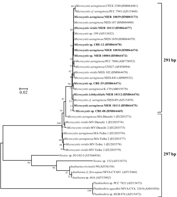
mcy gene cluster 중 가장 많은 수의 염기서열이 알려진 유 전자는 mcyA이다. GenBank에 올려진 mcyA 유전자 염기서열 은 517개(2010년 4월 1일 기준)이며, Microcystis 속에 대한 결과가 전체의 81%로 가장 많다. mcyA 유전자의 condensation region을 대상으로 개발된 PCR primer인 mcyA-Cd 1F/mcyA-Cd 1R (7)로 증폭된 염기서열은 359개로 전체의 69%이다. 현재 까지 알려진 염기서열에 따르면, 이 primer set를 사용하여 증 폭하였을 때 Anabaena, Planktothrix 및 Nostoc 속으로부터 297 bp의 산물이 만들어지며, Microcystis sp.의 경우에는 291 또는 297 bp가 만들어진다(Fig. 1). PCR 산물의 크기가 297 bp인 Microcystis 속은 미국의 Lake Erie, Lake Ontario, Klamath River 및 San Francisco Bay와 중국의 Dianchi Lake를 비롯한 담수생태계에서 발견되었다(8, 22, 30, 31). 국내 호소에서 분 리된 9종의 Microcystis 속의 경우 모두 291 bp의 PCR 산물을
Microcystis aeruginosa UTEX 2388 (HM064481) Microcystis cf. aeruginosa PCC 7941 (AJ515460) Microcystis aeruginosa NIER 10039 (HM003173) Microcystis aeruginosa NIES 107 (HM004480) Microcystis viridis NIER 10113 (HM064477) Microcystis sp. 199 (AJ515452)
Microcystis aeruginosa NIES 1058 (HM064479) Microcystis sp. CBE-12 (HM064470)
Microcystis aeruginosa NIER 10038 (HM064474) Microcystis sp. NIER 10004 (HM064472)
Microcystis aeruginosa PCC 7806 (AM778952) Microcystis aeruginosa UV027 (AF458094) Microcystis viridis NIES 102 (HM064478) Microcystis aeruginosa NIES-843 (AP009552) Microcystis sp. CBE-29 (HM064471) Microcystis aeruginosa K-139 (AB019578) Microcystis ichthyoblabe NIER 10112 (HM064476) Microcystis cf. aeruginosa NIES-89 (AJ515459) Microcystis aeruginosa NIER 10111 (HM064475)
Microcystis sp. CBE-08 (HM064469) Microcystis aeruginosa MA-Dianchi 1 (EU203573) Microcystis viridis MV-Dianchi 1 (EU203574)
Microcystis viridis MV-Dianchi 2 (EU203575) Microcystis aeruginosa MA-Taihu 1 (EU203576) Microcystis aeruginosa MA-Taihu 2 (EU203577)
Microcystis viridis MV-Taihu 1 (EU203578) Microcystis viridis MV-Taihu 2 (EU203579) Nostoc sp. IO-102-I (AY566856)
Nostoc sp. 152 (AJ515475) Anabaena circinalis 90 (AJ536156)
Anabaena cf. flos-aquae NIVA-CYA83 (AJ515466) Anabaena sp. 66A (AJ515462)
Planktothrix sp. PCC 7821 (AJ515473)
Planktothrix agardhii NIVA-CYA 126/8 (AJ441056) Planktothrix sp. HUB 076 (AJ515472)
68
66 53 44
16
34
32 62
23 17 53 96
58
42 48
43
40 39
100
100 96
96 87
72
0.02
291 bp
297 bp
Fig. 1. Phylogenetic tree based on the partial sequences of mcyA gene in microcystin-producing cyanobacteria. The tree was constructed by using the neighbor-joining method. Strains isolated from the Korean freshwaters were indicated in bold-face. Local bootstrap probabilities are indicated at nodes.
생성하였다(Fig. 1).
297 bp 산물의 경우 291 bp에 비해 2개의 아미노산이 많으 며, Microcystis 속 남조세균의 경우 297 bp 산물에 첨가된 아 미노산은 Trp-Ala 또는 Phe-Ala이다(7, 14, 31). 아직까지 아 미노산의 삽입이 효소의 구조 및 활성에 미치는 영향에 대한 연구가 이루어지지 않았다. 최근의 연구에서, 명확한 결론을 얻지는 못하였으나, 배양된 남조세균에서 mcyA와 mcyB의 염 기서열 다양성이 microcystin의 종류에 영향을 미칠 수 있는 가능성이 제시되었다(22).
297 bp의 산물이 만들어지는 수생태계 시료나 남조세균에 서 나타나는 microcystins의 종류를 분석한 결과 독성이 강한
microcystin-LR의 농도가 높았다(8, 22). 하지만, 국내에서 분 리된 9종의 Microcystis 속 남조세균의 경우, microcystin-LR 을 만드는 종은 없었으며, 모든 종에서 microcystin-RR 또는 -YR이 생산되었다(Table 1). 이러한 사실은 mcyA의 염기서열 의 다양성이 microcystins의 종류를 결정하는데 관여할 가능성 이 있음을 의미한다.
수시료에서 mcyA 유전자 염기서열의 분석
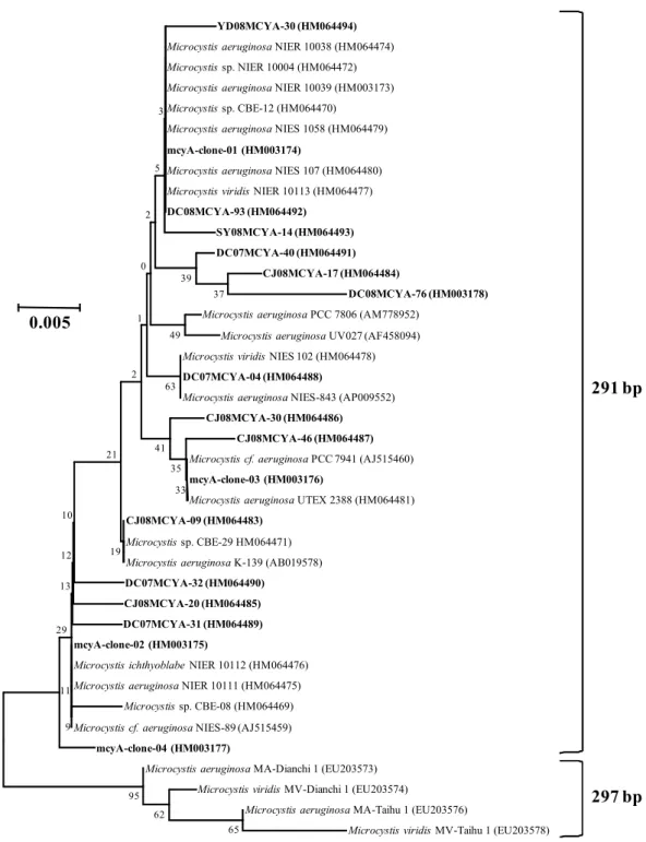
국내 5개의 호수에서 채취한 시료에서 mcyA 유전자 염기서 열을 분석한 결과, mcyA-Cd 1F/mcyA-Cd 1R을 사용하여 PCR 을 수행하였을 때 291 bp 길이의 산물이 생성되었으며, 모든
YD08MCYA-30 (HM064494) Microcystis aeruginosa NIER 10038 (HM064474) Microcystis sp. NIER 10004 (HM064472) Microcystis aeruginosa NIER 10039 (HM003173) Microcystis sp. CBE-12 (HM064470)
Microcystis aeruginosa NIES 1058 (HM064479) mcyA-clone-01 (HM003174)
Microcystis aeruginosa NIES 107 (HM064480) Microcystis viridis NIER 10113 (HM064477) DC08MCYA-93 (HM064492)
SY08MCYA-14 (HM064493) DC07MCYA-40 (HM064491)
CJ08MCYA-17 (HM064484)
DC08MCYA-76 (HM003178) Microcystis aeruginosa PCC 7806 (AM778952)
Microcystis aeruginosa UV027 (AF458094) Microcystis viridis NIES 102 (HM064478) DC07MCYA-04 (HM064488)
Microcystis aeruginosa NIES-843 (AP009552) CJ08MCYA-30 (HM064486)
CJ08MCYA-46 (HM064487) Microcystis cf. aeruginosa PCC 7941 (AJ515460) mcyA-clone-03 (HM003176)
Microcystis aeruginosa UTEX 2388 (HM064481) CJ08MCYA-09 (HM064483)
Microcystis sp. CBE-29 HM064471) Microcystis aeruginosa K-139 (AB019578) DC07MCYA-32 (HM064490)
CJ08MCYA-20 (HM064485) DC07MCYA-31 (HM064489) mcyA-clone-02 (HM003175)
Microcystis ichthyoblabe NIER 10112 (HM064476) Microcystis aeruginosa NIER 10111 (HM064475)
Microcystis sp. CBE-08 (HM064469) Microcystis cf. aeruginosa NIES-89 (AJ515459)
mcyA-clone-04 (HM003177)
Microcystis aeruginosa MA-Dianchi 1 (EU203573) Microcystis viridis MV-Dianchi 1 (EU203574)
Microcystis aeruginosa MA-Taihu 1 (EU203576)
Microcystis viridis MV-Taihu 1 (EU203578) 65
62 95
37
33 35
39
9 11 29
13 12 10
41 49
63
19 21
2 1
0 2
5 3
0.005
291 bp
297 bp
Fig. 2. Phylogenetic tree based on the partial sequences of mcyA gene found in the Korean reservoirs and taken from the GenBank Database.
The tree was constructed by using the neighbor-joining method. The clones found in the targeted reservoirs were indicated in bold-face. Local bootstrap probabilities are indicated at nodes.
clone이 Microcystis 속과 유사성이 큰 것으로 나타났다(Fig.
2). 이러한 결과는 대상 지역에서 독소를 생산하는 Anabaena 속 또는 Planktothrix 속이 존재하지 않거나, 매우 적은 수가 존재함을 의미한다. 현미경으로 대상 시료를 관찰하였을 때, 용 담호 및 대청호 시료에서 다수의 Anabaena가 존재하는 것이 관 찰되었다. 또한 남조세균이 만드는 색소의 일종인 phycocyanin 유전자에 대한 PCR primer로 널리 사용되는 PCβF/PCαR (16)
을 사용하여 대상 시료를 분석하였을 때, Anabaena에 해당되 는 clone이 용담호와 대청호에서 각각 49%와 40%를 차지하였 다(결과 미제시).
Phycocyanin β subunit와 α subunit 사이의 intergenic spacer 의 길이는 남조세균에 따라 다르며, GenBank에 등록된 염기서 열을 분석하면 Microcystis 속의 경우 66 bp가 전체의 95%이 며, Anabaena 속의 경우에는 76-103 bp의 길이이며 61%가 99
Frequency (%)
0 20 40 60 80 100
mcyA-clone-01 mcyA-clone-02 mcyA-clone-03
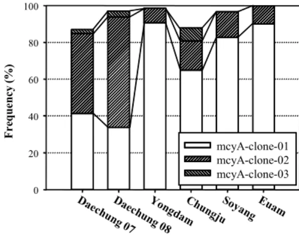
Fig. 3. Distribution dynamics of mcyA gene sequences in the Korean reservoirs. The remaining parts to 100% of the bars indicate the sum of clones occupied below 2% or those found in only one reservoir.
bp 길이를 나타낸다. 선행 연구에서, GenBank에 등록된 Anabaena 속의 mcyA 염기서열과 mcyA-Cd 1F/mcyA-Cd 1R의 염기 서열 을 비교한 결과, 일부 염기에서 차이가 있어, 이들 primer를 사 용하였을 때 Anabaena 속의 mcyA gene이 증폭되지 않을 가능 성이 제기되었다(14). 이를 확인하기 위하여 Anabaena 속의 mcyA 와 mcyB gene에 specific한 primer set를 제작하여 PCR을 수 행한 결과, 대상 시료에서 PCR 산물이 전혀 생성되지 않았다 (결과 미제시). 이러한 결과로 판단할 때, 본 연구에서 분석한 시료에서 microcystins를 생산하는 Anabaena 속은 없거나 매 우 적은 농도로 존재하는 것으로 보인다. 본 연구의 대상호소 는 5개이며, 특정 시점에 채취한 시료를 대상으로 분석한 것이 기 때문에, 국내 수생태계에 microcystins를 생성하는 Anabaena 속의 존재 여부에 대한 것은 확실하지 않다.
대상 시료에서 특정 clone의 빈도를 조사한 결과, 모든 시료 에서 두 종류의 clone이 우점하는 것으로 나타났다(Fig. 3).
Microcystins의 농도가 낮은 의암호, 소양호, 용담호 시료의 경 우 clone의 종류가 2-3개로 다양성이 매우 낮았다. 현미경으로 관찰하였을 때, Microcystis 속 이외의 다른 남조세균이 거의 관찰되지 않은 충주호 시료에서는 microcystins의 농도는 14.2 μg/L로 크게 높지 않았으나, clone의 종류는 9개로 분석한 시 료 중 가장 많은 수를 나타내었다(Fig. 3). 대청호의 경우, 조류 대발생이 일어난 2008년 시료에서 clone의 종류가 5개인 반면, 조류대발생이 끝난 시점인 2007년 10월에 채취한 시료에서는 8종의 clone이 관찰되었다. 또한, 각 clone의 비율이 두 시료에 서 상이하게 나타났다. 이러한 결과는 조류대발생 시기에 특정 한 균주의 성장이 촉진되었기 때문으로 사료된다.
소양호를 제외한 지역에서 비슷한 농도의 엽록소 a가 관찰 되었으나, 대청호의 경우 microcystins의 농도가 매우 높은 것 은, 독소 생성 남조세균의 수가 많거나 microcystins의 생성률 이 높은 Microcystis가 대청호에 우점하였기 때문일 가능성이 있다. 대청호를 제외한 시료와 대청호 시료에서 가장 우점하는 clone의 종류가 상이하였다. 즉, 대청호를 제외한 시료에서는
mcyA-clone-01이 가장 비율이 높았으나, 대청호에서는 mcyA- clone-02가 가장 높은 비율이었다(Fig. 3). 따라서, 대청호의 microcystins의 높은 농도가 다른 호소와 달리 독소 생성능이 뛰어난 균주에 기인할 가능성이 있으며, 이에 대한 후속연구가 필요한 것으로 사료된다.
실시간 PCR법을 이용하여 목표 미생물을 계수하는 연구가 많이 이루어 졌으며, 독소 생산 Microcystis의 경우에도 실시간 PCR법을 이용하여 이를 계수하기 위한 여러 방법이 제안되었 다(6, 28). 국내 호소에서 mcyA의 염기서열이 매우 보존되어 있는 것으로 나타났으며, 이는 본 연구에서 증폭한 유전자 부 분이 실시간 PCR을 통한 독성 Microcystis의 계수를 위한 방 법에 활용될 수 있음을 제시한다.
본 연구에서 국내 대형 호수를 대상으로 조류 독소인 microcystins의 생합성에 관여하는 유전자의 염기서열 다양성 을 분석하였다. 국내 호소에서 분리된 Microcystis 속과 수시료 에서 mcyA 유전자를 분석한 결과, 외국의 호소와 달리 다양성 이 매우 낮은 것으로 나타났다. 국내 수생태계에서 제한된 종 류의 microcystins가 검출되는 결과와 비교할 때, 이는 mcy 유 전자의 낮은 다양성에 기인할 가능성이 있다. 또한 독소의 농 도에 따라 우점하는 균주의 mcyA 유전자가 다르게 나타났다.
이러한 결과로 판단할 때, mcyA 유전자가 microcystins의 종류 및 독소 합성능에 중요한 역할을 수행할 가능성이 있으며, 이 를 규명하기 위한 후속 연구가 필요할 것으로 보인다.
적요
국내에서 분리된 독소생산 Microcystis 속과 조류 대발생 시 기에 대청호, 충주호, 용담호, 소양호, 및 의암호에서 채취한 시료에서 조류 독소인 microcystins의 생합성에 관여하는 mcyA 유전자의 염기서열 다양성을 분석하였다. GenBank에 등록된 mcyA 염기서열과 비교한 결과, 국내 호소에서 분리된 Microcystis 속의 mcyA 유전자 염기서열은 매우 낮은 다양성을 나타내었 다. 환경 시료 분석 결과, 2-3종류의 clone이 전체의 87-100%
를 차지하였으며, Anabaena 속이나 Planktothrix 속의 mcyA 유전자와 유사한 염기서열은 발견되지 않았다. 이러한 결과로 볼 때, 대상 호소에서 microcystins는 주로 Microcystis 속에 의해 생산되며, 독소 생산에 관여하는 mcyA 유전자의 염기서 열은 보존되어 있는 것으로 판단된다.
감사의 말
이 논문은 2008년도 충북대학교 학술연구지원사업의 연구 비 지원에 의하여 연구되었으며, 이에 감사드립니다.
참고문헌
1. Ahn, C.Y., H.S. Kim, B.D. Yoon, and H.M. Oh. 2003. Influence of rainfall on cyanobacterial bloom in Daechung Reservoir. Kor.
J. Limnol. 36, 413-419.
2. American Public Health Association. 2005. Standard Methods for the Examination of Water and Wastewater, 21th edition. American Public Health Association, Washington, D.C., USA.
3. Cho, J.E., S.W. Bang, and M.S. Han. 2004. Development of oligonucleotide primers for the detection of harmful Microcystis in water. Bull. Environ. Contam. Toxicol. 72, 655-662.
4. Dittmann, E. and T. Börner. 2005. Genetic contributions to the risk assessment of microcystin in the environment. Toxicol. Appl.
Pharm. 203, 192-200.
5. Falconer, I., J. Bartram, I. Chorus, T. Kuiper-Goodman, H.
Utkilen, M. Burch, and G.A. Codd. 1999. Safe levels and safe practices, pp. 161-182. In I. Chorus and J. Bartram (eds.), Toxic Cyanobacteria in water, A guide to their public health conse- quences, monitoring and management. Spon Press, London, UK.
6. Furukawa, K., N. Noda, S. Tsuneda, T. Saito, T. Itayama, and Y.
Inamori. 2006. Highly sensitive real-time PCR assay for quantifi- cation of toxic cyanobacteria based on microcystin synthetase A gene. J. Biosci. Bioeng. 102, 90-96.
7. Hisbergues, M., G. Christiansen, L. Rouhiainen, K. Sivonen, and T. Börner. 2003. PCR-based identification of microcystin-producing genotypes of different cyanobacterial genera. Arch. Microbiol.
180, 402-410.
8. Hotto, A.M., M.F. Satchwell, and G.L. Boyer. 2007. Molecular characterization of potential microcystin-producing cyanobacteria in Lake Ontario embayments and nearshore waters. Appl. Environ.
Microbiol. 73, 4570-4578.
9. Kardinaal, W.E.A. and P.M. Visser. 2005. Dynamics of cyano- bacterial toxins, pp. 41-64. In J. Huisman, H.C.P. Matthijs, and P.M. Visser (eds.), Harmful Cyanobacteria. Springer, Dordrecht, The Netherlands.
10. Kim, B., H.S. Kim, H.D. Park, K. Choi, and J.G. Park. 1999.
Microcystin content of cyanobacterial cells in Korean reservoirs and their toxicity. Korean J. Limnol. 32, 288-294.
11. Larkin, M.A., G. Blackshields, N.P. Brown, R. Chenna, P.A.
McGettigan, H. McWilliam, F. Valentin, I.M. Wallace, A. Wilm, R. Lopez, J.D. Thompson, T.J. Gibson, and D.G. Higgins. 2007.
ClustalW2 and ClustalX version 2. Bioinformatics 23, 2947-2948.
12. Lee, K.L., W.H Jheong, J.M. Kim, and H.S. Kim. 2007.
Detection of toxigenicity of cyanobacteria by molecular method.
Kor. J. Limnol. 40, 149-154.
13. Lee, H.K., J.H. Kim, S.A. Yoo, T.S. Ahn, C.K. Kim, and D.H.
Lee. 2003. Primer evaluation for the detection of toxigenic Microsystis by PCR. Kor. J. Microbiol. 39, 166-174.
14. Lee, H.S., K.H. Oh, and Y.C. Cho. 2008. Isolation of cyanobacteria producing microcystin from lakes. Kor. J. Microbiol. 44, 251-257.
15. Maatouk, I., N. Bouaich, D. Fontan, and Y. Lev. 2002. Seasonal variation of microcystin concentrations in the Saint-Caprais Reservoir (France) and their removal in a small full-scale treatment plant.
Water Res. 36, 2891-2897.
16. Neilan, B.A., D. Jacobs, and A.E. Goodman. 1995. Genetic diversity and phylogeny of toxic cyanobacteria determined by DNA polymorphisms within the phycocyanin locus. Appl. Environ.
Microbiol. 61, 3875-3883.
17. Park, J.G. 2005. Developmental characteristic of cyanobacterial bloom in Lake Daecheong. Korean J. Environ. Biol. 23, 304-314.
18. Park, H.D., B. Kim, E. Kim, and T. Okino. 1998. Hepatotoxic
microcystins and neurotoxic anatoxin-a in cyanobacterial blooms from Korean lakes. Environ. Toxicol. Water Qual. 13, 225-234.
19. Pouria, S., A. Deandrade, J. Barbosa, R.L. Carvalcanti, V.T.S.
Barreto, C.J. Ward, W. Preiser, G.K. Poon, G.H. Neild, and G.A.
Codd. 1998. Fatal microcystin intoxification in haemodialysis unit in Caruaru, Brazil. Lancet 352, 21-26.
20. Rantala, A., D.P. Fewer, M. Hisbergues, L. Rouhiainen, J.
Vaitomaa, T. Börner, and K. Sivonen. 2004. Phylogenetic evidence for the early evolution of microcystin synthesis. Proc. Nat. Acad.
Sci. USA 101, 568-573.
21. Richardson, L.L., R. Sekar, J.L. Myers, M. Gantar, J.D. Voss, L.
Kaczmarsky, E.R. Remily, G.L. Boyer, and P.V. Zimba. 2007.
The presence of the cyanobacterial toxin microcystin in black band disease of corals. FEMS Microbiol. Lett. 272, 182-187.
22. Rinta-Kanto, J.M., M.A. Saxton, J.M. DeBruyn, J.L. Smith, C.H.
Marvin, K.A. Krieger, G.S. Sayler, G.L. Boyer, and S.W.
Wilhelm. 2009. The diversity and distribution of toxigenic Microcystis spp. in present day and archived pelagic and sediment samples from Lake Erie. Harmful Algae 8, 385-394.
23. Rinta-Kanto, J.M. and S.W. Wilhelm. 2006. Diversity of microcystin-producing cyanobacteria in spatially isolated regions of Lake Erie. Appl. Environ. Microbiol. 72, 5083-5085.
24. Rochelle, P.A., J.C. Fry, R.J. Parkes, and A.J. Weightman. 1992.
DNA extraction for 16S rRNA gene analysis to determine genetic diversity in deep sediment communities. FEMS Microbiol. Lett.
79, 59-65.
25. Saker, M.L., M. Vale, D. Kramer, and V.M. Vasconcelos. 2007.
Molecular techniques for the early warning of toxic cyanobacteria blooms in freshwater lakes and rivers. Appl. Microbiol.
Biotechnol. 75, 441-449.
26. Shirai, M., K. Matumaru, A. Ohotake, Y. Takamura, T. Aida, and M. Nakano. 1989. Development of a solid medium for growth and isolation of axenic Microcystis strains (cyanobacteria). Appl.
Environ. Microbiol. 55, 2569-2571.
27. Tamura, K., J. Dudley, M. Nei, and S. Kumar. 2007. MEGA4:
molecular evolutionary genetics analysis (MEGA) software version 4.0. Mol. Biol. Evol. 24, 1596-1599.
28. Vaitomaa, J., K. Rantala, P. Rouhiainen, L. Mokelke, and K.
Sivonen. 2003. Quantitative real-time PCR for determination of microcystin synthetase E copy numbers for Microcystis and Anabaena in lakes. Appl. Environ. Microbiol. 69, 7289-7297.
29. Welker, M. and H. Von Dhren. 2006. Cyanobacterial peptides- nature's own combinatorial biosynthesis. FEMS Microbiol. Rev.
30, 530-563.
30. Xu, Y., Z. Wu, B. Yu, X. Peng, G. Yu, Z. Wei, G. Wang, and R. Li. 2008. Non-microcystin producing Microcystis wesenbergii (Komarek) Komarek (Cyanobacteria) representing a main water- bloom-forming species in Chinese waters. Environ. Pollut. 156, 162-167.
31. Yilmaz, M., E.J. Phlips, and D. Tillett. 2009. Improved methods for the isolation of cyanobacterial DNA from environmental samples. J. Phycol. 45, 517-521.
32. Zurawell, R.W., H. Chen, J.M. Burke, and E.E. Prepas. 2005.
Hepatotoxic cyanobacteria: a review of the biological importance of microcystins in fresh water environments. J. Toxicol. Environ.
Health Part B 8, 1-37.