http://bx.doi.org/10.7783/KJMCS.2012.20.4.245
Dieldrin에 의한 DNA와 세포 손상에 대한 오가피 추출물의 억제효과
류아름·김지혜·이미영
†순천향대학교 의료생명공학과
Suppressive Effect of Acanthopanax sessiliflorus Extract on the DNA and Cell Damage by Dieldrin
A Reum Ryu, Ji Hae Kim and Mi Young Lee
†Department of Medical Biotechnology, Soonchunhyang University, Asan 336-600, Korea.
ABSTRACT : Dieldrin, one of the organochlorine pesticides (OCPs), induced the damages in neuroblastoma cells and DNA damages in lymphocytes. The ethanol extracts of
A. sessiliflorusleaves were examined for the suppressive effects on the dieldrin-induced cell damages. Moreover, the extract was used to test whether it might inhibit the oxidative DNA damage of lymphocytes using Comet assay. The cell and DNA damage by dieldrin were suppressed
in vitroupon treating
A. sessiliflorusextract. This result suggests that
A. sessiliflorusextract might be useful to reduce dieldrin toxicity
.Key Words
: Dieldrin, Acanthopanax, Cell damage, DNA damage
INTRODUCTION
Organochlorine pesticides (OCPs) are synthetic chemicals used several decades in agriculture (Hara, 2001; Kataoka
et al
., 2010). Dieldrin (1,2,3,4,10,10-hexachloro-6,7-epoxy- 1,4,4a,5,6,7,8,8a-octahydro-1,4,5,8-dimethano-naphthalene) belongs to a class of OCPs which control insects, weeds, fungi and so on (Diamanti-Kandarakis
et al., 2009;
Gonzalez
et al., 2003; Rowland
et al., 2011). Dieldrin made by a Diels-Alder reaction included hexachlorocyclopentadiene group (Fig. 1), and this group showed toxicidal activity, while epoxidated dieldrin triggered toxicity decrease (Kaushik and Kaushik, 2007).
Dieldrin stimulated lipid peroxidation in cell membrane and induced ROS generation which inhibit mitochondrial oxidative phosphorylation in dopaminergic cells (Kitazawa
et al
., 2001; Slotkin and Seidler, 2010). Bioaccumulated dieldrin was known to be a potential etiological agent of Parkinson’s disease and increased risk of breast cancer (Rowland
et al., 2011; Fleming
et al., 1994; Høyer
et al., 1998). While dieldrin is hydrophobic, so very hard to be dissolved in water, dieldrin has been detected in many
rivers in U.S. (Jorgenson, 2001). It has been regarded as a priority pollutant in water by US EPA. Because of its hydrophobicity, it is not easily digested and accumulated in human body (Jorgenson, 2001). However, lipophilic dieldrin could be stored in lipid portions such as body fat, liver, blood serum and breast milk and it is harmful when feed to infant (Schlaud
et al., 1995; Lopez-Espinosa
et al
., 2007).
Acanthopanax
has been reported to have treatment effect of many diseases such as gastric ulcers, chronic bronchitis, chronic renal failure, rheumatics, hypertension and ischemic heart diseases (Choi
et al., 2002; Lin
et al., 2008).
Triterpenoids were isolated from the leaves of
Acanthopanaxspecies. Especially 3,4-seco-lupine-type triterpenoids, named as chiisanogenin, chiisanoside, 24-hydroxychiisanogenin, and 22a-hydroxychiisanogenin were isolated from the leaves of
Acanthopanax divaritacus
var.
albeofructus(Araliaceae) (Sawada
et al., 1993; Bae
et al., 2001). Chiisanoside is a major component of
Acanthopanaxwith anti-inflammatory, anti-hepatotoxic, anti-diabetic and antiviral effects and inhibitory effect on mitogen-induced lymphocyte proliferation (Bae
et al., 2001; Kim
et al., 1999; Jung
et al., 2005).
†Corresponding author: (Phone) +82-41-530-1355 (E-mail) [email protected]
Received 2012 July 26 / 1st Revised 2012 July 31 / Accepted 2012 August 7
Recently, extracts of these plants were reported to prevent bradykinesia in the Parkinson’s disease models (Fujikawa
et al
., 2005) and increase the levels of antioxidant enzymes such as superoxide dismutase, catalase and glutathione peroxidase in high fat diet models (Hong
et al., 2009).
The toxicity of dieldrin resulted in the damage of human neuroblastoma cells and DNA damage of lymphocytes
in vitro.
However, the dieldrin-induced cytotoxicity and oxidative DNA damage were suppressed by
Acanthopanax sessiliflorusextract
in vitroin this investigation.
MATERIALS AND METHODS
1. Sample preparation and dieldrin treatment
The ethanol (70%) extracts of
Acanthopanax sessiliflorusleaves were provided by Sushin Ogapy Co., Ltd (Cheonan- City, Chungnam, Korea). The human neuroblastoma cell line SH-SY5Y was purchased from ATCC and maintained in Dulbecco’s modified essential medium (DMEM) supplemented with 10% heat inactivated FBS and 1%
penicillin-streptomycin at 37°C in atmosphere of 5% CO
2. Dieldrin purchased from Sigma-Aldrich was dissolved in PBS (phosphate buffered saline) and diluted to 25, 50, 75, 100 and 125 uM using DMEM medium. Dieldrin was directly treated to the culture plate for 24 h.
2. Trypan blue exclusion assay
To determine the cytotoxicity of dieldrin on the SH- SY5Y cells, a trypan blue exclusion assay (Shi
et al., 2010) was performed. SH-SY5Y cells were seeded 1 × 10
5cells/well in a 24 well microplate and treated with 25, 50, 75, 100 or 125 uM dieldrin with
Acanthopanaxextract for 24 h. In order to determine the inhibitory effect of
Acanthopanaxextract on the cytotoxicity by dieldrin, the cell viability was measured by trypan blue exclusion assay.
3. Lymphocyte preparation
Whole blood was collected from 200 ~ 250 g SD male rats (Orient Bio Inc. Seongnam, Korea). Mixture of 300 ul whole blood and 700
㎕PBS was layered onto 300
㎕of Histopaque 1077 (Sigma-Aldrich, St. Louis, MO). After centrifugation at 1,450 rpm for 5 min at 20°C, the lymphocytes were collected from buffy-coat layer and washed with PBS. For determination of DNA damage, dieldrin dissolved in DMSO was diluted to 100, 200 and
400 uM in PBS and then incubated for 1 h on ice. For treatment of lymphocytes with
Acanthopanaxextract, the lymphocytes were incubated with 3 and 5
㎍/
㎖ Acanthopanaxextract for 30 min at 37°C, and then treated with 400 uM dieldrin for 1 h on ice.
4. Determination of lymphocytes DNA damage by comet assay
The alkaline comet assay was performed according to Singh
et al. (1988) for evaluation of dieldrin-induced DNA damage and antioxidative effect of
Acanthopanaxextract. The lymphocytes were mixed with 75
㎕of 0.7%
low-melting-point agarose. And then the slides were pre- coated with 1% normal-melting-point agarose and solidified for 30 min at 4°C. After solidified, the slides were covered with 100
㎕of 0.7% low-melting-point agarose.
After the agarose was solidified, slides was immersed in lysis solution (2.5 M NaCl, 100 mM EDTA, 10 mM Tris, 1% Triton X-100 and 10% DMSO) for 1 h at 4°C in the dark condition. To unwind DNA, the slides were placed in an electrophoresis tank containing electrophoresis buffer (300 mM, NaOH and 10 mM Ma
2EDTA (pH 13.0)) for 20 min. Electrophoresis was performed at 25 V/300 mA for 20 min at 4°C. The slides were washed with neutralizing buffer (0.4 M Tris · HCl pH 7.5) three times for 5 min at 4°C and fixed with crude ethanol for 5 min. Slides were dried for 15 min, and then stored in slide box at 4°C.
5. Image analysis
The slides were stained with 20
㎕of ethidium bromide (50 uM) and measured using a fluorescence microscope (Leica, Wetzlar, Germany) in the dark and viewed with a CCD camera (Hitachi, Japan). And then image was analyzed using Komet 5.5 software (Kinetic Imaging, UK).
To quantify DNA damage in the comet assay, the olive tail moment was calculated as (Tail.mean-Head.mean)
× Tail% DNA/100. Total 100 cells were randomly captured and the comets were examined from each slide. All measurements were made in duplicate and performed from three independent experiments.
6. Statistics
All comet data were analyzed using the SPSS package
for Windows version 13 (SPSS Inc., Chicago, IL). The
mean values of DNA damage (Olive tail moment) for
each treatment were compared using one way analysis of variance (ANOVA) followed by Duncan’s multiple range test.
P< 0.05 was considered significant.
RESULTS AND DISCUSSION
1. Suppression of dieldrin-induced cell damage by
Acanthopanax
Figure 1 shows the structure of organochlorine pesticide dieldrin (1,2,3,4,10,10-hexachloro-6,7-epoxy-1,4,4a,5,6,7,8,8a- octahydro-1,4,5,8-dimethano-naphthalene).
Dieldrin induced oxidative stress in the cells by triggering generation of reactive oxygen species (ROS).
Moreover, current hypotheses linked long-term environmental exposure of humans to persistent organochlorine pesticides to the development of neurodegenerative disorders, such as Parkinson’s disease. Primary adverse neurological effects of these insecticides are directed at inhibition of GABA(A) and glycine receptors. Thus, dieldrin can be used as a critical parameter of neuronal function and survival.
(Heusinkveld and Westerink, 2012).
The cytotoxicity by dieldrin was measured by trypan blue exclusion assay as shown in Figure 2. Various concentrations of dieldrin were treated in neuroblastoma cells of SH-SY5Y cells for 24 h, and dieldrin exposure reduced the cell viability in a concentration-dependent manner. The exposure of SH-SY5Y cells to 25 and 50 uM dieldrin resulted in approximately 20 and 30%
decrease in cell viability, respectively.
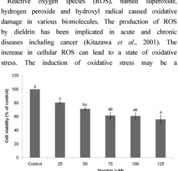
The inhibitory effect of
A. sessiliflorusleaf extract on the toxicity of dieldrin in SH-SY5Y cells was shown in
Figure 3. 25 uM dieldrin resulted in approximately 20%
decrease in cell viability. Upon treating 3.125
㎍/
㎖of the extract, the maximum concentration without cytotoxicity against SH-SY5Y cells, however, about 9% of the live cells increased. This result shows that
A. sessiliflorusextracts reduce the cell damage by dieldrin
in vitro. This result also raises the possibility that
A. sessiliflorusextract might protect the nerve cells against organochlorine pesticide dieldrin.
2. Suppression of dieldrin-induced DNA damage by
Acanthopanax
Reactive oxygen species (ROS), named superoxide, hydrogen peroxide and hydroxyl radical caused oxidative damage in various biomolecules. The production of ROS by dieldrin has been implicated in acute and chronic diseases including cancer (Kitazawa
et al., 2001). The increase in cellular ROS can lead to a state of oxidative stress. The induction of oxidative stress may be a
Fig. 1. Structure of dieldrin.
Fig. 2. Measurements of cytotoxicity of dieldrin on SH-SY5Y cells by trypan blue exclusion assay.
Values with different letters are significantly different. (P < 0.05)Fig. 3. Inhibitory effect of
A. sessiliflorusleaf extract on the
toxicity of dieldrin against SH-SY5Y cells.
Values with different letters are significantly different. (P < 0.05)mechanism by which dieldrin selectively induce toxic effects in vivo. Oxidative damage is considered as a major type of endogenous damage leading to a variety of diseases (Kim
et al., 2011; Lim
et al., 2011). Superoxides are transformed into hydrogen peroxide by superoxide dismutase, and hydrogen peroxide is degraded into H
2O by catalase. However, some of the hydrogen peroxide is transformed into hydroxyl radicals in response to oxidative damage. Therefore, antioxidants might be great benefits against oxidative damage by means of removing the generated ROS directly or indirectly.
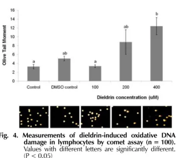
The single-cell electrophoresis (comet) assay was used for assessment of oxidative DNA damage and rapid assay for the detection of DNA damage at the individual cell level (Noroozi
et al., 1998; Hartley
et al., 1999). Dieldrin- induced DNA damage in lymphocytes was shown in Fig.
4. The olive tail moment in a comet assay at 400 uM dieldrin was about 12.41 ± 1.92, compared with 5.08 ± 0.51 in the DMSO-treated control. This result shows that DNA damage by dieldrin increased about 2.5-fold than by DMSO control as judged by olive tail moment. However, the oxidative damage by dieldrin was weaker than by other herbicides such as 2,4-D, 2,4,5-T and paraquat. The olive tail moment at 50 uM 2,4-D, 50 uM 2,4,5-T and paraquat, much lower dose than 400 uM dieldrin, enhanced the DNA damage measured by olive tail moment about 4 fold, 4 fold and 5 fold, respectively, when compared with the DMSO-treated control (Seo
et al., 2010).
Figure 5 shows the suppressive effect of
A. sessiliflorusextract on lymphocytes DNA damage by dieldrin. The olive tail moments at 3 and 5
㎍/
㎖of
Acanthopanaxextract-treated group were 10.61 ± 2.30 and 13.11 ± 1.51, respectively. The extract suppressed the oxidative DNA damage by dieldrin prominently at this experimental condition. When general antioxidant vitamin C and hydroxyl radical scavenger catalase were added to the system, the tail lengths of the DNA were recovered almost to the control levels, indicating that catalase and vitamin C notably blocked the DNA damage by dieldrin.
Therefore, ROS produced by dieldrin was scavenged by the presence of catalase or vitamin C.
Acanthopanax
was reported to have suppressive effect on the oxidative cell damage (Kim
et al., 2000; Lee
et al., 2008).
Acanthopanaxextracts enhanced antioxidant enzymes such as superoxide dismutase, catalase and glutathione peroxidase GPx1 to protect against oxidative stress catalyze by H
2O
2and other peroxides (Wang
et al., 2010). In this study, dieldrin-induced oxidative DNA damage and cell damage were greatly reduced by
A. sessiliflorusextract
in vitro. This raises the possibility that
A. sessiliflorusleaf extract might scavenge ROS generated by dieldrin to protect the oxidative damages. Therefore, the data provide the possible therapeutic values of
A. sessiliflorusextract on the dieldrin-induced DNA and cell damage.
ACKNOWLEDGEMENTS
This work was supported in part by SoonChunHyang University research grant.
Fig. 4. Measurements of dieldrin-induced oxidative DNA damage in lymphocytes by comet assay (n = 100).
Values with different letters are significantly different.
(P < 0.05)
Fig. 5. Suppressive effect of
A.
sessiliflorusextract on dieldrin- induced oxidative DNA damage in lymphocytes (n=100).
Values with different letters are significantly different.
(P < 0.05)