Copyrightⓒ 2011, The Korean Academy of Oral Biology
83
Journal of Oral Biology
Involvement of Crosstalk Between cAMP and cGMP in Synaptic Plasticity in the Substantia Gelatinosa Neurons
Tae-Hyung Kim1#, Gehoon Chung1#, Seok-Beom Park2, Won-Young Chey2, Sung Jun Jung3, Joong Soo Kim1, and Seog Bae Oh1*
1Department of Neurobiology and Physiology, School of Dentistry, Seoul National University, Seoul 110-749, Korea
2Department of Physiology, School of Medicine, Kangwon National University, Chuncheon 200-701, Korea
3Department of Physiology, School of Medicine, Hanyang University, Seoul 133-791, Korea (received April 13, 2011 ; revised April 26, 2011 ; accepted April 29, 2011)
Substantia gelatinosa (SG) neurons receive synaptic inputs from primary afferent Aδ- and C-fibers, where nociceptive information is integrated and modulated by numerous neuro- transmitters or neuromodulators. A number of studies were dedicated to the molecular mechanism underlying the modu- lation of excitability or synaptic plasticity in SG neurons and revealed that second messengers, such as cAMP and cGMP, play an important role. Recently, cAMP and cGMP were shown to downregulate each other in heart muscle cells.
However, involvement of the crosstalk between cAMP and cGMP in neurons is yet to be addressed. Therefore, we investigated whether interaction between cAMP and cGMP modulates synaptic plasticity in SG neurons using slice patch- clamp recording from rats. Synaptic activity was measured by excitatory post-synaptic currents (EPSCs) elicited by stim- ulation onto dorsal root entry zone. Application of 1 mM of 8- bromoadenosine 3,5-cyclic monophosphate (8-Br-cAMP) or 8-bromoguanosine 3,5-cyclic monophosphate (8-Br-cGMP) for 15 minutes increased EPSCs, which were maintained for 30 minutes. However, simultaneous application of 8-Br- cAMP and 8-Br-cGMP failed to increase EPSCs, which suggested antagonistic cross-talk between two second mes- sengers. Application of 3-isobutyl-1-methylxanthine (IBMX) that prevents degradation of cAMP and cGMP by blocking phosphodiesterase (PDE) increased EPSCs. Co-application of cAMP/cGMP along with IBMX induced additional increase in EPSCs. These results suggest that second messen-
gers, cAMP and cGMP, might contribute to development of chronic pain through the mutual regulation of the signal transduction.
Keywords: substantia gelatinosa, spinal cord slice, patch clamp, cAMP, cGMP
서 론
만성통증(chronic pain)은 신경병성 통증, 염증성 통증, 당뇨병에 의한 신경병성 통증 등 다양한 원인에 의해 발 생하며 그 치료가 힘든 것으로 알려져 있다. 최근 만성통 증에 대한 연구가 활발히 진행되면서 세포 수준에서의 통 증정보 신호전달기전을 규명하고자 하였으나 이를 뒷받침 할 기초 지식의 부재 및 연구방법의 제한 등에 의해 만성 통증의 발생 및 제어기전에 대한 연구 발전이 어려운 상 태이다. 따라서 만성통증과 관련된 신호전달계 규명은 만 성통증 치료를 위해 반드시 해결되어야 하는 부분이다.
일반적으로 말초 조직이나 신경 손상 시 자발적 통증 및, 통각과민, 이질통 등 급·만성통증이 발생한다(Campbell et al. 1979; LaMotte et al. 1983; Willis, 2002). 다양한 연구를 통하여 여러 가지 치료법이 제안되었으나 효과적인 치료방법이 아직 없으며 이는 만성통증의 발생기전에 대한 정확한 이해가 부족하기 때문이다(Ma et al. 2002; Mills et al. 2002). 최근까지의 많은 임상 및 동물 연구들에 의 하면 만성통증은 신경 시냅스의 변화에 의해 이뤄지며 이 는 정상 개체와는 다른 시냅스를 이룬다. 이러한 관점에 서 중추성 감작이 만성통증과 관련이 있는 것으로 보고되 었으며, 특히 해마와 소뇌의 중추신경계에서 관찰된 시냅
#These authors contributed equally to this work.
*Corresponding author: Seog Bae Oh, D.D.S., Ph.D., National Research Lab for Pain and Department of Neurobiology and Physiology, School of Dentistry, Seoul National University, 28 Yeongun, Jongno, Seoul 110-749, Korea
Tel.: +82-2-740-8656, Fax.: +82-2-762-5107 Email: odolbae@snu.ac.kr
스 가소성(long-term potentiation or depression)이 중추성 감작의 시냅스 기전으로 해석하고 있다. 따라서 시냅스 가 소성 관점에서 만성통증에 대한 연구는 만성통증 발생기 전 확립을 위해서 필수적이 할 수 있다.
Bliss는 해마(hippocampus) 에서 시냅스 활동이 장기적으 로 변할 수 있음을 보여주고 이를 학습과 기억의 기본과 정으로 파악하였다(Bliss et al., 1973; Bliss et al., 1993).
시냅스 가소성은 presynaptic terminal로부터 glutamate등의 신경전달물질이 유리되는 것이 변하거나 postsynaptic 세 포에 존재하는 이온통로의 활성이 다양한 경로를 통해 변 화함으로써 세포의 반응성이 달라져 일어나는 것으로 알려 져 있다. 현재까지 기억 및 학습기전으로 알려진 장기 시냅 스 가소성은 해마를 포함하여, 중추의 여러 부위에서 관찰 되고 있으나, 척수수준에서의 통증정보처리 기전과 관련해 서 기억 및 학습기전의 하나인 LTP (long-term potentiation), LTD (long-term depression)의 관점에서 접근한 연구결과 는 현재까지 미비한 상태이다. 이러한 장기가소성 변화는 해마와 소뇌 이외에도 중추 여러 곳에서 관찰되며 최근에 는 체성감각 정보가 중추에 들어가 처음 처리되는 척수 후 각의 시냅스 가소성이 보고되었다(Randic et al., 1993; Willis, 1997; Moore et al., 2000; Pockett, 1994; Rygh et al., 2002).
통증은 dynamic한 과정으로 조직손상에 대한 정보를 전 달할 뿐 아니라 손상에 따른 지속적인 구심흥분의 중추유 입은 중추에 시냅스 활동을 장기적으로 변화시킨 일종의 기억현상에 의하여 조직손상이 제거된 후에도 만성적으로 통증을 유발된다. 만성통증, 통각과민(hyperalgesia)의 발생 및 유지 기전을 척수 시냅스 가소성(LTP, LTD)과 연관시 켜 이해하려는 노력이 이루어져, 이에 대한 기전으로 NMDA 수용체와의 관련성을 제시하였으며(Randic, 1996; Sandkuhler et al., 1997; Ikeda et al., 1999), Willis 등은 만성통증 의 병인이라 생각되어지는 중추성 감작현상이 시냅스 가 소성과의 관련성이 있음을 시사하였다(Willis, 1997). 또한 Sandkuhler, Svendsen 등을 포함한 여러 연구자들에 의해 척수에서의 LTP 또는 LTD 현상이 중추감작(central sensiti- zation) 및 급, 만성 통증을 유발하는 기전으로 해석하였다 (Sandkuhler et al., 2000; Rygh et al. 2002). 그러나 척수 에서 시냅스 가소성은 동일한 강축자극에 의해서 다양한 형태의 시냅스 가소성(LTP, LTD, No plasticity)이 보고되 었으나 이에 대한 해석은 이뤄지지 않았으며, 현재까지 알 려진 척수 통증정보처리 기전에서 일어나는 LTP현상은 glutamate에 의한 시냅스 후 세포에 존재하는 NMDA, non- NMDA 그리고 metabotropic glutamate 수용체의 활성화, 전압의존성 Ca2+이온통로를 통한 Ca2+의 유입, 다양한 세 포내 신호전달기전 - PKC, CaMKII, PKA, PKG, NO - 등 에 의해 이뤄지며 이러한 현상이 중추성 감작이라고 보고 하였다(Svendsen et al., 1997; Willis, 1997).
현재까지 알려진 바에 의하면 해마 등의 시냅스 가소성 은 세포내 Ca2+의 농도에 의존적으로 그 방향성이 결정된
다. 세포내 Ca2+의 농도가 급격하게 높아지면 그 시냅스는 LTP 현상을 보이는데 이는 세포내 protein kinase등이 활 성화 되어 인산화 반응에 의해 방향이 결정되는 반면 Ca2+
의 농도가 점진적으로 상승하게 되면 오히려 LTD현상을 보이는데 이는 세포내 탈인산화 반응이 활성화에 의한 것 으로 알려져 있다. 이와 관련된 시냅스 가소성의 기전에 대한 연구는 다양하게 이뤄졌으며 2차 전령계도 시냅스 가 소성에 중요한 인자이다. 세포내 2차 전령계는 시냅스 가 소성의 방향을 결정하는데 cAMP와 cGMP가 대표적 물 질로, cAMP의 경우 바다달팽이의 plasticity에 있어서 중 요한 역할을 한다고 알려져 있다. 여러 보고들에 의하면 cAMP의 세포내 증가는 해마에서 LTP를 보인다고 알려져 있으며 cAMP는 통증반응에 있어서 중요한 역할을 하여 hyperalgesia 등을 유발하는 인자로 보고되었다(Hoeger- Bement et al.; 2003; Parada et al., 2005). cGMP 또한 시 냅스 가소성에 영향을 주는 2차 전령계로 일반적으로 NO 에 의해 활성화 되며 이때 생성된 cGMP는 PKG를 활성화 하여 시냅스 가소성에 영향을 준다. cGMP는 기억과 학습 과정에 작용하여 LTP 현상을 유발하며, 또한 통증에 있어 서 hyperalgesia를 유발하는데 중요한 역할을 하는 것으로 알려져 있다(Tao et al., 2000; Tegeder, 2004; Feil et al., 2005; Kang et al., 2009). 하지만, 2차 전령계의 각각의 기 능 및 효과에 대하여 많은 보고가 있으나, 그들 간의 crosstalk 에 대한 연구는 거의 없는 실정이다.
따라서 이번 연구에서는 척수 통증신경 시냅스 수준에서 대표적인 2차 전령계인 cAMP와 cGMP가 척수 시냅스 가 소성에 어떠한 영향을 주는지, 시냅스 가소성에 영향을 준 다면 두 2차 전령계가 서로 관련되어 어떠한 영향을 주는 지를 척수절편을 이용한 전기 생리학적 방법을 이용하여 알아보았다. 척수 통증시냅스 가소성에서 세포내 신호전달 계(cAMP, cGMP)간의 crosstalk 기전을 규명하여 향후 척 수 감각계의 시냅스 가소성 관련 신호전달계 규명에 기초 적인 자료를 제공하고자 하였다.
실험재료 및 방법
척수절편 준비
본 연구는 서울대학교 실험동물위원회의 승인을 얻었으 며, 의식이 있는 동물의 실험에 관한 통증연구학회의 윤리 적 규정을 준수하였다. 생후 10-14일 된 흰쥐(Sprague- Dawley rat)를 50% CO2로 마취한 상태에서 순간적으로 희 생시켰다. 요수(lumbar segment)와 흉수(thoracic segment) 척추부위를 떼어내 얼음으로 차게 한 인공 뇌척수액(artificial cerebrospinal fluid, aCSF(mM); NaCl 126, KCl 3, CaCl2 2.5, MgSO4 1.5, NaHCO3 26.2, NaH2PO4 1, glucose 10, pH 7.4)에 옮겼다. 수술 현미경 시야에서 척추궁 제거술 (laminectomy)를 하여 척수만을 분리한 후 요천수 팽대부
(L5, 6)에서 0.5 cm정도 길이의 절편을 얻었다. 조직 절편 기(Vibratome, Campen, 752M, UK)의 stage에 옮겨 순간 접착제를 이용하여 분리한 절편을 고정하고 agar block으 로 뒤를 받쳐 칼날에 척수가 밀리지 않도록 하였다. 이때 얼린 aCSF를 띄워 절편의 온도를 가능한 낮추도록 노력하 였다(< 4oC). 진동 속도는 최대 그리고 칼날의 진행속도는 최저(1~0.5)로 하여 350 µm씩 절편을 얻었다. 32oC에서 5%
CO2, 95% O2로 포화된 aCSF에서 1시간 이상 회복시킨 후 현미경 위에 장치한 기록 챔버로 옮겼다. 절편은 명주실로 만든 그물로 움직이지 않도록 고정한 후에 5% CO2, 95%
O2로 포화된 aCSF를 지속적으로 흘렸다. 세포 밖 용액은 여러 관을 하나로 모아 그 끝을 세포에서 500 µm 위치에 놓은 후 필요한 관만을 열어 교환하여 주는 Y-tube 법을 사용하였다.
척수절편 막전압 고정 및 전류기록
Normaski DIC법을 이용하여 수침용 정립현미경(Olympus, BX50WI, Japan)하에서 SG neuron을 확인하였다(Edwards et al.,1989). 또한 대물렌즈는 물에 잠길 수 있는 수침 렌 즈로서 세포와 렌즈사이에 관류액이 흐르면서 전극을 위치 시킬 수 있는 충분한 초점거리(3.2 mm)를 제공하며 세포와 전극을 관찰하면서 전극을 세포에 접근시킬 수 있었다. 실 제 세포에 전극을 접근시키는 과정은 보다 분명한 영상을 관찰하기 위하여 흑백 CCD 카메라(NC300A, Takenaka sys- tem, Japan)를 이용한 videomicroscopy 하에서 시행하였다.
전극은 borosilicate 유리 capillary (Kimax 51, Kimble, USA)를 Narishige puller (PP-83; Narishige, Tokyo, Japan) 으로 제작하며, 전극의 저항을 3-4 MΩ 되도록 하였다. 전 극 내는 K-gluconate 123 mM; KCl 14 mM; sodium glu- conate 2 mM; EGTA 1 mM; HEPES 10 mM; pH 7.3 (adjusted with KOH)와 같은 조성을 갖는 세포 내 용액 으로 채웠다. 세포가 조직 내에 들어있기 때문에 전극 내 에 양압(positive pressure)을 가하면서 세포에 접근하고 세 포에 닿으면 음압을 가하여 giga-seal을 형성하였다.
막전압 고정 및 전류기록은 EPC-9 막전압고정 신호증폭 기(HEKA, Lambrecht, Germany)를 사용하여 수행하였다.
Pulse & Pulsefit (HEKA, Germany)을 이용하여 일련의 실험수행 명령을 발생시키고 얻어진 전기신호의 저장, 분 석 및 제도를 하였다. 모든 기록은 상온(22oC)에서 수행하 였다. 척수후근 유입 부분(dorsal root entry zone)을 자극 하기 위하여, theta-유리 전극(Clark Electromedical Instru- ments, England)을 이용하여 bipolar 전극을 제작하였다.
이 전극을 현미경 하에서 자극부위에 접근시킨 후 음압을 걸어 일정하여 유지되도록 하였다. 자극기(WPI, USA)를 통하여 자극 전류를 가하였으며 이 전류에 의해 유발되는 SG neuron의 자극 유발 흥분성 시냅스후 전류(evoked EPSCs)를 기록하였고, 발생된 전류의 양상을 분석하여 단
일 시냅스를 가진 SG neuron만을 선택하였다. 자극 방법 으로는 eEPSC를 발생시키기 위한 시험자극으로 1분 간격 으로 역치전류의 1.5배 크기의 전류를 척수후근 유입부에 가하였다.
약물적용
실험에 쓰인 모든 약물은 Sigma Aldrich (Sigma-Aldrich Corp, St. Louis, MO, USA)에서 구입하였고, stock solution 으로 만들어 −20oC에 보관 후 실험 당일날 최종 농도로 희석하여 사용하였다. cAMP와 cGMP의 막투과성 analogue 인 8-Bromoadenosine 3,5-cyclic monophosphate (8-Br- cAMP)와 8-Bromoguanosine 3,5-cyclic monophosphate (8-Br-cGMP)의 최종 농도는 1 mM이었고, phosphodiesterase inhibitor인 3-isobutyl-1-methylxanthine (IBMX)의 최종 농 도는 20 µM이었다.
실험통계
대조군과 실험군 사이에 통계적으로 유의한 차이가 존 재하는지 여부는 paired 혹은 non-paired t-test를 시행하 여 확인하였으며 이때 95% 유의 수준으로 그 유의도를 판정하였다. 통계 자료의 평균값은 평균값 ± 표준오차(S.E.M) 로 표시하였다.
결 과
cAMP에 의한 척수 아교질 신경세포의 시냅스후전류 의 크기 증가
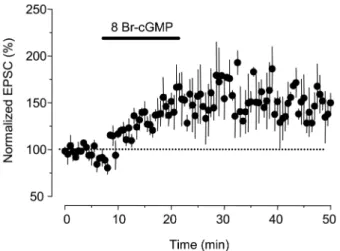
척수 절편의 척수 후근 유입부를 전기자극하여 발생하는 시냅스후전류(EPSC)를 기록 하였으며 이때 발생하는 시냅 스후전류의 정점의 크기를 측정하여 비교 분석하였다. 일 정한 시냅스후전류의 크기가 관찰되면 control value를 위 하여 10분간 그 크기를 기록하였으며, 1 mM 8-Br-cAMP 를 15분간 처치하였다. 그 결과 EPSC의 크기는 증가하였 으며 또한 washout 이후에도 EPSC의 크기가 지속적으로 증가하여 약 150%의 증가된 상태로 유지되었다. 그 유지 시간은 30분 이상 지속되는 long term change를 보였다 (Fig. 1, n = 7). 그러나 짧은 시간의 cAMP 처치는 약물 투 여 동안에만 EPSC의 크기 증가 관찰되었으며, washout 이후에는 control value와 유사한 반응을 보였다(data not shown). 기록 동안 기록전극의 저항은 큰 변화가 관찰되 지 않았으며, cAMP washout 이후 다른 전기적 특성의 변화가 관찰되지 않았다.
cGMP에 의한 척수 아교질 신경세포의 시냅스후전류 의 크기 증가
똑같은 조건에서 1 mM 8-Br-cGMP를 15분간 처치한 결
과 EPSC의 크기는 증가하였으며 또한 washout 이후에 도 EPSC의 크기가 지속적으로 증가하여 약 170%의 증 가된 상태로 유지되었다. 그 유지 시간은 30분 이상 지속 되는 long term change를 보였다(Fig. 2, n = 7). 그러나 짧은 시간의 cGMP 처치는 약물 투여 동안에만 EPSC의 크기 증가 관찰되었으며, washout 이후에는 control value 와 유사한 반응을 보였다(data not shown). 기록동안 기 록전극의 저항은 큰 변화가 관찰되지 않았으며, cGMP washout 이후 다른 전기적 특성의 변화가 관찰되지 않았 다. 따라서 cAMP, cGMP는 각각 15분 이상 처치하면 시 냅스 가소성(LTP)가 발생함을 알 수 있다.
1 mM 8-Br-cAMP와 1 mM 8-Br-cGMP의 동시 처치에 의한 척수 아교질 신경세포의 시냅스 전류의 크기 변화 위 실험에서 EPSC의 반응을 증가시켰던 두 약물 1 mM 8-Br-cAMP와 1 mM 8-Br-cGMP를 동시에 15분간 처치하 면 EPSC의 반응이 통계적으로 유의하지 않는 수준에서 약 간의 증가(약120%)가 관찰되었다(Fig. 3, n = 7). 또한 두 약물을 투여하는 동안에도 큰 변화가 관찰되지 않았다. 이 러한 결과는 각각의 약물 작용이 시냅스 전류의 증가를 나 타내지만 두 약물이 동시에 투여되면 그 반응을 서로 길 항하는 세포내 작용기전이 있음을 시사한다. 이와 같은 신 호전달계 상호작용 기전이 신경 시냅스 가소성에 적용될 가능성이 있다. 이를 위해 다음 실험에서 관련된 신호전달 경로를 알아보고자 하였다.
Phosphodiesterase 차단 후 8-Br-cGMP/8-Br-cAMP ratio (1:1)에 따른 시냅스 가소성의 변화
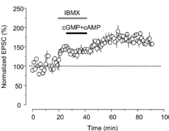
cAMP와 cGMP의 세포내 신호전달 경로에서 각 물질들 을 분해하는 효소 phosphodiesterase (PDE)를 차단한 후 cAMP와 cGMP를 처치하면 각 약물들의 반응을 차단하는 효소가 차단되어 있으므로 시냅스후전류의 증가를 관찰할 수 있으리라 가정할 수 있다. 따라서 이를 확인하기 위하 여 PDE 차단제인 3-isobutyl-1-methylxanthine (IBMX) 20 µM을 5분간 전처치하면 일시적인 EPSC의 증가를 관찰할 수 있었으며(약 140%), 이 조건에서 8-Br-cGMP/8-Br- cAMP (1:1)를 15분간 처치하면 EPSC의 크기는 더욱 증 가되었고(약 170%) 이러한 시냅스 가소성은 시간 경과에 따라 지속적으로 증가하여 50분 이상 유지되는 양상을 보 였다(Fig. 4, n = 7).
Fig. 1. Modulation of synaptic plasticity by 8-Br-cAMP in spinal substantia gelatinosa neurons. EPSCs were recorded for 10 min- utes and normalized to 100%. Application of 8-Br-cAMP (1µM), a membrane permeable analogue of cAMP, for 15 minutes induced a gradual increase in EPSCs, which lasted for 30 minutes. Dots are averaged EPSCs collected from 7 different cells.
Fig. 2. Modulation of synaptic plasticity by 8-Br-cGMP in spinal substantia gelatinosa neurons. EPSCs were recorded for 10 min- utes and normalized to 100%. Application of 8-Br-cGMP (1µM), a membrane permeable analogue of cGMP, for 15 minutes induced a gradual increase in EPSCs, which lasted for 30 minutes. Dots are averaged EPSCs collected from 7 different cells.
Fig. 3. Failure of synaptic plasticity by co-application of 8-Br- cAMP/8-Br-cGMP (1:1) in spinal substantia gelatinosa neurons.
EPSCs were recorded for 10 minutes and normalized to 100%. Sta- tistically significant changes in amplitudes of EPSCs were not observed for 30 minutes after simultaneous application of 8-Br- cAMP/8-Br-cGMP (1µM/1 µM) for 15 minutes. Dots are aver- aged EPSCs collected from 7 different cells.
고 찰
이번 연구의 목적은 척수 통증시냅스 가소성에서 세포내 신호전달계(cAMP, cGMP)간의 상호연관성이 어떠한 영향 을 주는지와 이와 관련된 crosstalk 기전을 규명하여 다양 한 형태로 관찰되는 만성통증의 발생기전에 대한 새로운 재해석을 하고자 하는 것이다. 이 연구를 통해서 얻은 결 과를 정리하면, 1) cAMP 또는 cGMP는 각각 척수 통증 시냅스 효율을 강화시키는 작용을 통해 과감작 통증을 유 발할 수 있고, 2) cAMP와 cGMP의 동시 투여는 척수 통 증 시냅스 효율 조절에 있어서 서로의 효과를 길항하는 작용을 가지고 있으며, 3) 이러한 cAMP와 cGMP의 상호 길항작용에 PDE가 중요한 역할을 함을 알 수 있었다. 이 상의 결과로부터 과감작 통증이 유발되는 상황에서 2차 전 령계의 상호 작용을 통해 통증의 효율이 조절됨을 유추할 수 있으며, 이러한 상호관련 작용기전은 신호전달계 내의 효율을 조절하는 내재적 요소를 지니고 있는 것으로 사료된다.
척수의 Rexed's lamina II에 존재하는 척수 아교질세포는 통각 정보가 통합되고 조절되는 중추 신경계의 일차 관문 으로 상위 중추로 통각정보를 전달한다(Kumazawa et al., 1978; Light et al., 1979). 척수 아교질세포는 주로 Aδ와 C 섬유 같은 통각정보에 선택적인 일차감각 신경섬유와 시 냅스를 이루고(Light et al., 1979; Sugiura et al., 1989;
Yoshimura et al., 1989), 말초에서 가해진 유해자극에 반 응 한다(Woolf et al., 1983). 척수아교질은 그 생리적인 중요성에 상응하여 많은 종류의 신경전달물질 혹은 조절 물질에 의하여 그 흥분성 및 시냅스 가소성이 조절 된다 (Yoshimura et al., 1990; Cerne et al., 1992). 말초 조직
이 손상되면 일차감각 C-섬유가 반복적으로 흥분하고, 이 흥분을 받은 척수 아교질세포의 감수성이 증가하여 같은 자극에도 더 많은 통증을 느끼게 되는데 이를 통각과민 (hyperalgesia)이라고 한다(Torebjork et al., 1992; Woolf et al., 1994). 일차감각 신경세포와 척수 아교질세포 사이 의 시냅스 효율이 시냅스 활동에 의존적으로 변하는 시냅 스 가소성이 바로 통각과민의 기전을 이해하는 열쇠가 된 다. 척수 아교질세포의 시냅스 가소성에 관한 연구 중 해 마에서 기억과 학습을 설명하는 시냅스 가소성 현상인 long- term potentiation (LTP)이 척수 아교질세포의 시냅스에서 도 관찰되었으며(Bliss et al., 1993; Randic et al., 1993) 실제로 조직을 손상시켰을 경우에도 C-섬유를 전기적으로 자극하였을 경우와 마찬가지로 척수 아교질 세포와 같은 척수후각 신경세포의 흥분발사나 C-fiber evoked potential 이 증가되는 LTP를 관찰하였다(Rygh et al., 1999). 이러 한 관점은 LTP가 만성 통증을 시발하고 유지하는 기전이 며, 이 기전을 바로 이해하면 통각과민이 동반되는 만성 통증을 예방하거나 치유할 수 있다는 접근이다.
이번 연구에 있어서 그 중요성은 Yin-Yang theory(음양 이론)에 따른 신호전달 조절과 그와 관련된 신호전달물질 로서 cAMP와 cGMP의 가능성이다. 2차 전령계의 각각의 기능에 대하여 많은 보고가 있으나, 그들 간의 crosstalk에 대한 연구는 거의 없는 실정이다. 그러나 심장세포에서 If 전류는 cyclic nucleotide (cAMP, cGMP)에 의해 증가되나 그들은 서로의 작용을 길항하는 현상을 관찰하였다(Yoo et al., 1998). 심장근 세포에서 If 전류는 cGMP와 cAMP 각 각에 의해 증가되지만 각 약물들의 반응은 각각의 반응을 Fig. 4. Modulation of synaptic plasticity by IBMX and co-applica-
tion of 8-Br-cGMP/8-Br-cAMP (1:1) in spinal substantia gelatinosa neurons. EPSCs were recorded for 10 minutes and normalized to 100%. Application of 3-isobutyl-1-methylxanthine (IBMX, 20µM, 5 minutes) that prevents cAMP/cGMP from degradation by inhibi- tion of phosphodiesterase increased EPSCs. Co-application of 8-Br- cAMP/8-Br-cGMP (1µM/1 µM) for 15 minutes in the presence of IBMX induced additional increase in EPSCs, which lasted for 30 minutes. Dots are averaged EPSCs collected from 7 different cells.
Fig. 5. Schematic diagram of modulation of synaptic plasticity by crosstalk between second messengers, cAMP and cGMP. Each of cAMP and cGMP may induce hypersensitive nociception by mod- ulating synaptic plasticity. However, co-application of cAMP and cGMP antagonizes each other by activation of degradation enzyme, phosphodiesterase (PDE). Therefore, simultaneous acti- vation of both cAMP and cGMP may interfere with synaptic plas- ticity and inhibition of PDE may result in increase of synaptic potentiation. This mechanism might contribute to the modulation of nociception in spinal cord. NO: nitric oxide, mAch: metabotro- pic acetylcholine receptor, PKA, PKC, PKE, PKG: protein kinase A, C, E, G, respectively.
서로 차단하는 성상을 가지고 있다. 이러한 결과는 세포의 특성과 시냅스라는 부분에서는 차이가 있지만 신호전달계 라는 관점에서 이번 연구결과가 잘 일치한다. 이러한 관점 에서 Wen 등은 음양이론에 근거하여 신호전달계에 따른 신경말단의 성장방향이 결정됨을 보고하였고(Wen et al.
2006), Nishiyama등은 신경세포에 존재하는 Ca2+이온통로 가 cAMP/cGMP ratio에 따라 조절되어 신경세포의 성장 방향이 달라진다고 보고하였다(Nishiyama et al., 2003;
Henley et al., 2004). 이러한 현상은 시냅스 가소성 및 발 달에 영향을 줄 수 있으리라 기대되나 아직 보고는 없었 다. 그러나 이번 연구에서 확인한 바에 의하면, 각각의 신 호전달계는 LTP를 유발시키나, 서로 공존하는 경우에 각각 의 작용을 길항하여 반응이 오히려 감소하거나 사라지게 되 는 이중적 조절 구조를 가지고 있는 것으로 사료된다. Fig.
5는 세포내 인산화효소와 2차 전령계 cAMP, cGMP에 따 라 시냅스 가소성의 방향이 결정됨을 도식적으로 그린 그 림이다. 세포내 Ca2+이 중등도로 증가하면 LTD가 사라지며 오히려 시냅스가소성이 발생하지 않는 구간이 있으나 이에 대한 해석은 논란이 있으며 이번 연구결과에서처럼 cAMP/
cGMP ratio로 이 부분에 대한 작용기전을 해석할 수 있으 리라 기대된다(Fig. 5).
이러한 결과를 종합해 보면 각각의 2차 전령계는 통증 유발에 있어서 중요한 역할을 하며 또한 두 2차 전령계가 활성화 되는 상황에서는 서로의 신호전달경로를 조절함으 로써 통증조절을 하고 있음을 의미한다. 이처럼 척수 통증 조절은 분자적 수준에서 서로의 신호전달 과정을 조절하는 음양이론이 적용되고 있으며 이에 근거한 시냅스 가소성 및 통증조절에 대한 재해석을 위한 기초 자료로서 그 의 미가 있다. 또한 기억과 학습의 기전을 척수 수준에 도입 함으로써 만성통증과 관련된 통증정보처리 기전에서 시냅 스 가소성에 대한 정확한 이해를 할 수 있으며 만성통증 의 발생기전에 대한 음양이론에 근거한 새로운 재해석은 이후 통증연구의 발전에 기여할 것으로 사료되며 통증지식 증진에 도움이 되리라 사료된다.
감사의 글
이 논문은 2006년 서울대학교 발전기금의 지원과 2008 년 정부(교육과학기술부) 한국연구재단의 기초의과학연구 센터육성사업(Medical science and engineering Research Center: MRC)의 지원(Grant 2010-0029510)을 받아 수행 된 연구임
참고문헌
Bliss TV, Collingridge GL. A synaptic model of memory: long-
term potentiation in the hippocampus. Nature. 1993;361 (6407):31-9.
Bliss TV, Lomo T. Long-lasting potentiation of synaptic transmission in the dentate area of the anaesthetized rabbit following stimulation of the perforant path. J Physiol. 1973;
232(2):331-56.
Campbell JN, Meyer RA, LaMotte RH. Sensitization of myelinated nociceptive afferents that innervate monkey hand. J Neurophysiol. 1979;42:1669-1679.
Cerne R, Jiang M, Randic M. Cyclic adenosine 3'5'- monophosphate potentiates excitatory amino acid and synaptic responses of rat spinal dorsal horn neurons. Brain Res. 1992;596:111-123.
Edwards FA, Konnerth A, Sakmann B, Takahashi T. A thin slice preparation for patch clamp recordings from neurons of the mammalian central nervous system. Pflugers Arch. 1989;414 (5):600-612.
Feil R, Hofmann F, Kleppisch T. Function of cGMP-dependent protein kinases in the nervous system. Rev Neurosci. 2005;
16(1):23-41.
Henley J, Poo MM. Guiding neuronal growth cones using Ca2+ signals. Trends Cell Biol. 2004;14(6):320-30.
Hoeger-Bement MK, Sluka KA. Phosphorylation of CREB and mechanical hyperalgesia is reversed by blockade of the cAMP pathway in a time-dependent manner after repeated intramus- cular acid injections. J Neurosci. 2003;23(13):5437-45.
Ikeda H, Heinke B, Ruscheweyh R, Sandkuhler J. Synaptic plasticity in spinal lamina I projection neurons that mediate hyperalgesia. Science. 2003;299(5610):1237-40.
Kang YM, Lee MK, Yang GY, Bae YC, Ahn DK. Participation of nitric oxide pathways in interleukin 1β-induced mechanical allodynia in the orogicial area of rats. Int J Oral Biol. 2009;
34(1):1-6.
LaMotte RH, Thalhammer JG. Torebjork HE, Robinson CJ.
Peripheral neural correlates of magnitude of cutaneous pain and hyperalgesia: a comparison of neural events in monkey with sensory judgements in human. J Neurophysiol 1983;
50;1-26.
Light AR, Trevino DL, Perl ER. Morphological features of functionally defined neurons in the marginal zone and substantia gelatinosa of the spinal dorsal horn. J Comp Neurol. 1979;186:151-172.
Ma W, Du W, Eisenach JC. Role for both spinal cord COX-1 and COX-2 in maintenance of mechanical hypersensitivity following peripheral nerve injury. Brain Res. 2002;937(1- 2):94-9.
Mills CD, Johnson KM, Hulsebosch CE. Group I metabotropic glutamate receptors in spinal cord injury: roles in neuroprotection and thedevelopment of chronic central pain. J Neurotrauma. 2002;19(1):23-42.
Moore KA, Baba H, Woolf CJ. Synaptic transmission and plasticity in the superficial dorsal horn. Prog Brain Res.
2000;129:63-80.
Nishiyama M, Hoshino A, Tsai L, Henley JR, Goshima Y, Tessier-Lavigne M, Poo MM, Hong K. Cyclic AMP/GMP- dependent modulation of Ca2+ channels sets the polarity of nerve growth-cone turning. Nature. 2003;423(6943):990-5.
Parada CA, Reichling DB, Levine JD. Chronic hyperalgesic
priming in the rat involves a novel interaction between cAMP and PKCepsilon second messenger pathways. Pain. 2005;
113(1-2):185-90.
Pockett S. Spinal cord synaptic plasticity and chronic pain.
Anesth Analg. 1995;80(1);173-9.
Randic M. Plasticity of excitatory synaptic transmission in the spinal cord dorsal horn. Prog Brain Res. 1996;113:463-506.
Randic M, Jiang MC, Cerne R. Long-term potentiation and long-term depression of primary afferent neurotransmission in the rat spinal cord. J Neurosci. 1993;13:5228-5241.
Rygh LJ, Tjolsen A, Hole K, Svendsen F. Cellular memory in spinal nociceptive circuitry. Scand J Psychol. 43(2);153-9, 2002.
Rygh LJ, Svendsen F, Hole K, Tjolsen A. Natural noxious stimulation can induce long-term increase of spinal nocicep- tive responses. Pain. 1999;82:305-310.
Sandkuhler J, Benrath J, Brechtel C, Ruscheweyh R, Heinke B. Synaptic mechanisms of hyperalgesia. Prog Brain Res.
2000;129:81-100.
Sandkuhler J, Chen JG, Cheng G, Randic M. Low-frequency stimulation of afferent Adelta-fibers induces long-term depres- sion at primary afferent synapses with substantia gelatinosa neurons in the rat. J Neurosci. 1997;17(16):6483-91.
Sugiura Y, Terui N, Hosoya Y. Difference in distribution of central terminals between visceral and somatic unmyelinated (C) primary afferent fibers. J Neurophysiol. 1989; 62(4):834- 840.
Svendsen F, Tjolsen A, Gjerstad J, Hole K. Long term potentiation of single WDR neurons in spinalized rats. Brain Res. 1999;816:487-492.
Tao YX, Hassan A, Haddad E, Johns RA. Expression and action of cyclic GMP-dependent protein kinase Ialpha in inflammatory hyperalgesia in rat spinal cord. Neuroscience.
2000;95(2):525-33.
Tegeder I, Del Turco D, Schmidtko A, Sausbier M, Feil R, Hofmann F, Deller T, Ruth P, Geisslinger G. Reduced inflammatory hyperalgesia with preservation of acute thermal nociception in mice lacking cGMP-dependent protein kinase I. Proc Natl Acad Sci U S A. 2004 ;101(9):3253-7.
Torebjork HE, Lundberg LE, LaMotte RH. Central changes in processing of mechanoreceptive input in capsaicin-induced secondary hyperalgesia in humans. J Physiol (Lond). 1992;
448:765-80.
Wen Z, Zheng JQ. Directional guidance of nerve growth cones. Curr Opin Neurobiol. 2006;16(1):52-58.
Willis WD Jr. Is central sensitization of nociceptive transmission in the spinal cord a variety of long-term potentiation?
Neuroreport. 1997;8(16):3427-30.
Willis WD. Long-term potentiation in spinothalamic neurons.
Brain Res Brain Res Rev. 2002;40(1-3):202-14.
Woolf CJ, Doubell TP. The pathophysiology of chronic pain- increased sensitivity to low threshold A beta-fiber inputs.
Curr Opin Neurobiol. 1994;4:525-534.
Woolf CJ, Fitzgerald M. The properties of neurones recorded in the superficial dorsal horn of the rat spinal cord. J Comp Neurol. 1983;221(3):313-328.
Yoo S, Lee SH, Choi BH, Yeom JB, Ho WK, Earm YE. Dual effect of nitric oxide on the hyperpolarization-activated inward current (I(f)) in sino-atrial node cells of the rabbit. J Mol Cell Cardiol. 1998;30(12):2729-38.
Yoshimura M, Jessell T. Amino acid-mediated EPSPs at primary afferent synapses with substantia gelatinosa neurones in the rat spinal cord. J Physiol (Lond). 1990; 430:315-35.
Yoshimura M, Jessell T. Membrane properties of rat substantia gelatinosa neurons in vitro. J Neurophysiol. 1989;62:109-18.