DOI: https://doi.org/10.3339/jkspn.2020.24.1.53 ISSN 2384-0250 (online)
Group B Streptococcal Renal Abscess in a 17-Year-Old Girl with Type 1 Diabetes Mellitus
Streptococcus agalactiae or group B streptococcus (GBS) is associated with infec- tions in neonates and pregnant women. Herein, we describe a rare case of GBS renal abscess with peritonitis and pleural effusion in a 17-year-old girl with type 1 diabetes mellitus. The girl was admitted due to fever and right flank pain. Labora- tory findings included leukocytosis and increased C-reactive protein level and erythrocyte sedimentation rate. Her serum glucose level was 484 mg/dL. Urinalysis showed no pyuria. Renal sonography revealed parenchymal swelling in the right kidney. The patient was administered intravenous cefotaxime. Urine and blood cultures were negative. Fever seemed to improve, but the following day, she com- plained of abdominal pain and fever. Antibiotic was switched to imipenem, and abdominal and pelvic CT revealed a ruptured right renal abscess, peritonitis, and bilateral pleural effusion with atelectasis. Pigtail catheter drainage of the abscess was performed. Culture from the abscess was positive for GBS, and fever subsided 2 days after the drainage. She was discharged with oral cefixime. The clinical course of urinary tract infections (UTIs) can be atypical in patients with diabetes, and GBS can be a cause of UTIs. Prompt diagnosis and management are necessary to pre- vent complications in patients showing atypical courses.
Key words: Streptococcus agalactiae, Diabetes mellitus, Urinary tract infection, Pleural effusion
Kyeong Eun Oh, M.D.
Hyung Eun Yim, M.D., Ph.D.
Kee Hwan Yoo, M.D., Ph.D.
Department of Pediatrics, Korea University College of Medicine, Seoul, Korea
Corresponding author:
Hyung Eun Yim, MD, PhD
Department of Pediatrics, Korea University Ansan Hospital, Korea University College of Medicine, 123 Jeokgeum-ro, Danwon- gu, Ansan 15355, Republic of Korea Tel: +82-31-412-5096
Fax: +82-31-405-8591 E-mail: [email protected] Received: 5 March 2020 Revised: 27 March 2020 Accepted: 3 April 2020
This is an open-access article distributed under the terms of the Creative Commons Attribu tion Non-Commercial License (http://
crea tivecom mons.org/licenses/by-nc/4.0/) which permits unrestricted non-commercial use, distribution, and reproduction in any medium, provided the original work is properly cited.
Copyright © 2020 The Korean Society of Pediatric Nephrology
Introduction
Streptococcus agalactiae or group B streptococcus (GBS) is a Gram-positive pathogen that is typically associated with infections in neonates and pregnant women1). This organism is responsible for about 2–3% of all urinary tract in- fections (UTIs), and only a few cases of GBS renal abscesses have been reported
2,3). Moreover, diabetes mellitus (DM) is a major risk factor for complicated UTIs and serious GBS infections4,5). Herein, we describe a rare case of GBS renal abscess accompanied by peritonitis and bilateral pleural effusion in a 17-year-old girl with uncontrolled type 1 DM.
Case report
A 17-year-old girl was admitted due to fever and flank pain. She had been diagnosed with type I DM 3 years prior, but had stopped taking insulin and
self-monitoring her blood sugar levels. She had experienced 2 days of fever and right flank pain without other urinary symptoms. Her body temperature was 38.8℃, and her blood pressure was 120/70 mmHg. Right costovertebral angle tenderness was noted. The laboratory findings included leukocytosis (white blood cell count of 16,980/μL, neutro- phil-dominant) and an elevated C-reactive protein level and erythrocyte sedimentation rate (15.4 mg/dL and 58 mm/
hr, respectively). The patient’s plasma and urine neutrophil gelatinase-associated lipocalin levels were within the normal range, but her serum glucose level was 484 mg/dL, and he- moglobin A1c level, 14.6%. Urinalysis showed a pH of 5.5, a specific gravity of 1.040, glucosuria and ketonuria but no proteinuria, hematuria, or pyuria. Blood gas analysis showed a pH of 7.44, pCO2 of 26 mmHg, and HCO3– of 17.8 mmol/
L. Initial renal sonography revealed diffuse wall thickening of the urinary bladder and hyperechoic parenchymal swel- ling in the right kidney. A dimercaptosuccinic acid renal scan was performed and revealed cortical defects in the right kidney. She was administered intravenous cefotaxime and insulin. The initial urine and blood cultures showed negative results. The patient’s high fever and flank pain appeared to improve after treatment was initiated, and fever was not noted on hospital day 6. On the following day, however, she once again complained of abdominal pain and fever. No pyuria or hematuria was found on the urinalysis.
White blood cell count was decreased from 16,980/μL to 6,090/μL and C-reactive protein level was also decreased
from 15.38 mg/dL to 2.87 mg/dL. By contrast, erythrocyte sedimentation rate level was increased from 58 mm/hr to 87 mm/hr.
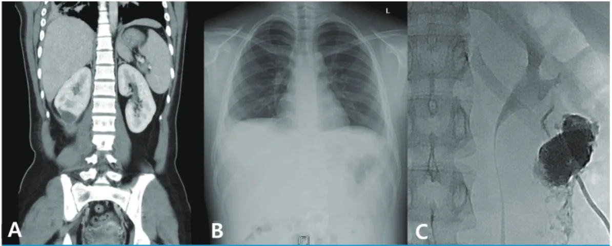
Antibiotic resistance was suspected, so her antibiotic was switched to imipenem on hospital day 7. Abdominal and pelvic computed tomography (APCT) revealed a ruptured right renal and perinephric abscess, diffuse peritoneal thic- kening, ascites, and bilateral pleural effusion with passive atelectasis in basal segment of both lungs (Fig. 1A). On hospital day 8, pigtail catheter drainage was performed and pus taken from the abscess was cultured (Fig. 1B). The abscess culture was positive for GBS. GBS was sensitive penicillin, cefotaxime, and imipenem. We maintained imi- penem and the patient’s fever subsided 2 days after the dra- inage of the abscess. Follow up sonography demonstrated no abscess pocket and tubogram of the pigtail catheter revealed no remaining connection to the vascular system (Fig. 1C, Fig. 2A). On hospital day 18, she was discharged with oral cefixime and subcutaneous insulin regimens.
One month later, APCT showed improvement of the right renal abscess with cortical scarring at the lower pole of the right kidney (Fig. 2B).
Discussion
This is a rare case of a GBS renal abscess accompanied by peritonitis and bilateral pleural effusion in a 17-year-old girl with uncontrolled type 1 DM. Although she showed
B C A
Fig. 1. Imaging studies demonstrating a ruptured right renal abscess with peritonitis, pleural effusion and catheter drainage of the abscess. (A) Abdomen pelvis CT demonstrating a ruptured right renal abscess, peritonitis, and bilateral pleural effusion with passive atelectasis in both basal lungs. (B) Chest X-ray showing bilateral pleural effusion. (C) Pigtail catheter drainage of the right renal abscess.
initial clinical improvement upon intravenous administra- tion of a third generation cephalosporin, her condition then deteriorated and APCT revealed a ruptured renal abscess with peritonitis and bilateral pleural effusion. After chan- ging to a broad-spectrum β-lactam antibiotic and perfor- ming pigtail catheter drainage, the patient’s fever subsided and she was discharged from the hospital.
GBS is common cause of perinatal infections in neonates and pregnant women1), and a few cases of GBS-associated UTIs or renal abscesses have been reported3). DM is known to be a major risk factor for serious GBS infections5). The most common sites of infection are the skin, soft tissue, bones, joints, urinary tract, lungs, peritoneum, and genital tract6). Patients with DM are also vulnerable to UTIs be- cause the antimicrobial defense mechanisms of the kidney are insulin-dependent. Moreover, high glucose concentra- tions in the urine of DM patients may promote the growth of pathogenic bacteria7). Therefore, patients with DM are at a higher risk of developing serious UTIs8). In many studies, Escherichia coli was shown to be the most common pathogen responsible for UTIs among diabetic patients.
One study described gram-negative bacteria such as Kleb- siella, Proteus, and Pseudomonas also commonly cause UTIs in diabetic patients9). On the other hand, another study reported that less common organisms such as coa- gulase-negative Staphylococci and Enterococcus species
can cause UTIs in diabetic patients and showed that the multi-drug resistance rate was higher in diabetic patients with UTI than in patients without underlying DM10). Our patient was a 17-year-old girl with DM, and GBS was found to be a pathogen underlying the complicated UTI. Her uncontrolled blood sugar levels may have increased glucose concentrations in the urine and impaired antimicrobial defense of her urinary system, leading to the development of serious renal infection.
Our patient presented with fever and costovertebral angle tenderness, but the initial urinalysis and urine culture re- vealed negative results. She didn’t take any medicine inclu- ding antibiotics before she came to our hospital. She was administered intravenous cefotaxime to treat the UTI, but redeveloped fever. APCT was reperformed and revealed a ruptured right renal abscess, peritonitis, and bilateral ple- ural effusion with passive atelectasis in both basal lungs.
Why were the initial urinalysis and urine culture negative in this patient? One study reported that 25–30% of women with UTI symptoms have a negative urine culture because the infection does not affect the collecting system or involve obstruction of the collecting system11). Moreover, in a small randomized controlled trial involving women with UTI symptoms but a negative dipstick urine test result, anti- biotic treatment significantly reduced the median time to dysuria resolution compared to placebo12). In our patient,
A B
Fig. 2 Follow up imaging studies after treatment. (A) Tubogram with pigtail catheter demonstrating no link between the abscess pocket and the vascular system. (B) Follow up abdomen pelvis CT after 1 month showing improved right renal abscess with cortical scarring at right kidney lower pole.
abscess did not seem to have communication with the renal collecting system and the location of the renal abscess. Em- piric antibiotic treatment was initially effective, but ca- theter drainage and administration of antibiotic were ulti- mately required to successfully treat the ruptured renal abscess. In general, GBS is sensitive to penicillin and GBS that was detected in our patient was also sensitive to peni- cillin. We maintained imipenem to treat renal and peri- nephric abscess with diffuse peritonitis and bilateral pleural effusion effectively. There have been no cases of GBS renal abscess in children reported domestically or abroad. We found 1 case of GBS perinephric abscess in a 24-year-old woman with DM who experienced recurrent fever, flank pain, and increased urinary frequency for 1 month. Ulti- mately, she underwent percutaneous abscess drainage and was administered intravenous antibiotics to treat the GBS infection3). Therefore, GBS infection should be considered in patients with underlying diseases such as diabetes who exhibit atypical UTI manifestations.
A variety of intra-abdominal or pelvic disorders such as renal and/or perirenal abscesses and urinary tract obstruc- tion are associated with pleural effusion13). A case of pleural empyema and perinephric abscess was recently reported in a patient who presented with a large right‐sided pleural effusion14). In our case, bilateral pleural effusion and passive lung atelectasis were also noted alongside the ruptured renal abscess and peritonitis. The effusions were initially suspected to be of bacterial or tuberculous origin, but were ultimately deemed to be reactive effusions when the pati- ent’s clinical course was considered. Right-sided diffuse peritoneal thickening and pleural effusion were noted, so it is possible that the right perinephric abscess may have extended into the pleural cavity. Because the amount of ef- fusion was relatively small, we did not perform diagnostic or therapeutic thoracentesis. The pleural effusion and renal abscess improved concurrently. No pleural or peritoneal complications were noted following treatment. Here, we chose to report this case because GBS renal and perinephric abscess are rare in children and adolescents, and the seve- rity of the disease was such that it was accompanied by pe- ritonitis and pleural effusion.
In conclusion, renal abscesses are relatively uncommon in children but may need a prolonged antibiotic treatment, an increased length of hospital stay, and a high treatment
cost, and may be associated with life-threatening complica- tions15). The clinical course of UTIs can be atypical in pati- ents with diabetes, and GBS can be a relevant organism causing renal abscess. Prompt diagnosis and management are necessary to prevent complications in UTI patients showing atypical clinical courses.
Patient consent
This study was approved by the institutional review board (IRB), and informed consent was waived due to retrospec- tive study design and anonymous patient data2018AS0122).
Conflicts of interest
No potential conflict of interest relevant to this article was reported.
References
1. Ulett KB, Shuemaker JH, Benjamin WH, Tan CK, Ulett GC. Group B streptococcus cystitis presenting in a diabetic patient with a massive abdominopelvic abscess. J Med Case Rep 2012;6:237.
2. Leclercq SY, Sullivan MJ, Ipe DS, Smith JP, Cripps AW, Ulett GC.
Pathogenesis of Streptococcus urinary tract infection depends on bacterial strain and B-hemolysin/cytolysin that mediates cytotoxicity, cytokine synthesis, inflammation and virulence.
Nature 2012;6:29000.
3. Yoon KH, Lee JH, Han WJ, Cho MS, Kim JY, Park YS, et al. A peri- nephric abscess caused by Streptococcus agalactiae. Korean J Med 2009;76:220-3.
4. Mnif MF, Kamoun M, Kacem FH, Bouaziz Z, Charfi N, Mnif F, et al.
Complicated urinary tract infections associated with diabetes mellitus: Pathogenesis, diagnosis and management. Indian J Endocrinol Metab 2013;17:442-5.
5. Yanai H, Hamasaki H, Tsuda N, Adachi H, Yoshikawa R, Moriyama S, et al. Group B streptococcus infection and diabetes: A review.
J Microbiol Antimicrob 2012;4:1-5.
6. Batista RP, Ferreira CR. Streptococcus agalactiae septicemia in a patient with diabetes and hepatic cirrhosis. Autops Case Rep 2015;5:35-43.
7. Fünfstück R, Nicolle LE, Markolf H, Naber K. Urinary tract infection in patients with diabetes mellitus. Clin Nephrol 2012;77:40-8.
8. Zasloff M. Why are diabetics prone to kidney infections? J Clin
Invest 2018;128:5213-5 .
9. Hakeem LM, Bhattacharyya DN, Lafong C, Janjua KS, Serhan JT, Campbell IW. Diversity and complexity of urinary tract infection in diabetes mellitus. Br J Diabetes Vasc Dis 2009;9:119-25.
10. Woldemariam HK, Geleta DA, Tulu KD, Aber NA, Legese MH, Fenta GM, et al. Common uropathogens and their antibiotic suscepti- bility pattern among diabetic patients. BMC Infect Dis 2019;19:43.
11. Heytens S, De Sutter A, Coorevits L, Cools P, Boelens J, Van Simaey L, et al. Women with symptoms of a urinary tract infection but a negative urine culture: PCR-based quantification of Escherichia coli suggests infection in most cases. Clin Microbiol Infect 2017;
23:647-52.
12. Mangin D, Toop L, Chambers S, Fletcher L. Response to antibiotics
of women with symptoms of urinary tract infection but negative dipstick urine test results: double blind randomised controlled trial. BMJ 2005;331:143.
13. Wang IK, Chuang FR, Chang HY, Lin CL, Yang CT. Acute pyelone- phritis associated with transudative pleural effusion in a middle- aged woman without urinary tract obstruction. Med Princ Pract 2006;15:309-11.
14. Tan PSC, Badiei A, Fitzgerald DB, Kuok YJ, Lee YCG. Pleural empy- ema in a patient with a perinephric abscess and diaphragmatic defect. Respirol Case Rep 2019;7:e00400.
15. Chen CY, Kuo HT, Chang YJ, Wu KH, Yang WC, Wu HP. Clinical assessment of children with renal abscesses presenting to the pediatric emergency department. BMC Pediatrics 2016;16:189.