Clinical effect and standardization of indocyanine green angiography in the laparoscopic colorectal surgery
10
0
0
전체 글
(2) 114 total mesorectal excision, male patient, stapler characteristics, anemia, and nutrition [2]. Most of these variables are unmodifiable patient-related risk factors. However, there are some modifiable risk factors that are gradually becoming possible to overcome with technological advances. Intestinal perfusion status has become a modifiable risk factor, and surgeons can evaluate and select anastomosis sites with optimal perfusion to perform safe colorectal anastomoses [3]. Ligation of the inferior mesenteric artery (IMA) has been considered an essential step for oncological dissection of the rectosigmoid colon. Several studies have reported that IMA ligation may cause hypoperfusion of the left-sided colon [4,5]. Almost half of anastomotic leaks are suspected to be associated with hypoperfusion. Subjective assessments have been used to evaluate blood f low in the gastrointestinal tract. This might have limited diagnostic accuracy dependent on the surgeon’s experience. Indocyanine green (ICG) angiography is a simple, noninvasive method that is carried out using a near-infrared (NIR) camera system. Currently, ICG angiography is rapidly gaining adoption in colorectal surgery. The clinical utility of ICG angiography for reducing anastomotic complications is under scientific investigation through several multicenter randomized clinical trials [6,7]. The expectations and interests of ICG angiography among surgeons are continually increasing. However, since ICG angiography has been carried out with various protocols, standardization is required [8,9]. In this review, we discuss the clinical utility and standardization of ICG angiography for colonic perfusion assessment.. MAIN SUBJECTS Prolonged debate about inferior mesenteric artery ligation and hypoperfusion IMA ligation in rectosigmoid colon cancer surgery is commonly performed for sufficient D3 lymph node dissection and tensionfree anastomosis through sufficient proximal colon lengthening. However, it may increase the risk of damage to the autonomic nerve plexus of the IMA root and ischemia of the left-sided colon. To overcome this risk, some surgeons perform low ligation of the IMA to preserve the left colic artery [10]. This procedure requires a high level of surgical expertise for vascular branch dissection. However, D3 lymph node dissection cannot be performed as an en bloc resection, and controversy remains over oncological safety for low IMA ligation even though these two procedures have not been shown to significantly affect oncologic outcomes [11]. The IMA could be a mesenteric anchoring point in the cases of short sigmoid colons and advanced rectosigmoid colon cancer. Therefore, high ligation is inevitably selected to obtain sufficient redundancy of the proximal colonic segment. Journal of Minimally Invasive Surgery Vol. 24 24.. No. 3, 2021. Gyung Mo Son et al.. When performing colorectal surgery, anastomotic complications are the most difficult hurdles faced by surgeons and patients. The large intestine has a different blood f low supply system on the right and left-sided colon. The marginal arterial arcade of the right and left-sided colon might be connected near the splenic f lexure, forming a collateral circulation between the superior mesenteric artery (SMA) and the IMA. The lower rectum is supplied with blood f low by the middle and lower rectal arteries branching from the internal iliac artery. Therefore, even when IMA ligation accompanies anterior resection, blood f low in the left colon can be maintained by the SMA. However, an anatomical cadaver study reported that in 20% of individuals, linkage of the marginal arcade forming the collateral circulation might be insufficient [12]. Hypoperfusion of the left side of the colon is a well-known phenomenon associated with IMA ligation. We reported on marginal arterial pressures measured through mesenteric artery cannulation, particularly looking at changes in colonic mesenteric artery pressure before and after the IMA ligation; we measured marginal arterial pressure before and after IMA clamping. In more than 10% of rectosigmoid cancer patients, marginal arterial pressure was significantly decreased by more than 30% [4]. Therefore, colonic hypoperfusion might be a rare but real event, potentially leading to ischemic colitis in some patients with poor collateral circulation. To prevent hypoperfusion-related anastomotic complications, this 10% of high-risk patients with ischemic changes must be identified. Experienced surgeons have traditionally practiced observing intestinal color, arterial pulsation, and colonic peristalsis, or bleeding at the cutting edge of the mesenteric artery. These subjective evaluations could be considered good clinical assessments by experienced surgeons. However, the accuracy and reliability of subjective evaluations could have a wide spectrum depending on surgical experience [13]. Accordingly, an objective and quantitative technique is needed to improve the accuracy and reliability of bowel perfusion status evaluations. Such an evaluation method should be noninvasive and easy to apply, and the results could be interpreted intuitively in real time by the surgeon. Additionally, the diagnostic technique used should be able to be safely applied to the patient for measuring colonic perfusion [14]. There are a variety of methods for evaluating blood f low. Arterial pressure can be measured through arterial cannulation via connections to blood pressure monitoring equipment in the operating room. [4,15]. Color Doppler ultrasonography is a safe, noninvasive procedure, which has the advantage of allowing repeated and safe evaluation of perfusion status in real time. However, there are technical limitations for evaluating very small blood vessels within 1–2 mm of the colonic wall [16]. Computed tomography (CT) angiography can be used for assessing the mesenteric arterial structure. CT resolution has been gradually.
(3) 115. ICG angiography for colorectal surgery. improving and now permits the evaluation of the arterial circuit using three-dimensional reconstruction. However, CT angiography has a limited resolution for visualization of the collateral marginal artery connection with very small vascular diameters of less than 1 mm at the splenic f lexure [4]. Therefore, a simple and noninvasive assessment method is needed for obtaining high-resolution images in real time for daily practice in the era of minimally invasive surgery.. Indocyanine green angiography and anastomotic leakage ICG is a f luorescent agent that was approved by the U.S. Food and Drug Administration in 1956. To date, it is the only f luorescent substance that can be administered to humans for clinical practice. Although ICG-related side effects are rare, allergic reactions may occur, particularly when a patient has a history of CT contrast allergy. ICG solutions contain a trace amount of iodine, which is a component of CT contrast solution [17]. Therefore, it is necessary to review a patient’s allergy history before performing ICG angiography; it is also important to monitor for allergic reactions during the examination. ICG becomes f luorescent at around 800 nm, which is within the NIR wavelength range. NIR spectroscopy was developed to detect ICG f luorescence, and it is possible to use this technology to visualize ICG in blood samples. This function is mainly used to evaluate the reserve liver function before liver transplantation or liver resection [18]. However, the equipment used for this investigation is still so large that the collected venous blood should be measured in a special laboratory for ICG concentration measurement. Since ICG binds to proteins, such as albumin and globulins in the blood, extravasation does not occur easily, which is useful for evaluating blood f low. ICG was first applied to cardiac function evaluation and ophthalmoscopic angiography. In the surgical field, a handheld NIR camera was developed to evaluate blood f low by detecting f luorescence from intravascular ICG. Recently, a laparoscopic NIR camera has been developed, making ICG angiography available for use in minimally invasive surgery [19]. With ICG angiography, a surgeon can directly observe the intestinal perfusion status with a f luorescence image. Changes in the f luorescence intensity (FI) are viewed along the path of blood f low to the target organ. Fluorescence images can be expressed in green, cyan, or blue on a black-and-white background, depending on the NIR camera system [20]. A f luorescence image can also be expressed as an overlay on the same image of a white light view. ICG angiography can be used to unequivocally reveal unfavorable perfusion zones and obtain quantitative parameters to predict the risk of complications associated with hypoperfusion. Transection line changes have been reported in 5% to 20% of patients with unfavorable perfusion segments identified by ICG. angiography. At our institution, the anastomotic complication rate reached nearly 10% before we instituted ICG angiography, and it rapidly decreased to 1% to 2% after we started evaluating colonic perfusion [3]. Many studies have demonstrated the clinical utility of ICG angiography for reducing anastomotic complications (Table 1) [3,5–7,13,14,19,21–41]. However, two multicenter randomized clinical trials reported that ICG angiography did not significantly reduce the incidence of anastomotic leakage [6,7]. Nevertheless, a meta-analysis determined that there were significantly fewer anastomotic complications in patients who underwent ICG angiography in the included studies (Table 2) [42–49]. One of the reasons for these contradictory results is that quantitative analysis and optimal standardized protocols are still lacking for ICG angiography. Most of the existing evidence has been obtained from smallscale, single-center studies, and the ICG angiography protocol has not been standardized. Additionally, the evaluation of ICG angiography has mostly relied on the subjective judgment of the surgeon, and few studies have applied quantitative analyses. For these reasons, it is necessary to establish a standardized ICG angiography protocol and develop a quantitative analysis protocol for objectively assessing FI.. Standardization of indocyanine green angiography protocol To date, various NIR devices and different angiography protocols have been applied at different institutions. Therefore, to compare and analyze image data from multiple institutions, we need to establish a standardized ICG angiography protocol to improve efficacy and accuracy. Additionally, qualitative perfusion parameters could improve the accuracy of a surgeon’s subjective judgment. The ability to detect changes in FI is a breakthrough in f luorescence imaging that allows tissue perfusion to be easily visualized [3]. However, in ICG angiography, FI is greatly affected by the external environment. Other quantitative parameters based on FI also depend on external conditions. For example, one study found the maximal FI level and increasing slope to vary significantly, inf luenced by various conditional factors, including perfusion status [9]. FI scale can be changed because it is quite sensitive to external environmental conditions. We previously applied various external conditions to obtain the best f luorescence images for several years. From these experiences, we found the optimal external environmental conditions for maximizing FI [9]. The distance between the camera and tissue was one of the most important determinants of brightness. In current ICG cameras for laparoscopy, the brightness changes sharply according to the distance when it is closer than 4 to 5 cm; when it is further than 4 to 5 cm, the brightness becomes relatively stable, which reduces the quality of evaluation of FI changwww.e-jmis.org.
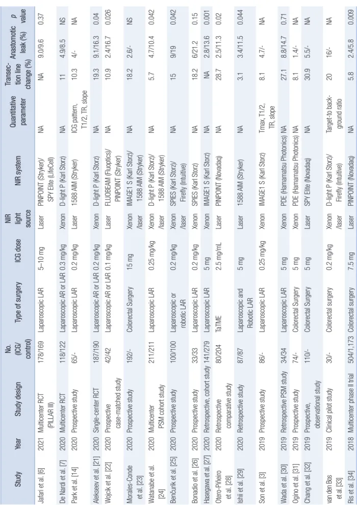
(4) Journal of Minimally Invasive Surgery Vol. 24 24.. No. 3, 2021. 2020 Prospective study. 2020 Multicenter PSM cohort study. Morales-Conde et al. [23]. Watanabe et al. [24]. 2020 Retrospective study. 2019 Prospective study. 2019 Retrospective PSM study. 2019 Prospective study. 2019 Prospective, observational study. 2019 Clinical pilot study. 2018 Multicenter phase II trial. Ishii et al. [29]. Son et al. [3]. Wada et al. [30]. Ogino et al. [31]. Chang et al. [32]. van den Bos et al. [33]. Ris et al. [34]. Colorectal surgery. Colorectal Surgery. Colorectal Surgery. Laparoscopic LAR. Laparoscopic LAR. 504/1,173 Colorectal surgery. 30/-. 110/-. 74/-. 34/34. 86/-. 87/87. Laparoscopic and Robotic LAR. Laparoscopic LAR TaTME. Laparoscopic LAR. Laparoscopic or robotic LAR. Laparoscopic LAR. Colorectal Surgery. 7.5 mg. 0.2 mg/kg. 5 mg. 5 mg. 5 mg. 0.25 mg/kg. 5 mg. 2.5 mg/mL. 5 mg. 0.2 mg/kg. 0.2 mg/kg. 0.25 mg/kg. 15 mg. Laparoscopic AR or LAR 0.1 mg/kg. 2020 Retrospective comparative study. 80/204. 0.2 mg/kg. Laparoscopic AR or LAR 0.2 mg/kg. Laparoscopic LAR. Otero-Piñeiro et al. [28]. 33/33. Bonadio et al. [26] 2020 Prospective study. 5–10 mg. ICG dose. Laparoscopic AR or LAR 0.3 mg/kg. Laparoscopic LAR. Type of surgery. Hasegawa et al. [27] 2020 Retrospective, cohort study 141/279. 100/100. Benčurik et al. [25] 2020 Prospective study. 211/211. 192/-. 42/42. 2020 Prospective case-matched study. Wojcik et al. [22]. 65/187/190. 2020 Prospective study. Park et al. [14]. 118/122. 178/169. No. (ICG/ control). Alekseev et al. [21] 2020 Single-center RCT. 2020 Multicenter RCT. De Nardi et al. [7]. Study design. 2021 Multicenter RCT (PILLAR III). Year. Jafari et al. [6]. Study. Table 1. Characteristics of studies and ICG angiography to reduce anastomotic leak. Laser. Xenon /laser. Laser. Xenon. Xenon. Xenon. Laser. Laser. Xenon. Xenon. Xenon /laser. Xenon /laser. Xenon /laser. Laser. Xenon. Laser. Xenon. Laser. NIR light source. Tmax, T1/2, TR, slope. NA. NA. NA. NA. NA. NA. NA. NA. NA. ICG pattern, T1/2, TR, slope. NA. NA. Quantitative parameter. PINPOINT (Novadaq). D-light P (Karl Storz)/ Firefly (Intuitive). SPY Elite (Novadaq). NA. Target-to background ratio. NA. PDE (Hamamatsu Photonics) NA. PDE (Hamamatsu Photonics) NA. IMAGE1 S (Karl Storz). 1588 AIM (Stryker). PINPOINT (Novadaq). IMAGE1 S (Karl Storz). SPIES (Karl Storz). SPIES (Karl Storz)/ Firefly (Intuitive). D-light P (Karl Storz)/ 1588 AIM (Stryker). IMAGE1 S (Karl Storz)/ 1588 AIM (Stryker). FLUOBEAM (Fluoptics)/ PINPOINT (Stryker). D-light P (Karl Storz). 1588 AIM (Stryker). D-light P (Karl Storz). PINPOINT (Stryker)/ SPY Elite (LifeCell). NIR system. 5.8. 20. 30.9. 8.1. 27.1. 8.1. 3.1. 28.7. NA. 18.2. 15. 5.7. 18.2. 10.9. 19.3. 10.3. 11. NA. 2.4/5.8. 16/-. 5.5/-. 1.4/-. 8.8/14.7. 4.7/-. 3.4/11.5. 2.5/11.3. 2.8/13.6. 6/21.2. 9/19. 4.7/10.4. 2.6/-. 2.4/16.7. 9.1/16.3. 4/-. 4.9/8.5. 9.0/9.6. 0.009. NA. NA. NA. 0.71. NA. 0.044. 0.02. 0.001. 0.15. 0.042. 0.042. NS. 0.026. 0.04. NA. NS. 0.37. TransecAnastomotic p tion line leak (%) value change (%). 116 Gyung Mo Son et al..
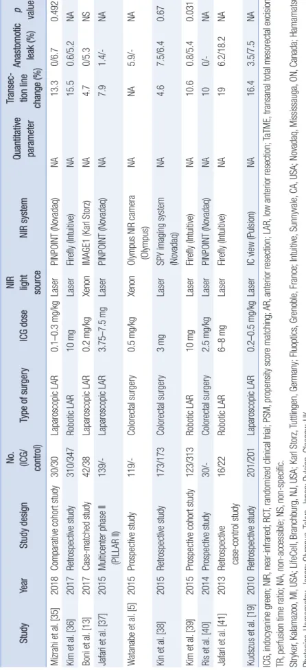
(5) 2017 Case-matched study. 2015 Multicenter phase II (PILLAR II). Jafari et al. [37]. 2015 Prospective cohort study. 2014 Prospective study. 2013 Retrospective case-control study. Kim et al. [39]. Ris et al. [40]. Jafari et al. [41]. 30/-. 201/201. 16/22 Laparoscopic LAR. Robotic LAR. Colorectal surgery. Robotic LAR. Colorectal surgery. Colorectal surgery. Laparoscopic LAR. Laparoscopic LAR. Robotic LAR. Laparoscopic LAR. Type of surgery. NIR light source. Laser. Laser. Laser. Laser. Xenon. Laser. Xenon. Laser. 0.2–0.5 mg/kg Laser. 6–8 mg. 2.5 mg/kg. 10 mg. 3 mg. 0.5 mg/kg. 3.75–7.5 mg. 0.2 mg/kg. 10 mg. 0.1–0.3 mg/kg Laser. ICG dose. IC view (Pulsion). Firefly (Intuitive). PINPOINT (Novadaq). Firefly (Intuitive). SPY imaging system (Novadaq). Olympus NIR camera (Olympus). PINPOINT (Novadaq). IMAGE1 (Karl Storz). Firefly (Intuitive). PINPOINT (Novadaq). NIR system. NA. NA. NA. NA. NA. NA. NA. NA. NA. NA. Quantitative parameter. 16.4. 19. 10. 10.6. 4.6. NA. 7.9. 4.7. 15.5. 13.3. 3.5/7.5. 6.2/18.2. 0/-. 0.8/5.4. 7.5/6.4. 5.9/-. 1.4/-. 0/5.3. 0.6/5.2. 0/6.7. NA. NA. NA. 0.031. 0.67. NA. NA. NS. NA. 0.492. TransecAnastomotic p tion line leak (%) value change (%). ICG, indocyanine green; NIR, near-infrared; RCT, randomized clinical trial; PSM, propensity score matching; AR, anterior resection; LAR, low anterior resection; TaTME, transanal total mesorectal excision; TR, perfusion time ratio; NA, non-accessible; NS, non-specific. Stryker, Kalamazoo, MI, USA; LifeCell, Branchburg, NJ, USA; Karl Storz, Tuttlingen, Germany; Fluoptics, Grenoble, France; Intuitive, Sunnyvale, CA, USA; Novadaq, Mississauga, ON, Canada; Hamamatsu Photonics, Hamamatsu, Japan; Olympus, Tokyo, Japan; Pulsion, Glasgow, UK.. Kudszus et al. [19] 2010 Retrospective study. 173/173. 2015 Retrospective study. Kin et al. [38] 123/313. 119/-. Watanabe et al. [5] 2015 Prospective study. 139/-. 42/38. 310/347. 2017 Retrospective study. Boni et al. [13]. No. (ICG/ control). Kim et al. [36]. Study design. 2018 Comparative cohort study 30/30. Year. Mizrahi et al. [35]. Study. Table 1. Continued. ICG angiography for colorectal surgery. 117. www.e-jmis.org.
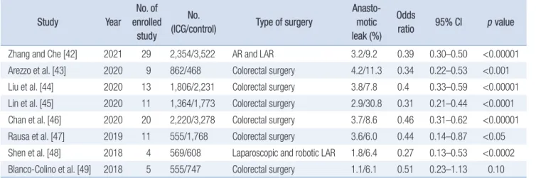
(6) 118. Gyung Mo Son et al.. Table 2. Meta-analysis for clinical effects of ICG angiography to reduce anastomotic leak. Study. Year. No. of No. enrolled (ICG/control) study. Zhang and Che [42]. 2021. 29. Arezzo et al. [43]. 2020. 9. Liu et al. [44]. 2020. Lin et al. [45]. Type of surgery. Anastomotic leak (%). Odds ratio. 95% CI. p value. 2,354/3,522. AR and LAR. 3.2/9.2. 0.39. 0.30–0.50. <0.00001. 862/468. Colorectal surgery. 4.2/11.3. 0.34. 0.22–0.53. <0.001. 13. 1,806/2,231. Colorectal surgery. 3.8/7.8. 0.4. 0.33–0.59. <0.00001. 2020. 11. 1,364/1,773. Colorectal surgery. 2.9/30.8. 0.31. 0.21–0.44. <0.0001. Chan et al. [46]. 2020. 20. 2,220/3,278. Colorectal surgery. 3.7/8.6. 0.46. 0.31–0.62. <0.00001. Rausa et al. [47]. 2019. 11. 555/1,768. Colorectal surgery. 3.6/6.0. 0.44. 0.14–0.87. <0.05. Shen et al. [48]. 2018. 4. 569/608. Laparoscopic and robotic LAR. 1.8/6.4. 0.27. 0.13–0.53. <0.0002. Blanco-Colino et al. [49]. 2018. 5. 555/747. Colorectal surgery. 1.1/6.1. 0.51. 0.23–1.13. 0.10. ICG, indocyanine green; AR, anterior resection; LAR, low anterior resection; CI, confidence interval.. es. Therefore, with laparoscopic ICG cameras, it is expected that the optimal f luorescence image can be obtained by measuring f luorescence at a distance of 4 to 5 cm. Additionally, it is helpful to obtain a f luorescence image of good quality by performing ICG angiography in the abdominal cavity or in as dark an environment as possible by turning off all room lights when capturing images from outside the abdominal cavity. Moreover, using a laser rather than a xenon lamp as a f luorescence emission source is advantageous for obtaining a f luorescence image of higher resolution and quality. Even with ICG laparoscopy using xenon lamps, the ICG-specific image mode can improve the quality of f luorescence images through image processing methods, such as red color inversion. Under these premises, when the conditions for f luorescence imaging are satisfied, we will be able to evaluate the colonic perfusion status using quantitative parameters, such as ascending slope or maximum FI. FI and persisting duration are affected by the dilution concentration and dose of the intravenous ICG injection. In our previous study for ICG angiography, a typical dilution concentration of 2.5 mg/mL was prepared, and an intravenous dose of 0.25 mg/ kg was applied according to the patient’s weight. In early ICG cameras using xenon lamps, blue f luorescence was visualized, so there was a resolution limitation in which the difference between the background and the blue color tone was not clear, so the appropriate ICG dose was set at 0.25 mg/kg. The initial ICG study of our research team also applied a dose of 0.2 to 0.25 mg/ kg using a laparoscopic NIR camera with a xenon lamp. As the f luorescence emission source for the laparoscopic camera is a laser, and with improvements in f luorescence imaging resolution, research on the optimal ICG dosage is required for repetitive ICG angiography. Optimized ICG angiography should be able to show a definite FI change. If necessary, repeated ICG angiography could be performed without disturbances from remnant f luorescence in the tissues. Therefore, the optimal ICG Journal of Minimally Invasive Surgery Vol. 24 24.. No. 3, 2021. dose should also be rapidly cleared by the liver, facilitating repeat ICG angiography. We conducted a study to find the optimal ICG capacity for quantitative f luorescence analysis using a laser NIR camera and compared the f luorescence images between standard (0.2 mg/kg) and experimental (0.01–0.05 mg/kg) dosage groups. In our recent study, sufficient quantitative perfusion evaluation was possible even with a low dose of 0.02 mg/kg, which is ten times lower than the conventional dose of 0.2 mg/kg (unpublished data).. Quantitative perfusion analysis After 10 to 30 seconds of intravenous ICG injection, f luorescence begins to appear in the colon wall. It is possible to evaluate perfusion status by changing FI to ensure that the perfusion is well preserved to the planned transection line. A time-f luorescence graph could be displayed by measuring changes in FI, allowing evaluation of various quantitative parameters. In the measurement of FI, the basic quantitative factors are the maximal level of FI (Fmax) and the interval to FImax (Tmax). Time scales include latency time (the initial f luorescence onset time), the interval to half of FImax (T1/2max), and perfusion time ratio (TR). The ascending slope can ref lect arterial f low conditions and can be calculated considering both FI and time scale (Fig. 1). Since the perfusion status of the small intestine can be used as a criterion for a good FI level, the perfusion status can also be evaluated by calculating the FI ratio of the small intestine and colon [30]. Among these variables, many studies have explored T1/2max. In cases of insufficient blood f low, T1/2max delays have been reported in a number of digestive organs, such as the small intestine and large intestine. We found that T1/2max and TR can ref lect blood f low conditions well [3]. In particular, T1/2max has the highest sensitivity for predicting anastomotic complications, and TR is.
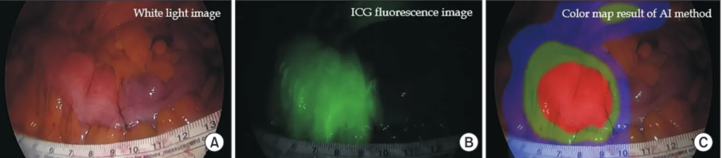
(7) 119. ICG angiography for colorectal surgery. analyzed as a variable with high specificity. Therefore, we proposed a step-by-step f low chart to improve diagnostic accuracy. At first, T1/2max can be used to screen for patients at risk of insufficient perfusion; second, TR can be used to predict the risk of anastomotic complications [3]. Based on the tissue perfusion status information, the surgeon will be able to perform safer colonic anastomoses by clearly identifying colonic segments with excellent blood f low. In particular, such quantitative perfusion evaluation would be of great help to a novice surgeon who lacks experience in subjective evaluation of tissue perfusion [50]. ICG angiography allows the surgeon to. 180. Fluorescence intensity. 160 140 120 100. Slope. 80 60 40 20 0. 5. Latency. 10. 15. 20 25 Time (sec). 30. 35. 40. Fig. 1. Quantitative parameters based on the time-fluorescence of indocyanine green angiography. This graph could be displayed by measuring changes in the fluorescence intensity (FI), allowing evaluation of various quantitative parameters. In the measurement of FI, the basic quantitative factors are the maximal level of FI (Fmax) and the interval to FImax (Tmax). Time scales include latency time (the initial fluorescence onset time), the interval to half of FImax (T1/2max), and perfusion time ratio (TR). The ascending slope can reflect arterial flow conditions and can be calculated considering both FI and time scale. Adapted from Ahn et al. [9], according to the Creative Commons License.. A. intuitively estimate tissue perfusion in real time. Eventually, ICG angiography may facilitate a reduction in anastomotic complications associated with poor colonic perfusion. For this, a safe and reliable ICG angiography protocol must be established. Additionally, it is necessary to develop and verify accurate analytical indicators for predicting the risk of complications and quantitatively evaluating the state of tissue microperfusion in real time. Experienced surgeons can empirically assess tissue perfusion without ICG angiography, but surgical beginners do not. However, manual evaluation by novice surgeons is subject to error. Accurate evaluation requires many years of practice to obtain sufficient experience. On the other hand, artificial intelligence (AI) can evaluate tissue perfusion with the highest accuracy via learning using numerous ICG angiography videos. The processed information can then be used to supplement the surgeon’s judgment. In the f luorescence graph used for quantitative analysis, a nonideal pattern may be observed due to the various blood f low pathways of the vascular structures and collateral circulation. In such a case, the quantitative parameter would be incorrectly recorded. This could be a hidden pitfall potentially leading to interpretation errors in perfusion evaluation. Additionally, environmental factors of ICG angiography such as NIR illuminance and lighting outside the shooting distance can affect the perfusion evaluation. Quantitative analysis has this limitation, but AI can be used to overcome it. AI, which has learned both patterns and various environmental factors, has superior analysis performance and stability relative to quantitative analysis. Real-time AI analysis can be used to quickly evaluate the state of the tissue microcirculation so that analysis results can be checked right in the operating room. The results can be expressed visually through a color map overlying the surgical field (Fig. 2). When the limited data for machine learning is solved in the future, it is expected that this will play an important role as a reliable assistant in the operating room [14].. B. C. Fig. 2. Colonic perfusion images. (A) White light image, (B) indocyanine green (ICG) fluorescence image, and (C) color map result of artificial intelligence (AI) analysis of ICG pattern. Real-time AI-based quantitative analysis can be used to evaluate the tissue microcirculation and express visually through a color map overlying the surgical field. The visual information can help the surgeons assess the perfusion status during laparoscopic surgery. Adapted from Park et al. [14], according to the Creative Commons License. www.e-jmis.org.
(8) 120. Gyung Mo Son et al.. PERSPECTIVES. NOTES. Currently, various f luorescence imaging devices, f luorescent drugs, hyperspectral image, and laser speckle contrast imaging systems are being rapidly developed. ICG f luorescence images can be obtained more delicately through the second window using a wavelength of 1,000 nm or more. This can increase the resolution of ICG f luorescence images and improve transmittance to achieve deeper penetration of f luorescence [51]. In addition to ICG, various f luorescent materials are currently being developed for clinical applications. Fluorescence imaging of the urinary system, as well as the biliary tract, can be performed using the different metabolic pathways applicable to f luorescent drugs [52]. Additionally, window f luorescence drugs have been developed for tissue-specific f luorescence imaging in the laboratory. Aside from ICG angiography, tissue perfusion status and oxygen saturation levels can be determined using hyperspectral imaging or laser speckle contrast imaging. This new imaging technique will be able to noninvasively evaluate in vivo physiological states using the physical properties of light without f luorescent drugs, such as ICG. A tissue-specific imaging technology has also been developed for ref lecting the physiological characteristics of tissues [53]. We are fortunate to be able to stand in the middle of a revolutionary era of f luorescence surgical technology and surgical navigation systems. Currently, surgeons have a new imaging device at their disposal, with which they can easily and safely apply ICG angiography in the operating room. By evaluating tissue microcirculation in real time, tissues with poor blood f low can be accurately identified. The risk factors of hypoperfusion can be predicted intraoperatively, and postoperative complications can be prevented. These advanced imaging technologies will improve the safety of colorectal surgery, which will eventually bring medical and economic benefits to doctors, patients, and health care systems. We believe that this seemingly small step in the operating room could prove to be a giant leap toward preventing anastomotic complications for colorectal cancer patients.. Authors’ contributions. CONCLUSION ICG angiography could be useful for detecting poorly perfused colonic segments and preventing anastomotic leakage after colorectal surgery. An optimal standardized ICG angiography protocol should be established to facilitate the implementation of reliable multicenter clinical trials to confirm the clinical benefits of ICG angiography. In the future, we envision AI-based quantitative analysis software being used to assess colonic perfusion during laparoscopic or robotic colorectal surgery.. Journal of Minimally Invasive Surgery Vol. 24 24.. No. 3, 2021. Conceptualization: GMS Formal Analysis: GMS, HMA, SHP Investigation: GMS Methodology: GMS, SHP, IYL, KRB Writing–Original Draft: GMS, HMA, SML, SHP Writing–Review & Editing: GMS All authors read and approved the final manuscript.. Conflict of interest All authors have no conf licts of interest to declare.. Funding/support This study was supported by a 2021 research grant from Pusan National University Yangsan Hospital.. ORCID Gyung Mo Son, https://orcid.org/0000-0002-8861-6293 Hong-min Ahn, https://orcid.org/0000-0001-9963-2021 In Young Lee, https://orcid.org/0000-0001-5954-6188 Sun Min Lee, https://orcid.org/0000-0002-2896-3365 Sang-Ho Park, https://orcid.org/0000-0001-6380-301X Kwang-Ryul Baek, https://orcid.org/0000-0002-2928-2043. REFERENCES 1. Yeung TM. Fluorescence imaging in colorectal surgery. Surg Endosc 2021;35:4956-4963. 2. Park JS, Choi GS, Kim SH, et al. Multicenter analysis of risk factors for anastomotic leakage after laparoscopic rectal cancer excision: the Korean laparoscopic colorectal surgery study group. Ann Surg 2013;257:665-671. 3. Son GM, Kwon MS, Kim Y, Kim J, Kim SH, Lee JW. Quantitative analysis of colon perfusion pattern using indocyanine green (ICG) angiography in laparoscopic colorectal surgery. Surg Endosc 2019;33:1640-1649. 4. Son GM, Kim TU, Park BS, et al. Colonic hypoperfusion following ligation of the inferior mesenteric artery in rectosigmoid colon cancer patients. Ann Surg Treat Res 2019;97:74-82. 5. Watanabe J, Ota M, Suwa Y, et al. Evaluation of the intestinal blood flow near the rectosigmoid junction using the indocyanine green fluorescence method in a colorectal cancer surgery. Int J Colorectal Dis 2015;30:329-335. 6. Jafari MD, Pigazzi A, McLemore EC, et al. Perfusion Assessment.
(9) ICG angiography for colorectal surgery. 7.. 8.. 9.. 10.. 11.. 12.. 13.. 14.. 15.. 16.. 17.. 18.. 19.. in Left-Sided/Low Anterior Resection (PILLAR III): a randomized, controlled, parallel, multicenter study assessing perfusion outcomes with PINPOINT near-infrared fluorescence imaging in low anterior resection. Dis Colon Rectum 2021;64:995-1002. De Nardi P, Elmore U, Maggi G, et al. Intraoperative angiography with indocyanine green to assess anastomosis perfusion in patients undergoing laparoscopic colorectal resection: results of a multicenter randomized controlled trial. Surg Endosc 2020;34:53-60. Baiocchi GL, Guercioni G, Vettoretto N, et al. ICG fluorescence imaging in colorectal surgery: a snapshot from the ICRAL study group. BMC Surg 2021;21:190. Ahn HM, Son GM, Lee IY, Park SH, Kim NS, Baek KR. Optimization of indocyanine green angiography for colon perfusion during laparoscopic colorectal surgery. Colorectal Dis 2021;23:1848-1859. Fujii S, Ishibe A, Ota M, et al. Short-term and long-term results of a randomized study comparing high tie and low tie inferior mesenteric artery ligation in laparoscopic rectal anterior resection: subanalysis of the HTLT (High tie vs. low tie) study. Surg Endosc 2019;33:1100-1110. Matsuda K, Yokoyama S, Hotta T, et al. Oncological outcomes following rectal cancer surgery with high or low ligation of the inferior mesenteric artery. Gastrointest Tumors 2017;4:45-52. Brandt LJ, Feuerstadt P, Longstreth GF, Boley SJ; American College of Gastroenterology. ACG clinical guideline: epidemiology, risk factors, patterns of presentation, diagnosis, and management of colon ischemia (CI). Am J Gastroenterol 2015;110:18-44. Boni L, Fingerhut A, Marzorati A, Rausei S, Dionigi G, Cassinotti E. Indocyanine green fluorescence angiography during laparoscopic low anterior resection: results of a case-matched study. Surg Endosc 2017;31:1836-1840. Park SH, Park HM, Baek KR, Ahn HM, Lee IY, Son GM. Artificial intelligence based real-time microcirculation analysis system for laparoscopic colorectal surgery. World J Gastroenterol 2020;26:69456962. Guo Y, Wang D, He L, et al. Marginal artery stump pressure in left colic artery-preserving rectal cancer surgery: a clinical trial. ANZ J Surg 2017;87:576-581. Seike K, Koda K, Saito N, et al. Laser Doppler assessment of the influence of division at the root of the inferior mesenteric artery on anastomotic blood flow in rectosigmoid cancer surgery. Int J Colorectal Dis 2007;22:689-697. Kim M, Lee S, Park JC, et al. Anaphylactic shock after indocyanine green video angiography during cerebrovascular surgery. World Neurosurg 2020;133:74-79. Zheng J, Xie W, Huang Y, Zhu Y, Jiang L. The technique of 3D reconstruction combining with biochemistry to build an equivalent formula of indocyanine green (ICG) clearance test to assess the liver reserve function. BMC Surg 2020;20:283. Kudszus S, Roesel C, Schachtrupp A, Höer JJ. Intraoperative laser fluorescence angiography in colorectal surgery: a noninvasive analysis to reduce the rate of anastomotic leakage. Langenbecks Arch Surg. 121 2010;395:1025-1030. 20. Ahn HM, Son GM, Lee IY, et al. Optimal ICG dosage of preoperative colonoscopic tattooing for fluorescence-guided laparoscopic colorectal surgery. Surg Endosc 2021 Feb 26 [Epub]. https://doi.org/10.1007/ s00464-021-08382-5. 21. Alekseev M, Rybakov E, Shelygin Y, Chernyshov S, Zarodnyuk I. A study investigating the perfusion of colorectal anastomoses using fluorescence angiography: results of the FLAG randomized trial. Colorectal Dis 2020;22:1147-1153. 22. Wojcik M, Doussot A, Manfredelli S, et al. Intra-operative fluorescence angiography is reproducible and reduces the rate of anastomotic leak after colorectal resection for cancer: a prospective casematched study. Colorectal Dis 2020;22:1263-1270. 23. Morales-Conde S, Alarcón I, Yang T, et al. Fluorescence angiography with indocyanine green (ICG) to evaluate anastomosis in colorectal surgery: where does it have more value? Surg Endosc 2020;34:38973907. 24. Watanabe J, Ishibe A, Suwa Y, et al. Indocyanine green fluorescence imaging to reduce the risk of anastomotic leakage in laparoscopic low anterior resection for rectal cancer: a propensity score-matched cohort study. Surg Endosc 2020;34:202-208. 25. Benčurik V, Škrovina M, Martínek L, et al. Intraoperative fluorescence angiography and risk factors of anastomotic leakage in miniinvasive low rectal resections. Surg Endosc 2021;35:5015-5023. 26. Bonadio L, Iacuzzo C, Cosola D, et al. Indocyanine green-enhanced fluorangiography (ICGf) in laparoscopic extraperitoneal rectal cancer resection. Updates Surg 2020;72:477-482. 27. Hasegawa H, Tsukada Y, Wakabayashi M, et al. Impact of intraoperative indocyanine green fluorescence angiography on anastomotic leakage after laparoscopic sphincter-sparing surgery for malignant rectal tumors. Int J Colorectal Dis 2020;35:471-480. 28. Otero-Piñeiro AM, de Lacy FB, Van Laarhoven JJ, et al. The impact of fluorescence angiography on anastomotic leak rate following transanal total mesorectal excision for rectal cancer: a comparative study. Surg Endosc 2021;35:754-762. 29. Ishii M, Hamabe A, Okita K, et al. Efficacy of indocyanine green fluorescence angiography in preventing anastomotic leakage after laparoscopic colorectal cancer surgery. Int J Colorectal Dis 2020;35:269275. 30. Wada T, Kawada K, Hoshino N, et al. The effects of intraoperative ICG fluorescence angiography in laparoscopic low anterior resection: a propensity score-matched study. Int J Clin Oncol 2019;24:394-402. 31. Ogino T, Hata T, Kawada J, et al. The risk factor of anastomotic hypoperfusion in colorectal surgery. J Surg Res 2019;244:265-271. 32. Chang YK, Foo CC, Yip J, et al. The impact of indocyanine-green fluorescence angiogram on colorectal resection. Surgeon 2019;17:270276. 33. van den Bos J, Jongen ACHM, Melenhorst J, Breukink SO, Lenaerts K, Schols RM, Bouvy ND, Stassen LP. Near-infrared fluorescence image-guidance in anastomotic colorectal cancer surgery and its rela-. www.e-jmis.org.
(10) 122. 34.. 35.. 36.. 37.. 38.. 39.. 40.. 41.. 42.. 43.. 44.. tion to serum markers of anastomotic leakage: a clinical pilot study. Surg Endosc 2019;33:3766-3774. Ris F, Liot E, Buchs NC, et al. Multicentre phase II trial of near-infrared imaging in elective colorectal surgery. Br J Surg 2018;105:13591367. Mizrahi I, Abu-Gazala M, Rickles AS, et al. Indocyanine green fluorescence angiography during low anterior resection for low rectal cancer: results of a comparative cohort study. Tech Coloproctol 2018;22:535-540. Kim JC, Lee JL, Park SH. Interpretative guidelines and possible indications for indocyanine green fluorescence imaging in robot-assisted sphincter-saving operations. Dis Colon Rectum 2017;60:376-384. Jafari MD, Wexner SD, Martz JE, et al. Perfusion assessment in laparoscopic left-sided/anterior resection (PILLAR II): a multi-institutional study. J Am Coll Surg 2015;220:82-92. Kin C, Vo H, Welton L, Welton M. Equivocal effect of intraoperative fluorescence angiography on colorectal anastomotic leaks. Dis Colon Rectum 2015;58:582-587. Kim JC, Lee JL, Yoon YS, Alotaibi AM, Kim J. Utility of indocyaninegreen fluorescent imaging during robot-assisted sphincter-saving surgery on rectal cancer patients. Int J Med Robot 2016;12:710-717. Ris F, Hompes R, Cunningham C, et al. Near-infrared (NIR) perfusion angiography in minimally invasive colorectal surgery. Surg Endosc 2014;28:2221-2226. Jafari MD, Lee KH, Halabi WJ, et al. The use of indocyanine green fluorescence to assess anastomotic perfusion during robotic assisted laparoscopic rectal surgery. Surg Endosc 2013;27:3003-3008. Zhang W, Che X. Effect of indocyanine green fluorescence angiography on preventing anastomotic leakage after colorectal surgery: a meta-analysis. Surg Today 2021;51:1415-1428. Arezzo A, Bonino MA, Ris F, et al. Intraoperative use of fluorescence with indocyanine green reduces anastomotic leak rates in rectal cancer surgery: an individual participant data analysis. Surg Endosc 2020;34:4281-4290. Liu D, Liang L, Liu L, Zhu Z. Does intraoperative indocyanine green. Journal of Minimally Invasive Surgery Vol. 24 24.. No. 3, 2021. Gyung Mo Son et al.. 45.. 46.. 47.. 48.. 49.. 50.. 51.. 52.. 53.. fluorescence angiography decrease the incidence of anastomotic leakage in colorectal surgery?: a systematic review and meta-analysis. Int J Colorectal Dis 2021;36:57-66. Lin J, Zheng B, Lin S, Chen Z, Chen S. The efficacy of intraoperative ICG fluorescence angiography on anastomotic leak after resection for colorectal cancer: a meta-analysis. Int J Colorectal Dis 2021;36:27-39. Chan DK, Lee SK, Ang JJ. Indocyanine green fluorescence angiography decreases the risk of colorectal anastomotic leakage: systematic review and meta-analysis. Surgery 2020;168:1128-1137. Rausa E, Zappa MA, Kelly ME, et al. A standardized use of intraoperative anastomotic testing in colorectal surgery in the new millennium: is technology taking over?: a systematic review and network meta-analysis. Tech Coloproctol 2019;23:625-631. Shen R, Zhang Y, Wang T. Indocyanine green fluorescence angiography and the incidence of anastomotic leak after colorectal resection for colorectal cancer: a meta-analysis. Dis Colon Rectum 2018;61:1228-1234. Blanco-Colino R, Espin-Basany E. Intraoperative use of ICG fluorescence imaging to reduce the risk of anastomotic leakage in colorectal surgery: a systematic review and meta-analysis. Tech Coloproctol 2018;22:15-23. Kim DH, Son GM, Kwon MS, Baek SH, Park BS, Kim HS. Educational benefits of intraoperative indocyanine green angiography for surgical beginners during laparoscopic colorectal surgery. J Minim Invasive Surg 2018;21:25-30. Ma Z, Zhang M, Yue J, et al. Near-infrared IIb fluorescence imaging of vascular regeneration with dynamic tissue perfusion measurement and high spatial resolution. Adv Funct Mater 2018;28:1803417. Fushiki H, Yoshikawa T, Matsuda T, Sato T, Suwa A. Preclinical development and validation of ASP5354: a near-infrared fluorescent agent for intraoperative ureter visualization. Mol Imaging Biol 2021 May 11 [Epub]. https://doi.org/10.1007/s11307-021-01613-0. Jansen-Winkeln B, Holfert N, Köhler H, et al. Determination of the transection margin during colorectal resection with hyperspectral imaging (HSI). Int J Colorectal Dis 2019;34:731-739..
(11)
수치
관련 문서
In this study, to investigate how applying a critical pathway to stomach cancer patients affects their recovery and treatment, the clinical effect of the critical
Patient had laparoscopic surgery on the adnexal tumor and excised tissue was removed through Douglas pouch incision by single surgeon.. Results: The mean age
Kameoka, “Sentinel lymph node biopsy for breast cancer patients using fluorescence navigation with indocyanine green,” World Journal of Surgical Oncology, Vol..
During the course of the green clothes attitude education program, this study identified that the green clothes attitude education program makes positive
To promote green jobs through the expansion of green industry, chapter 2(II) examines the concept, objective, promotion field, and scope of green industry; the creation
UNEP, Green Jobs: Towards decent work in a sustainable, low-carbon world, 2008.. Green Growth and the Countermeasure of Energy Legislative System. 38) Hahm,
“Act of Low Carbon Green Growth (bill)” defines a term of the ‘green growth’ as “harmony out of economic growth and the environment” which reduce climate
Definition of patients presenting a high risk of developing peritoneal carcino- matosis after curative surgery for colorectal cancer: a systematic review..
