358 http://www.aard.or.kr
서 론
광과민성(photosensitivity) 피부염은 피부에 발생하는 약물유해 반응(adverse drug reaction)의 8%를 차지할 정도로 비교적 흔하게 볼 수 있는 질환이다.1 여러 가지 약물 중에서도 항생제, 이뇨제, 비 스테로이드성 항염증제가 광과민성피부염을 자주 일으키는 것으 로 보고되었다.2 피부발진이 얼굴, 목, 팔 등의 햇빛에 노출되는 피부 에 국한되어 있고 환자가 특정한 약물을 복용하고 있는 경우에 약 물에 의한 광과민성피부염을 의심하게 된다.
체내에 분포하고 있는 약물 또는 그 대사물이 주로 자외선 A로 부터 에너지를 흡수하여 활성화되어 광과민성을 일으키게 된다.3 이는 발생기전에 의해서 광독성(phototoxicity) 또는 광알레르기 (photoallergy)로 나뉘어진다. 광독성 반응에서는 산소를 매개로 막지질과 단백질의 산화가 발생하거나 전자 또는 수소 이온의 이동
으로 생성된 자유기(free radical)가 생체분자와 반응을 일으킨다.
반면에 광알레르기 반응에서는 약물이 합텐(hapten)으로 작용하 여 단백질과 상호작용에 의해서 T세포 매개 과민반응이 일어나게 된다.4
Dronedarone은 새로운 항부정맥 약물로써 심방세동의 치료에 사용되며, amiodarone과 같은 benzofuran 유도체이다.5 Droneda- rone은 amiodarone과 달리 갑상선 독성을 일으키는 요오드기가 제외되어 있으며, 보다 친수성을 갖는 methane-sulfonamyl 기가 포 함되어 있어서 조직 내에 머무는 시간이 감소하게 된다(Fig. 1).6 따 라서 dronedarone은 이러한 약물 구조의 차이로 인해 amiodarone 에 비해서 전반적인 부작용이 적게 발생하게 된다. 현재까지 drone- darone에 의해서 발생한 약물유해반응은 매우 드물게 보고되었 다. 이에 저자들은 dronedarone에 의해 발생한 광과민성피부염 증 례에 대해서 보고하고자 한다.
Allergy Asthma Respir Dis 5(6):358-360, November 2017 https://doi.org/10.4168/aard.2017.5.6.358
pISSN: 2288-0402 eISSN: 2288-0410
CASE REPORT
Correspondence to: Yoo Seob Shin https://orcid.org/0000-0002-9855-3185
Department of Allergy and Clinical Immunology, Ajou University School of Medicine, 206 World cup-ro, Yeongtong-gu, Suwon 16499, Korea
Tel: +82-31-219-5155, Fax: +82-31-219-4265, E-mail: [email protected] Received: February 28, 2017 Revised: May 2, 2017 Accepted: May 4, 2017
© 2017 The Korean Academy of Pediatric Allergy and Respiratory Disease The Korean Academy of Asthma, Allergy and Clinical Immunology This is an Open Access article distributed under the terms of the Creative Commons Attribution Non-Commercial License (http://creativecommons.org/licenses/by-nc/4.0/).
Dronedarone에 의해 발생한 광과민성피부염 1예
이지호,1 김소민,2 정창규,1 박해심,1 신유섭1 아주대학교병원 1알레르기내과, 2피부과
Photosensitivity caused by dronedarone: A case report
Ji-Ho Lee,1 So-Min Kim,2 Chang-Gyu Jung,1 Hae-Sim Park,1 Yoo Seob Shin1
Departments of 1Allergy and Clinical Immunology, 2Dermatology, Ajou University School of Medicine, Suwon, Korea
Dronedarone is a new antiarrhythmic drug for the treatment of nonpermanent atrial fibrillation. Compared with amiodarone, it is regarded as a safe medication due to its structural differences. In this report, we describe a 56-year-old man who developed photo- sensitivity due to dronedarone. He presented with itchy skin rashes for 1 week. Maculopapular exanthema was localized on the neck, both arms, and both hands, with sparing of the other parts of the body. Dronedarone was prescribed 4 weeks ago when atrial fibrillation occurred. After development of skin rashes, dronedarone was discontinued, and systemic steroid, antihistamine, and topical corticosteroid were administered for 1 week, with improvement in skin rashes. The photopatch test was performed with an- tiarrhythmic drugs, including dronedarone, amiodarone, and flecainide, 4 weeks after withdrawal of dronedarone. Positive reactions were recorded only to dronedarone at the site exposed to ultraviolet A. He was diagnosed with dronedarone-induced photosensi- tivity and advised to change the antiarrhythmic medication to others. There have been a few case reports on photosensitivity reac- tions due to dronedarone, which were diagnosed only by clinical suspicion. However, we suspected photosensitivity and proved it by the photopatch test. Photosensitivity should be considered in patients having skin rashes on the exposed area and taking antiar- rhythmic medication, including dronedarone. (Allergy Asthma Respir Dis 2017;5:358-360)
Keywords: Dronedarone, Photosensitivity, Photopatch test
1 / 1 CROSSMARK_logo_3_Test
2017-03-16 https://crossmark-cdn.crossref.org/widget/v2.0/logos/CROSSMARK_Color_square.svg
이지호 외 • Dronedarone에 의한 광과민성피부염 Allergy Asthma Respir Dis
https://doi.org/10.4168/aard.2017.5.6.358 359 증 례
환자: 56세, 남자
주소: 1주일 전부터 발생한 피부발진과 가려움
현병력: 환자는 손등, 아래팔과 목에 국한된 피부발진과 가려움 이 1주일 전부터 발생하였으며 이후 발진은 위치나 형태의 변화 없 이 지속되고 다른 신체 부위에서는 발생하지 않았다. 간헐적인 가슴 두근거림 증상이 지속되어 본원 심장 내과에서 24시간 심전도 검사 를 시행하였으며 발작성 심방세동이 진단되어 피부발진이 시작되 기 1개월 전부터 dronedarone 400 mg을 하루 2회 복용 중이었다.
과거력: 10년 전 협심증을 진단받고 aspirin, diltiazem 복용 중이 었으며, 2년 전 통풍이 진단되어 febuxostat을 계속해서 복용 중이 었다. 10년 전 겨울철에 악화되는 콧물, 재채기 등의 증상으로 알레 르기비염 치료를 받은 과거력이 있으나 이후 특별한 증상은 없었다.
그 외 약물 또는 식품알레르기 질환의 병력은 관찰되지 않았다.
가족력: 없음 직업: 교사
사회력: 음주와 흡연의 과거력은 없었다.
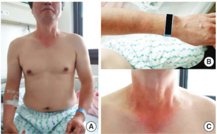
신체검사 소견: 내원 시 활력징후는 혈압 115/70 mmHg, 맥박 수 분당 60회, 호흡 수 분당 20회, 체온 36.6°C였으며 의식은 명료하였 다. 피부 진찰에서 햇빛에 노출되는 부분인 양 손등, 아래팔, 목에 반구진발진(maculopapular rash)이 관찰되었다(Fig. 2). 하지만 옷 에 의해서 가려지는 부위인 위팔, 몸통과 등에서는 이상 소견이 관 찰되지 않았다. 두경부 진찰에서 점막 침범 소견은 관찰되지 않았 으며 흉부 청진이나 복부 검사에서 특이 소견은 없었다.
검사실 소견: 흉부 및 부비동 엑스선 사진은 정상이었다. 혈액검 사에서 백혈구 8,900/µL, 혈색소 16.4 g/dL, 혈소판 166,000/µL, 호 산구 분율 3.3% (절대치 107/µL)였고, 혈청 총 면역글로불린 E (im- munoglobulin E)의 농도는 68 kU/mL였다.
치료 및 경과: 알레르기내과 외래에서 H1 항히스타민과 경구 스 테로이드, 국소 스테로이드를 처방하였고, 원인으로 판단되는 dronedarone 복용을 중단하도록 권고하였다. 하지만 피부발진이 호전되지 않아 2일 후 입원하였으며 스테로이드를 정맥으로 투여
하고 다른 치료는 유지하였다. 5일간 치료 후에 현저하게 피부발진 이 호전되고 가려움이 감소하여 퇴원하였다. 1주 후 외래에 방문하 였으며 피부발진은 모두 사라진 상태로 알레르기 약물을 중단하였 다. 피부과에 협진 의뢰하여 2주 후에 광첩포검사(photopatch test) 를 시행하였다.
광첩포검사: 원인으로 판단되는 dronedarone뿐만 아니라 다른 항부정맥 약물인 amiodarone과 flecainide를 포함시켰다. 먼저 세 가지 약물을 미세하게 가루 내어 petrolatum과 1:9의 비율로 섞은 후 이를 aluminum chamber (SmartPractice, Phoenix, AZ, USA) 에 넣었다. 왼쪽 등과 오른쪽 등에 대칭으로 각각 3개의 첩포를 붙 인 후 48시간 뒤에 모두 제거하고, 첫 판독을 하였을 때 특이 소견 은 보이지 않았다. 자외선 조사기(PUVA 800, Waldmann, Germa- ny)를 이용하여 오른쪽은 가리고 왼쪽에만 자외선A 10 J/cm2을 조 사하였다. 자외선 조사 48시간, 96시간 후에 판독하였을 때 96시간 후에 자외선A를 조사하고 dronedarone 첩포를 했던 곳에서만 양 성 반응이 나타났다(Fig. 3A). 그 외 다른 약물의 첩포를 붙인 위치 Fig. 1. Chemical structure of dronedarone (A) and amiodarone (B).
A
B
Fig. 2. Maculopapular eruptions on the upper body with a photodistributed pat- tern (A), localized in the forearm and the dorsal area of the hand (B), and the neck with V-neck sign (C) at presentation.
A
B
C
Fig. 3. Photopatch test results on day 4 showing an erythematous change (1+) with ultraviolet (UVA) (A), and a negative response at the nonirradiated control site (B). 1, dronedarone; 2, amiodarone; 3, flecainide.
A B
Lee JH, et al. • Dronedarone induced photosensitivity Allergy Asthma Respir Dis
360 https://doi.org/10.4168/aard.2017.5.6.358
에서는 자외선A의 조사와 관계 없이 특이 소견이 나타나지 않았으 며 dronedarone을 첩포하고 자외선을 조사하지 않은 오른쪽 등에 서도 이상 소견은 보이지 않았다(Fig. 3B).
환자 교육 및 추적 관찰: 따라서 광과민성피부염의 원인으로 밝 혀진 dronedarone을 회피하도록 하였고 심방세동의 치료를 위해 서 안전한 약으로 판명된 다른 항부정맥제의 사용을 권고하였다.
환자는 flecanide로 변경하여 복용하기 시작하였고 부작용 없이 심 장내과 추적 관찰 중이다.
고 찰
몇 가지 심각한 사례가 보고되었으나, dronedarone과 관련된 대 부분의 약물유해반응은 경미한 오심, 구토, 설사 등의 소화기계 증 상으로 알려져 있다.6,7 광과민성피부염의 증례 2예가 보고되었으나 객관적인 검사로 증명된 경우는 보고된 바가 없다.8,9 이 환자에서는 원인 약제인 dronedaron을 광첩포검사를 이용하여 진단한 것이 이전의 증례와 차이점이다.
광독성과 광알레르기는 발생기전의 차이뿐만 아니라 각각 특징 적인 임상 증상을 나타낸다. 광독성 반응이 보다 흔하게 관찰되며 환자는 주로 일광화상(sunburn)과 유사한 작열감을 호소한다. 원 인 약물을 처음으로 복용한 경우에도 발생 가능하고 햇빛에 노출 된 지 수시간 만에 나타날 수도 있다. 또한 색소침착을 자주 남기게 된다. 광알레르기 반응은 주로 가려운 증상과 습진(eczema)을 동 반하게 되고 보통 햇빛에 노출되고 24시간 후에 증상이 나타나기 시작한다. 알레르기 반응이므로 이전에 같은 약물을 복용한 과거 력이나 구조가 유사한 약물의 노출이 필요하다. 원인 약물을 중단 하면 색소침착을 남기지 않고 회복된다.2
광검사(phototesting)는 최소홍반선량(minimal erythema dose) 을 정하기 위한 목적으로 시행하며, 최소홍반선량은 인지 가능한 홍반을 발생하게 하는 최소한의 자외선량을 의미한다.10 원인 약물 을 복용하고 있는 중에 최소홍반선량이 정상 대조군보다 감소하 고, 약제를 중단 후 이 값이 정상으로 회복된다면 광과민성이 있다 고 진단할 수 있다. 광첩포검사는 광알레르기를 진단하기 위한 검 사로써, 피부에 바르는 국소 제제나 자외선 차단제의 화합물에 의 해서 발생하는 광알레르기 접촉피부염의 진단에 주로 이용된다.11 하지만 전신으로 투여하는 약제에 대해서는 아직 유효성이 입증되 지 않았다.3
이 증례에서 나타난 광과민성 반응의 발생기전을 정확하게 확인 하기 위해서는 광검사를 먼저 시행하여 최소홍반선량을 확인하고, 이보다 낮은 용량의 자외선을 조사하여 광첩포검사를 시행하는 것 이 옳은 것으로 판단된다. 하지만 이 증례에서는 광검사를 먼저 시 행하지 않았고 일반적인 용량인 5 J/cm2보다 많은 10 J/cm2의 자외 선A를 조사하였다. 따라서 광첩포검사 중에 나타난 양성 반응은 피 부에 접촉한 약물에 의해서 국소적으로 발생한 광독성 반응, 즉 위 양성의 가능성을 배제할 수 없다. 다른 문헌에서는 dronedarone에 의한 광과민성피부염을 광독성 반응으로 기술하고 있으나 아직 객 관적인 증거가 부족하다.9 또한 이번 증례의 경우와 같이 광첩포검 사에서 광알레르기 반응을 확인할 수 있는 양성 반응이 나타났으 므로 광알레르기 반응이 관여할 가능성을 염두에 두어야 하겠다.
결론적으로 저자들은 광첩포검사를 통해서 확진된 dronedar- one에 의해 발생한 광과민성피부염을 보고하는 바이다.
REFERENCES
1. Selvaag E. Clinical drug photosensitivity. A retrospective analysis of re- ports to the Norwegian Adverse Drug Reactions Committee from the years 1970-1994. Photodermatol Photoimmunol Photomed 1997;13:21- 2. Glatz M, Hofbauer GF. Phototoxic and photoallergic cutaneous drug re-3.
actions. Chem Immunol Allergy 2012;97:167-79.
3. Monteiro AF, Rato M, Martins C. Drug-induced photosensitivity: photo- allergic and phototoxic reactions. Clin Dermatol 2016;34:571-81.
4. Onoue S, Seto Y, Sato H, Nishida H, Hirota M, Ashikaga T, et al. Chemi- cal photoallergy: photobiochemical mechanisms, classification, and risk assessments. J Dermatol Sci 2017;85:4-11.
5. Vamos M, Hohnloser SH. Amiodarone and dronedarone: an update.
Trends Cardiovasc Med 2016;26:597-602.
6. De Ferrari GM, Dusi V. Drug safety evaluation of dronedarone in atrial fibrillation. Expert Opin Drug Saf 2012;11:1023-45.
7. Gecks T, Prochnau D, Franz M, Jung C, Kühnert H, Schliemann S, et al.
Toxic epidermal necrolysis during dronedarone treatment: first report of a severe serious adverse event of a new antiarrhythmic drug. Cardiovasc Toxicol 2015;15:399-401.
8. Kuo S, Menon K, Kundu RV. Photosensitivity reaction from dronedarone for atrial fibrillation. Cutis 2014;94:E10-1.
9. Ladizinski B, Elpern DJ. Dronaderone-induced phototoxicity. J Drugs Dermatol 2013;12:946-7.
10. Kutlubay Z, Sevim A, Engin B, Tüzün Y. Photodermatoses, including phototoxic and photoallergic reactions (internal and external). Clin Der- matol 2014;32:73-9.
11. Wilm A, Berneburg M. Photoallergy. J Dtsch Dermatol Ges 2015;13:7- 12.