213
<원례보저>
Characterization and comparison of the pathogenicity of viscerotropic velogenic Newcastle disease virus isolates in Korea
Jae-Hong Kim
1,*, Haan-Woo Sung
2, Il-Hwan Kim
1, Eun-Kyoung Lee
3, Kang-Seuk Choi
3, Daniel Jack King
41
Laboratory of Avian Diseases, Research Institute for Veterinary Science, College of Veterinary Medicine, Seoul National University, Seoul 151-742, Korea
2
College of Veterinary Medicine, Kangwon National University, Chuncheon 200-701, Korea
3
Animal, Plant and Fisheries Quarantine and Inspection Agency, Anyang 430-757, Korea
4
Southeast Poultry Research Laboratory, USDA-ARS, Athens, GA 30605, USA (Received: October 12, 2012; Accepted: October 19, 2012)
Abstract : A total of 18 Newcastle disease virus (NDV) isolates that were recovered from 1949 through 1997 were characterized and pathotyped. All viruses were highly virulent as determined by intracerebral pathogenicity indices
≥ 1.81 in day-old. These pathotypes are typical for viscerotropic velogenic NDV (VVNDV) pathotype viruses. Some
differences were observed for the chicken red blood cell elution rate and thermostability of the hemagglutinin at 56oC.Three antigenic groups were identified by a hemagglutination-inhibition assay using NDV monoclonal antibodies. And the predominant gross lesions were as follows: discharge from the nasal cavity, tracheal mucus, petechial hemorrhage in the heart fat, kidney urates and hemorrhage with or without necrosis in the gastrointestinal tract. Severe hemorrhagic or necrotic lesions were also noted in the lymphoid organs and were localized primarily in the spleen and cecal tonsil.
However, differences in the occurrence and frequency of the gross lesions were observed between the virus strains.
Among them, NDV strains that induced neurological symptoms belonged only to genotype VI. This strain had spread throughout Korea during the late 1980s to the 1990s, which suggests that specific VVNDVs genotypes might result in neurological symptoms.
Keywords : avian paramyxovirus, Newcastle disease, pathogenicity, velogenic neurotropic, viscerotropic velogenic
Introduction
Newcastle disease (ND) is a major disease of poultry that results in severe economic losses in the poultry industry worldwide. Ever since it was first reported in 1927, pan- zootic viscerotropic velogenic ND (VVND) has been observed throughout Asia, Africa and Central and South America [2, 4]. Since 1991, an increase in frequency of vel- ogenic ND epidemics in European countries has also been a serious problem [3]. Outbreaks of ND, primarily in pigeons and doves, caused by a variant ND virus (the ‘pigeon paramyxovirus type 1 (PMV-1) variant’) have been detected and observed to have spread throughout Europe [3, 11, 12].
Periodic VVND epizootics in Korea have occurred contin- ually every 3 to 5 years [19, 30] ever since the first outbreak that was reported in 1927. VVND was known to be present in the country as early as 1924 [4].
According to the severity of clinical signs that manifest in affected birds, ND virus (NDV) has been grouped into the following five pathotypes: viscerotropic velogenic NDV (VVNDV), neurotropic velogenic NDV (NVNDV), mesogenic
NDV, lentogenic NDV and asymptomatic enteric NDV [6].
Clinical manifestations of NDV vary based on the viral strain, host species, age of host, route of exposure to infec- tion and the environment [4, 8, 28]. As such, many attempts have been performed to characterize and differentiate NDV isolates [3, 5, 16, 17, 21, 22, 27] and to determine differ- ences in pathology among different NDV pathotypes [7, 8, 24, 25]. However, few studies have compared the pathoge- nicity of various VVNDV isolates based on pathological dif- ferences between the strains [7, 18].
VVNDV strains usually cause hemorrhagic ulceration with necrosis in the intestinal wall [18, 25], lymphoreticular necrosis [8, 9, 13] and respiratory lesions [8, 9]. Further- more, there is thought to be considerable variation in patho- genicity depending on the VVNDV strain.
This study was conducted to characterize Korean NDV isolates and to compare the pathological differences of vari- ous VVNDV isolates that have been isolated in Korea since the 1980s.
*Corresponding author
Tel: +82-2-880-1250, Fax: +82-2-885-6614 E-mail: [email protected]
Materials and Methods
Viruses
Working stocks of 18 NDV isolates and reference strains, including vaccine strains, were prepared using 9- to 10-day- old embryonated specific-pathogen-free (SPF) chickens eggs.
The NDV isolates were isolated from commercial chicken flocks in Korea, primarily from chickens that had clinical signs of ND. The Korean (Kr)-D/84 and Kr-M/88 were iso- lated from clinical cases of ND infection in a peafowl and a quail, respectively.
Of the total 18 isolates, the following 11 were isolated at the National Veterinary Research Institute, Korea: Kr-48/82, Kr-1129/83, Kr-K/84, Kr-D/84, Kr-M/88, Kr-12A/89, Kr-163/
90, Kr-9/91, Kr-104/92, Kr-147/97 and Kr-KJW/49. Kr-KJW/
49 was isolated in 1949 and used as a reference challenge strain in Korea [26]. The following 7 isolates were isolated at the College of Veterinary Medicine, Seoul National Univer- sity: 92-76A, 92-76B, 93-58GG, 93-58GS, 94-44, 95-119 and 95-132. The 92-76A and 92-76B isolates originated from the same isolate, and each viral isolate was propagated in SPF eggs after the clones were selected. This was also true for the 93-58GG and 93-58GS isolates. The years of virus isolation have been indicated in a previous paper [26].
The NVNDV reference strain Texas-GB and vaccine strains B1, La Sota and Ulster 2C were obtained from the Southeast Poultry Research Laboratory (SEPRL), ARS, USDA, USA [22].
Eggs and chickens
Embryonated SPF eggs for virus propagation and titration were obtained from the SPF white leghorn flock from the SEPRL. Day-old SPF white leghorn chicks for the intracere- bral pathogenicity index (ICPI) test and 4-week-old SPF white leghorn chicks for the pathogenicity test were hatched from SPF eggs and housed in negative-pressure isolators in the SEPRL [20].
Monoclonal antibodies and antiserum
The AVS [31], B79, 10D11, and 15C4 [23] monoclonal antibodies (MAbs) were obtained from NVSL and used to differentiate the NDV strains by a hemagglutination-inhibi- tion (HI) test. The HI reactivity of the MAbs is as follows:
AVS only reacts with NDV lentogens, particularly B1 and La Sota [31], 10D11 reacts mostly with NDV mesogens and neurotropic velogens from U.S. poultry, 79 is broadly reac- tive with avian PMV-1 strains, including most pigeon NDV, and 15C4 is broadly reactive with PMV-1 strains, except for pigeon NDV isolates [23]. Four additional mouse MAbs (P10B8, P11C9, P15D7 and P3A11) were generated at the SEPRL by immunization using the VVNDV strain CA 1083 (Fontana). A polyclonal chicken anti-NDV serum was prepared by immunization of SPF white rock chickens with two doses of inactivated NDV La Sota virus. The polyclonal chicken anti-NDV serum was used as a positive serum in the HI test.
Hemagglutination and HI assays
The hemagglutination (HA) and HI tests were performed using conventional microtiter methods. For HI tests, serial twofold serum or MAb dilutions were made in phosphate- buffered saline (PBS), pH 7.2. Four HA units of test antigen were added to each dilution, and the test was then incubated at room temperature for 30 min. An equal volume of 0.5%
chicken erythrocytes in PBS was added as a test indicator.
The HI endpoint was determined as the last dilution that maintained complete inhibition of HA activity. An aliquot of each viral isolate was used as a live virus antigen to differen- tiate strains by the HI assay. MAbs were originally diluted 1/
10 in PBS. Therefore, HI titers that were less than 1 : 20 were considered negative, titers of 1 : 40 were indeterminate and titers equal to or greater than 1 : 80 were considered pos- itive. Indeterminate reactions were considered negative if no increase in the titer was determined after a retest.
A La Sota virus that was inactivated with β-propiolacton was used as the antigen in HI test for chicken sera that were collected from the survivors of the ICPI and pathogenicity test.
Elution rate and HA thermostability
The stability of the virus attachment to chicken erythro- cytes was determined the elution rate at 4
oC. The thermosta- bility of the virus hemagglutinin at 56
oC was determined by standard procedures [14]. The HA titer was assayed after 0, 5, 10, 15, 30, 60 and 120 min of incubation at 56
oC. The thermostability time was determined as the longest treatment period that the sample maintained a residual HA titer equal to or greater than 1 : 8.
ICPI test
The ICPI test in one-day-old white leghorn chicks was per- formed as described previously [1]. Hatchmates that were inoculated with virus diluent by the same route of infection served as negative controls. NDV infection of survivors after the ICPI assay was confirmed by an HI test of chicken sera that was collected from all birds at the end of the 8-day observation period after inoculation.
Pathogenicity test
Clinical signs, mortality and gross pathological findings after inoculation with a NDV isolate by eye drop method were measured in 4-week-old SPF white leghorn and white rock chickens. For two weeks, chickens were observed daily for clinical signs of depression, conjunctivitis and eyelid swelling, respiratory symptoms, diarrhea and neurological symptoms, including incoordination, tremor, torticollis and paralysis. All birds that died during the experiment or that were euthanized at the end of the experimentation time frame were necropsied for the evaluation of pathological lesion.
NDV infection of the chickens that survived was confirmed
by a HI test of the sera collected from all birds at the end of
the 14 day observation period after inoculation.
Hatchmates that were inoculated with the NVNDV Texas GB strain by the same route served as a positive control.
Hatchmates infected with a NDV B1 strain and non-inocu- lated birds served as negative controls for the pathogenicity evaluations.
Reverse transcription-polymerase chain reaction and gene sequencing
Viral genomic RNA was extracted from infective allantoic fluid using the Viral Gene-spin kit (iNtRON biotechnology, Korea), according to the manufacturer’s instructions. Com- plementary DNA fragments from the N-terminal region of the F gene, including the fusion protein cleavage site and the matrix protein gene, were amplified using the AccuPower Reverse transcription-polymerase chain reaction (RT/PCR) PreMix kit (Bioneer, Korea) and a previously described primer set (M1055 and F508) [26]. The amplified DNA fragments
were purified using the Gel purification kit (iNtRON biotech- nology, Korea). The DNA sequence was determined using the Dye Terminator Cycle sequencing method and analyzed using the ABI 377 Autosequencer (PE Applied Biosystems, USA).
Sequence analysis
Assembly of sequencing contigs and translation of the nucleotide sequence into protein sequence were performed using the VectorNTI suite 10 program (Invitrogen, USA). Using the CLUSTAL W multiple alignment algorithm, sequence data were aligned by the MegAlign program in the Lasergene package (DNASTAR, USA). Sequences were aligned using Clustal-X [32]. Phylogenetic trees were constructed using the neighbor-joining method within Clustal X with 1,000 boot- strapping replicates. TreeView [29] was used to display the phylogenetic trees.
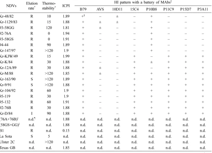
Table 1. Biological and physical characteristics of NDV isolates
NDVs Elution rate*
Thermo-
stability† ICPI HI pattern with a battery of MAbs‡
B79 AVS 10D11 15C4 P10B8 P11C9 P15D7 P3A11
Kr-48/82 R 10 1.89 +§ – ± – + – – –
Kr-1129/83 R 15 1.88 + ± ± – + – – –
93-58GG R 120 1.81 + ± – – + – – –
92-76A R 0 1.94 + – – – + – – –
93-58GS R 0 1.91 + – – – + – – –
94-44 R 90 1.89 + – – – + – – –
Kr-147/97 R >120 1.9 + – – – + – – –
Kr-KJW/49 R 15 1.99 + – – + – + + +
Kr-K/84 R 30 1.88 + – – + ± + + +
Kr-12A/89 R 30 1.88 + ± – + – + + +
Kr-M/88 R >120 1.85 + ± – + + + + +
Kr-163/90 S >120 1.89 + – – + + + + +
Kr-9/91 S >120 1.88 + – – + + + + +
Kr-104/92 R 60 1.9 + – – + + + + +
95-119 R 30 1.9 + – – + + + + +
95-132 R 60 1.91 + – – + + + + +
92-76B R 30 1.88 + – – + + + + +
Kr-D/84 I 90 1.88 + – – + + + + +
(76A+76B)|| n.d.¶ n.d. 1.88 n.d. n.d. n.d. n.d. n.d. n.d. n.d. n.d.
(58GS+GG)|| n.d. n.d. 1.88 n.d. n.d. n.d. n.d. n.d. n.d. n.d. n.d.
B1 R n.d. 0.15 n.d. n.d. n.d. n.d. n.d. n.d. n.d. n.d.
La Sota S 5 n.d. n.d. n.d. n.d. n.d. n.d. n.d. n.d. n.d.
Ulster 2C n.d. >120 n.d. n.d. n.d. n.d. n.d. n.d. n.d. n.d. n.d.
Texas GB n.d. n.d. 1.85 n.d. n.d. n.d. n.d. n.d. n.d. n.d. n.d.
NDV: Newcastle disease virus, ICPI: intracerebral pathogenicity index, HI: hemagglutination-inhibition, MAb: monoclonal antibody. *Elu- tion rate given as rapid (R), intermediate (I), or slow (S). †Thermostability of hemagglutinin at 56oC given in min. ‡Reactivity of MAbs; B79:
all paramyxovirus type 1 (PMV-1) including most pigeon PMVs, AVS: lentogens, 10D11: most mesogens and neurotropic velogens from U.S. poultry, 15C4: all PMV-1 except pigeon PMV. §
±: HI titer 2 log
2, +: HI titer higher than 3 log2. MAbs were originally diluted in 1/10, therefore, HI titer lower or less than 1 indicates a negative reaction (–). ||76A+76B and 58GS+GG mean combined subclones with 92-76A and 92-76B, and 93-58GS and 93-58GG, respectively, in which each combination were mixed with an equal volume of the individual sub- clone. ¶not done.Results
Elution rate and thermostability of NDV isolates Of the 18 total isolates, a rapid elution rate was observed for 15 isolates, a slow rate for 2 isolates and an intermediate rate was observed for one isolate. The reference NDV vac- cine strains La Sota and B1 were observed to have a slow and rapid elution rate, respectively (Table 1).
The thermostability of the hemagglutinin of the NDV iso- lates varied from 0 to 120 min. Five of the isolates that had been isolated since 1988 were observed to have a thermosta- bility in excess of 120 min, which is similar to the thermo- stable vaccine strain Ulster 2C. Differences in the thermostability were evident between the 93-58GS (0 min) and the 93-58GG (120 min) NDV isolates, which were clone-picked from the same isolate.
Reactivity pattern to MAb
After analysis by the HI test, the 18 NDV isolates were observed to have three distinct binding patterns against 8 MAbs. All of the isolates were inhibited by the MAb B79 but not by the MAbs AVS or 10D11.
For the remaining 5 MAbs, 7 isolates were inhibited by only MAb P10B8, but these isolates were not inhibited by MAb 15C4. The MAb 15C4 reacted with all of the PMV-1 isolates except for the pigeon PMV. Three isolates, including the reference challenge virus in Korea Kr-KJW/49, were not inhibited by MAb 15C4 alone. The remaining 8 isolates were observed to have a strong inhibition reaction with the 5 MAbs (Table 1).
Pathotyping
All of the 18 isolates were of the velogenic pathotype. The isolates had a high pathogenicity, as indicated by an ICPI that ranged from 1.99 (Kr-KJW/49) to 1.81 (93-58GG). The ICPI of the B1 strain and the NVNDV Texas-GB reference strain were 0.15 and 1.85, respectively (Table 1).
Clinical symptoms
A majority of the 4-week-old white leghorn chicken groups that were inoculated with the viral isolates had indica- tions of severe depression, conjunctivitis and eyelid swelling with watery eye within 2 to 4 days post-inoculation. The incubation period ranged from 2 to 4 days. Infection with 92-
Table 2. Clinical symptoms and mortality following eye-drop inoculation of NDV isolates in 4-week-old SPF Leghorn chicks
NDVs* Genotype No. of the
inoculated
No. of the dead/
Day of death†
Clinical symptoms‡
Conjunctivitis Depression Neurological sign
Kr-48/82 V§ 5 5/6, 7 5 5 0
Kr-1129/83 V 5 5/4, 5 5 5 0
Kr-K/84 V 5 5/4, 5 5 5 0
Kr-D/84 VII 5 5/3, 4 5 5 0
Kr-M/88 VI 5 5/7, 8 5 5 0
Kr-12A/89 VI 5 5/5, 6 5 5 1/5
Kr-163/90 VI 5 5/4 5 5 0
Kr-9/91 VI 5 5/4, 6 5 5 0
Kr-104/92 VI 5 5/6, 7 5 5 1/5
92-76A VI 5 5/4, 5 5 0 2/3,5
92-76B VI 5 3/5, 6|| 5 2 2/5,13
93-58GS VI 5 5/4, 5 5 5 3/3,4
93-58GG VI 5 0|| 5 5 1/11
94-44 VI 5 5/5 5 5 2/4
95-119 VII 5 5/5-7 5 5 0
95-132 VII 5 5/5, 6 5 5 0
Kr-147/97 VI 5 5/5, 6 5 5 2/5
Kr-KJW/49 III 5 5/4, 5 5 5 0
B1 II 10 0|| 0 1 0
Texas GB II 10 10/5, 6 10 0 9/4,5
Control – 10 0 0 0 0
NDV: Newcastle disease virus, SPF: specific-pathogen-free.*Each group was inoculated with 105 EID50 of a NDV isolate by eye drop method and observed for 2 weeks. †Days post-inoculation when death occurred. ‡Conjunctivitis included swelling of the eyelid. Neurological signs were incoordination, tremor, paralysis of the leg or torticollis of the neck (No. of chicks/days post-inoculation when signs appeared).
§Underlined genotypes were described in the precious paper [26]. ||All survived chicks were seroconverted 2 weeks post-inoculation except a control group.
76A or the NVNDV Texas GB strain resulted in no conjunctivi- tis or eyelid swelling. Eight isolates that have been isolated since 1989 induced neurological symptoms of disease, including inco- ordination, head tremor, paralysis of the leg or torticollis of the neck. Among these 8 isolates, the 93-58GS was observed to have more dramatic neurotropic characteristics (Table 2).
With the exception of 92-76B and 93-58GG, all of the iso- lates induced 100 % mortality within 3 to 8 days post-inocu- lation. However, the day of death after inoculation varied considerably depending on the isolate. Chicks that were inoc- ulated with Kr-D/84 all died within 3 to 4 days after inocula- tion, but chickens infected with isolate Kr-M/88 died within 7 to 8 days after inoculation. A sharp cough and greenish diarrhea were frequently observed for chickens in several of the experimental groups.
For the group that was inoculated with the NVNDV Texas GB, 10 birds died within 5 to 6 days after inoculation and presented with neurological symptoms within 4 to 5 days after inoculation. Although there appeared to be an increase in the number of birds with neurological symptoms, this increase was a resulted of the lower mortality rate. Thus, as a consequence, there was an increase in the number of birds suffering from convalescent torticollis or paralysis.
Gross pathological findings
As shown in Table 3, the primary gross findings in the internal organs in almost all of the white leghorn birds inoc- ulated with the 18 NDV isolates were mucous nasal dis- charge, mucus in the trachea, petechial hemorrhage in the heart fat and kidney urates. The Texas GB strain also caused petechial hemorrhage in the heart fat.
In the respiratory tract, nasal discharge and tracheal mucus were commonly present in all of experimental groups. Tra- cheal mucus without nasal discharge was found in all of the birds inoculated with the NVNDV Texas GB. Tracheal red- dening, petechia, or necrosis along the tracheal ring was rarely observed. Hemorrhage in the lungs was frequently observed in birds that were inoculated with 4 isolates (kr- 163/90, Kr-9/91, 92-76A and kr-147/97). The air sac was observed to have a normal appearance after inoculation.
In the lymphoid organs, small, discrete necrotic foci in the spleen were observed. This was particularly evident for the Kr-163/90, Kr-9/91 and 95-119 isolates. Hemorrhage in the thymus was also a common lesion. However, the number of birds with thymus lesions was not as numerous as those that were observed to have spleen necrosis. Hemorrhage in the spleen and bursal edema was also observed on occasion.
Table 3. Gross findings in the internal organs in SPF Leghorn chicks inoculated with NDV isolates at 4 weeks of age
NDV* Trachea† Lung‡ Hemorrhage§ Spleen Kidney
urate
Edema||
L H P M T HE N BF Subcue
Kr-48/82 5 0 1 4 0 0 0 1 0 5 0 0
Kr-1129/83 5 0 5 5 0 0 2 0 1 5 0 0
Kr-K/84 5 1 2 4 0 0 2 0 2 5 0 0
Kr-D/84 5 0 1 2 0 0 0 0 4 1 0 0
Kr-M/88 2 0 0 2 0 0 1 0 0 4 0 0
Kr-12A/89 4 0 0 3 0 0 2 1 3 5 0 0
Kr-163/90 5 3 0 5 1 0 0 0 5 5 0 0
Kr-9/91 4 3 0 2 0 0 0 0 5 5 0 0
Kr-104/92 5 1 0 3 0 0 1 0 1 5 0 0
92-76A 4 3 0 2 1 0 1 0 1 5 0 0
92-76B 2 0 0 1 0 0 0 0 0 1 0 0
93-58GS 3 0 0 0 1 4 1 1 0 3 0 0
93-58GG 0 1 0 4¶ 0 0 0 2 0 0 0 0
94-44 5 1 2 3 1 0 1 1 4 5 1 3
95-119 5 0 1 4 2 0 1 0 5 5 4 0
95-132 5 0 0 3 0 0 1 0 1 5 1 0
Kr-147/97 5 3 2 3 1 1 2 0 1 4 0 0
Kr-KJW/49 5 1 0 2 0 0 0 0 0 5 0 0
Texas GB 9 0 0 4 0 0 0 0 0 0 0 0
SPF: specific-pathogen-free, NDV: Newcastle disease virus. *Five chicks were inoculated in each experimental group with 105 EID50 of a NDV isolate by eye drop method and were observed for necropsy for 2 weeks. For the Texas-GB group 10 chicks were used. †Tracheal lesions included mucus, reddening and/or necrosis. ‡Lesions in lungs included reddening and hemorrhage. §Hemorrhage (HE) and/or necro- sis (N) in the larynx (L), heart fat (H), pancreas (P), rib or thigh muscle (M), thymus (T). ||BF: bursa of fabricious, Subcue: subcutaneous tis- sue of the neck. ¶One chick showed hemorrhage only in the heart muscle, and the other three revealed flaccid heart muscles without hemorrhage.
However, the appearance of gross lesions in certain organs varied based on the NDV isolate. Hemorrhage in the larynx (Kr-1129/83), hemorrhage in the rib or thigh muscle (93- 58GS), severe necrotic foci in the spleen (Kr-163/90, Kr-9/91 and 95-119), BF edema (95-119) and subcutaneous edema in the neck (94-44) were clearly present when chickens were infected with certain isolates. Unusually flaccid hearts (3/5) were observed in only one of the groups. This group was inoculated with the 93-58GG isolate that did not develop sig- nificant lesions in any other organs. Small discrete hemor- rhages in the pancreas were occasionally observed, and thymus atrophy was rarely found in 3 birds from 3 different groups (Kr-1129/83, 92-76B and 93-58GG).
The gross lesions in the gastrointestinal (GI) tract in white leghorn chicks were severe hemorrhage in the proventricu- lus, duodenum, ileum and cecal tonsil, and ulcerative necro- sis associated with the Payer’s patch. Lesions in the jejunum were rare (Table 4). Some isolates resulted in hemorrhage in the gizzard and cloaca, distended small intestines, and hem- orrhage and necrotic foci in the colon. There were no gross lesions in birds that were inoculated with isolate 93-58GG,
except for hemorrhage in the cecal tonsil. The birds that were inoculated with Kr-48/82 presented only with hemorrhage in the proventriculus and gizzard without significant lesions in any other GI tract locations. The appearance of gross lesions in the GI tract varied considerably depending on the NDV isolate. With the Texas GB strain, there were slight hemor- rhages in the proventriculus and duodenum (3/5) but not in the cecal tonsil.
Phylogenetic analysis
The sequence of the F gene was compared by phyloge- netic analysis to genotype the following isolates: 92-76A and B, 93-58GS and GG, 94-44, 95-119, 95-132 and Kr-147/97.
Based on the topology of the tree, the recent Kr isolates could be classified into the following 3 genotypes: V, VI and VII. The isolates that were isolated early in 1980s belonged to genotype V, those isolated in the late 1980s to the early 1990s belonged to genotype VI, and those that were isolated in 1995 belonged to genotype VII. However, the peafowl iso- late Kr-D/84 was genotype VII (Table 2). The Korean refer- ence challenge strain, Kr-KJW/49, was genotype III.
Table 4. Gross findings in the gastrointestinal tract in SPF Leghorn chicks inoculated with NDV isolates at 4 weeks of age
NDV*
HE in proventri- culus and/or
gizzard
HE, N and/or D of small intestines
HE in cecal tonsil
HE and/or N in large intestines
Sub-total Duodenum Jejunum Ileum Colon
Cloaca HE
HE N HE N D HE N D HE N
Kr-48/82 4 (2)† 2‡ 0 1 0 0 0 0 0 1 0 0 0 0
Kr-1129/83 5 5 4 2 2 0 0 1 1 0 3 4 0 0
Kr-K/84 5 5 5 3 0 0 0 1 5 0 3 1 0 1
Kr-D/84 4 5 4 2 1 2 1 3 2 1 5 5 0 0
Kr-M/88 3 3 2 1 1 0 0 1 1 0 3 0 0 0
Kr-12A/89 5 5 3 5 1 2 0 2 3 0 5 2 0 0
Kr-163/90 5 (1) 5 2 5 1 0 1 1 3 1 5 2 0 0
Kr-9/91 5 (1) 5 4 5 0 1 1 2 5 1 5 1 2 0
Kr-104/92 3 (2) 3 1 2 0 0 1 2 2 1 2 1 0 1
92-76A 4 5 4 1 2 1 0 2 1 0 4 1 0 1
92-76B 2 1 0 0 0 0 1 1 1 1 2 1 0 3
93-58GS 2 (1) 4 4 0 2 0 0 3 0 0 5 1 0 0
93-58GG 0 0 0 0 0 0 0 0 0 0 3 0 0 0
94-44 5 (2) 5 5 3 0 0 0 5 3 0 5 3 0 1
95-119 5 4 3 3 1 1 1 2 2 1 3 4 1 1
95-132 5 (2) 4 2 2 0 0 0 1 4 0 3 1 1 1
Kr-147/97 5 (2) 4 4 4 2 2 0 3 4 0 5 3 1 2
Kr-KJW/49 5 5 5 0 0 0 2 3 2 0 3 1 0 0
Texas GB 3§ 3 3 0 0 0 0 0 0 0 0 1 0 0
SPF: specific-pathogen-free, NDV: Newcastle disease virus. HE: hemorrhage, N: necrosis, D: distention. *Five chicks were inoculated in each experimental group with 105 EID50 of a NDV isolate by eye drop method and were observed for necropsy for 2 weeks. For the Texas- GB group, 10 chicks were used. †No. of chicks with hemorrhage (No. of the gizzard hemorrhage in the parenthesis). ‡No. of chicks showed any pathological lesions in the small intestines. §Slight focal reddening.
Discussion
Because infection with NDV in chickens appeared to vary from unapparent to peracute disease resulting in high mortal- ity [4], it has recently been suggested that the epidemiology and pathogenicity of NDV is similar to that of an avian influ- enza virus [2].
The characterization and pathotyping of NDV have been essential and reliable tools for the establishment of control and eradication policies, and characterization depends prima- rily on the mean time of death, ICPI and intravenous patho- genicity test. Regardless of pathogenicity, there are several methods for characterizing the biological properties of NDV, such as the elution rate of agglutinated red blood cells, ther- mostability of the hemagglutinin and the ability to form a plaque [1, 4].
Almost all of 18 NDV isolates were observed to have a rapid elution rate. For the thermostability of hemagglutinin, thermostable isolates were more common than thermolabile isolates, which supports previous data where 13 out of 18 NDV isolates that were isolated from 1945 to the early 1960s were observed to have heat stable hemagglutinin (unpub- lished data, 1963). However, it has also been reported that heat stable hemagglutinin ( ≥ 30 min at 56
oC) was only found in NDV strains that were isolated since 1988 in 16 isolates that were collected from 1984 to 1991 in Korea [10]. In this experiment, strong thermostability ( ≥ 120 min) was observed for all isolates that were collected after 1988. The increased frequency of NDV with strong thermostability might have been involved in the recent, sporadic cases of ND in the sum- mer in Korea. ND had been very rare in the prior year before these sporadic cases were reported. Difference in the thermo- stability was found between the 2 subclones that were pre- pared by the clone-picking method: 93-58GG and 93-58GS.
It may be important that different clones in the same MAb group had different pathogenicity profiles in 4-week-old chicks.
Two other subclones prepared by the same method, 92- 76A and 92-76B, were observed to have markedly different HI pattern when compared against a battery of 8 MAbs.
Moreover, the 92-76A and 93-58 GS subclones resulted in syncytial cell formation in chicken embryo fibroblast cell cultures, while the 92-76B and 93-58GG subclones resulted in degenerative cytopathic effect. This result confirmed that field NDV isolates consisting of various subpopulations may be significantly different from each other [15].
Rapid elution rates were observed for the NDV Kr-KJW/
49 isolate, which is the reference challenge virus in use since 1950 in Korea.
As a result of the HI analysis using a battery of 8 MAbs, the 18 NDV isolates were divided into 3 distinct groups.
There were no isolates that fell into lentogens, mesogens or neurotropic velogens. These data were in accordance with the pathotyping analysis and pathogenicity test, which demon- strated no reaction with AVS for lentogens or with 10D11 for
most mesogens and neurotropic velogens isolated from U.S.
poultry [22, 23]. It was not thought that the weak HI reac- tion ( ± HI titer 2 log
2) observed for some isolates with AVS or 10D11 was a result of reacting against a common lento- gen or mesogen antigen, respectively. These results may be the result of nonspecific HA inhibition. Seven of the NDVs that were isolated from chickens were observed to have no reaction with 15C4, which was observed to react with all PMV-1 isolates except for the pigeon PMV.
The MAb reaction profiles for the NDVs may not reveal the exact antigenicity patterns because the MAbs used in the present study do not cover all antigenic groups for NDV.
Therefore, the study should be confirmed using a more var- ied MAb profile.
A majority of the 18 NDV isolates caused mortality accompanied by depression, conjunctivitis and swelling of the eyelid, as described previously [4, 13, 25]. The 93-58GG was determined to have an ICPI of 1.81 but caused no mor- tality or gastrointestinal lesions except for hemorrhage of the cecal tonsil. Because the chickens that were inoculated with this isolate lived for the duration of the experiment and were necropsied at the end of the 2-week observation period after inoculation, observable lesions may have become less severe or may had fully resolved at the time of necropsy. No rela- tionship between the day of death and ICPI were observed;
however, 2 isolates that had an ICPI less than 1.85 were observed to result in delayed mortality or no mortality.
Neurological symptoms, including incoordination, tremor, torticollis and paralysis [4], appeared in birds that were inoc- ulated with 7 isolates. In addition, the neurological symp- toms appeared in 2 distinct patterns according to the time after inoculation. Some isolates caused neurological symp- toms that were accompanied with other symptoms prior to the death of the chicken. Other isolates caused convalescent torticollis of the neck and paralysis of the leg after 10 days of inoculation. Isolates that were observed to have low pathoge- nicity were included in this latter group. However, the VVNDV isolates that resulted in intestinal pathological lesions were easily classified as having neurological symp- toms. Nevertheless, clinical symptoms and pathological lesions as a result of NDV were varied depending on the strains of virus, pathogenicity of the virus, the host species, susceptibil- ity of the host and the environment [4, 8, 28].
The predominant gross lesions that were induced by all of
the NDV isolates were conjunctivitis with hemorrhage in the
inner eyelid, nasal discharge, tracheal mucus, kidney urates,
severe necrosis in the spleen and hemorrhage of the heart fat,
proventriculus, duodenum and cecal tonsil. In addition, bur-
sal edema and subcutaneous edema in the neck were observed
for the 95-119 and 94-44 isolates, respectively. However, the
total numbers of cases that were observed to have lesions
was significantly higher in cecal tonsil (64/90) and duode-
num (53/90) when compared to other organs. As previously
described [25], VVNDVs primarily developed hemorrhage
with necrosis in the duodenum, ileum and cecum and hemor-
rhage in the mucosa and submucosa of the proventriculus and gizzard. NDVs have differences depending on the patho- types and genotypes VVNDVs, although further study is needed to confirm this hypothesis. VVNDV has been known to cause predominant GI, respiratory tract and lymphoid tis- sue lesions [13, 18], and the spleen is more susceptible to the necrotic effects of NDV [13].
There were considerable differences in clinical symptoms and gross findings between NVNDV and VVNDV. This was especially true for neurological symptoms [4, 8]. Compared to the VVNDVs, NVNDVs induced severe neurological symptoms prior to death, no conjunctivitis, no kidney urate and no visible necrosis or hemorrhage in the GI tract, except in the proventriculus and duodenum. Unexpectedly, in the present study, it was observed that NVNDV induced slight focal reddening in the proventriculus as well as hemorrhag- ing in the duodenum. Because the presence of hemorrhagic lesions in the intestine of infected chickens has been a crite- ria to distinguish VVNDV from NVNDV [9, 16], hemor- rhage in the proventriculus and duodenum could be eliminated from this classification scheme. All of the isolates that were tested in this experiment were classified as VVNDV. Never- theless, mesogens, NVNDV and lentogens may still be circu- lating in poultry farms in Korean because all of the isolates were obtained from chickens that became ill.
The Korean NDV isolates were classified into three differ- ent genotypes by phylogenetic analysis of F gene, V, VI and VII, as described previously [26]. Each of the three geno- types may correspond to ND epidemics that occurred in the 1980s, late 1980s to early 1990s and after 1995, respec- tively. Although there was little direct correlation between the NDV tissue tropism and the genotype, all of the isolates that induced neurological symptoms belonged to genotype VI. This genotype had spread nationwide during the late 1980s through the 1990s, and these data support a previous report [19, 26]. Therefore, specific genotypes of VVNDVs may cause neurological symptoms in chickens.
Acknowledgments
This research was supported by the Southeast Poultry Research Laboratory, ARS, USDA, USA.
References
1. Alexander DJ. Newcastle disease. In: Purchase HG, Arp LH, Domermuth CH, Pearson JE (eds.). A Laboratory Manual for the Isolation and Identification of Avian Pathogens. 3rd ed. pp. 114-120, American Association of Avian Pathologists, Kennett Square, 1989.
2. Alexander DJ. The epidemiology and control of avian influenza and Newcastle disease. J Comp Pathol 1995, 112, 105-126.
3. Alexander DJ. Newcastle disease in countries of the European Union. Avian Pathol 1995, 24, 3-10.
4. Alexander DJ. Newcastle disease and other avian
Paramyxoviridae infections. In: Calnek BW, Barnes HJ, Beard CW, McDougald LR, Saif YM (eds.). Diseases of Poultry. 10th ed. pp. 541-569, Iowa State University Press, Ames, 1997.
5. Alexander DJ, Manvell RJ, Lowings JP, Frost KM, Collins MS, Russell PH, Smith JE. Antigenic diversity and similarities detected in avian paramyxovirus type 1 (Newcastle disease virus) isolates using monoclonal antibodies.
Avian Pathol 1997, 26, 399-418.
6. Beard CW, Hanson RP. Newcastle disease. In: Hofstad MS, Barns HJ, Calnek BW, Reid WM, Yoder HW Jr.
(eds.). Disease of Poultry. 8th ed. pp. 452-470, Iowa State University Press, Ames, 1984.
7. Brown C, King DJ, Seal BS. Pathogenesis of Newcastle disease in chickens experimentally infected with viruses of different virulence. Vet Pathol 1999, 36, 125-132.
8. Campbell RSF. The pathogenesis and pathology of avian respiratory infections. Vet Bulletin 1986, 56, 521-543.
9. Cheville NF, Stone H, Riley J, Ritchie AE. Pathogenesis of virulent Newcastle disease in chickens. J Am Vet Med Assoc 1972, 161, 169-179.
10. Choi KS. Characterization of Korean isolates of Newcastle disease virus. Ph.D dissertation. Seoul National University, Seoul, 1994.
11. Collins MS, Strong I, Alexander DJ. Evaluation of the molecular basis of pathogenicity of the variant Newcastle disease viruses termed "pigeon PMV-1 viruses". Arch Virol 1994, 134, 403-411.
12. Collins MS, Strong I, Alexander DJ. Pathogenicity and phylogenetic evaluation of the variant Newcastle disease viruses termed "pigeon PMV-1 viruses" based on the nucleotide sequence of the fusion protein gene. Arch Virol 1996, 141, 635-647.
13. Hamid H, Campbell RSF, Parede L. Studies of the pathology of velogenic Newcastle disease: Virus infection in non-immune and immune birds. Avian Pathol 1991, 20, 561-575.
14. Hanson RP. Newcastle disease. In: Hitchner SB, Domermuth CH, Purchase HG, Williams JE (eds.). Isolation and Identification of Avian Pathogens. 2nd ed. pp. 63-66, American Association of Avian Pathologists, College Station, 1980.
15. Hanson RP. Heterogeneity within strains of Newcastle disease virus: key to survival. In: Alexander DJ (ed.).
Newcastle Disease. 1st ed. pp. 113-130, Kluwer Academic, Boston, 1988.
16. Hanson RP, Spalatin J, Jacobson GS. The viscerotropic pathotype of Newcastle disease virus. Avian Dis 1973, 17, 354-361.
17. Hodder AN, Liu ZY, Selleck PW, Corino GL, Shiell BJ, Grix DC, Morrow CJ, Gorman JJ. Characterization of field isolates of Newcastle disease virus using antipeptide antibodies. Avian Dis 1994, 38, 103-118.
18. Jungherr EL, Tyzzer EE, Brandly CA, Moses HE. The comparative pathology of fowl plague and Newcastle disease. Am J Vet Res 1946, 7, 250-288.
19. Kim JH, Song CS. Consideration of cause of recent severe outbreaks of Newcastle disease in Korea and a brief review of virological differences, serological diagnosis and administration of a vaccine. Korean J Poult Sci 1992, 19,
65-76.
20. King DJ. Influence of chicken breed on pathogenicity evaluation of velogenic neurotropic Newcastle disease virus isolates from cormorants and turkeys. Avian Dis 1996, 40, 210-217.
21. King DJ, Seal BS. Biological and molecular characterization of Newcastle disease virus isolates from surveillance of live bird markets in the northeastern United States. Avian Dis 1997, 41, 683-689.
22. King DJ, Seal BS. Biological and molecular characterization of Newcastle disease virus (NDV) field isolates with comparisons to reference NDV strains. Avian Dis 1998, 42, 507-516.
23. Lana DP, Snyder DB, King DJ, Marquardt WW.
Characterization of a battery of monoclonal antibodies for differentiation of Newcastle disease virus and pigeon paramyxovirus-1 strains. Avian Dis 1988, 32, 273-281.
24. Lancaster JE. Diagnosis of Newcastle disease. Vet Bulletin 1963, 33, 347-360.
25. Lancaster JE. Newcastle disease - pathogenesis and diagnosis. Worlds Poult Sci J 1981, 37, 26-33.
26. Lee YJ, Sung HW, Choi JG, Kim JH, Song CS.
Molecular epidemiology of Newcastle disease viruses isolated in South Korea using sequencing of the fusion
protein cleavage site region and phylogenetic relationships.
Avian Pathol 2004, 33, 482-491.
27. Marín MC, Villegas P, Bennett JD, Seal BS. Virus characterization and sequence of the fusion protein gene cleavage site of recent Newcastle disease virus field isolates from the southeastern United States and Puerto Rico. Avian Dis 1996, 40, 382-390.
28. McFerran JB, McCracken RM. Newcastle disease. In:
Alexander DJ (ed.). Newcastle Disease. 1st ed. pp. 161- 183, Kluwer Academic, Boston, 1988.
29. Page RD. TreeView: An application to display phylogenetic trees on personal computers. Comput Appl Biosci 1996, 12, 357-358.
30. Park KS. Epizootiological study on the outbreaks of Newcastle disease in Korea. Korean J Poult Sci 1979, 6, 38-46.
31. Srinivasappa GB, Snyder DB, Marquardt WW, King DJ. Isolation of a monoclonal antibody with specificity for commonly employed vaccine strains of Newcastle disease virus. Avian Dis 1986, 30, 562-567.
32. Thompson JD, Gibson TJ, Plewniak F, Jeanmougin F, Higgins DG. The CLUSTAL_X windows interface: flexible strategies for multiple sequence alignment aided by quality analysis tools. Nucleic Acids Res 1997, 25, 4876-4882.