© 2018 The Korean Ophthalmological Society
This is an Open Access article distributed under the terms of the Creative Commons Attribution Non-Commercial License (http://creativecommons.org/licenses /by-nc/3.0/) which permits unrestricted non-commercial use, distribution, and reproduction in any medium, provided the original work is properly cited.
Original Article
Predicting Factor of Visual Outcome in Unilateral Idiopathic Cataract Surgery in Patients Aged 3 to 10 Years
Jihyun Park1, Youn Gon Lee2, Kook Young Kim2, Byoung Yeop Kim2
1Daesung Eye Clinic, Bucheon, Korea
2Department of Cornea and Refractive Surgery, Kim’s Eye Hospital, Myung-Gok Eye Research Institute, Konyang University College of Medicine, Seoul, Korea
Purpose: To report the surgical results of unilateral pediatric cataracts from uncertain causes in relatively older children and to identify factors related to better visual outcomes.
Methods: We retrospectively evaluated the medical records of 39 patients who underwent surgery between the ages of 3 and 10 years for unilateral pediatric cataracts of no known cause. All patients underwent primary intraocular lens implantation and postoperative amblyopia treatment. A postoperative final visual acuity better than 20 / 30 was considered to be a good visual outcome.
Results: The mean age of patients was 6.0 ± 1.8 years at the time of surgery. The mean preoperative visual acuity was 1.07 ± 0.71 logarithm of the minimum angle of resolution (range, 0.15 to 3.00), while the mean final postoperative visual acuity was 0.47 ± 0.54 logarithm of the minimum angle of resolution (range, 0.00 to 2.00).
Of 39 patients, 18 (46.2%) achieved a good visual outcome. Only the preoperative visual acuity maintained a significant association with a good visual outcome according to our multivariate analysis (p = 0.040). A pre- operative visual acuity of 20 / 100 or better was found to increase the chance of achieving a good visual out- come by 13.79-fold (95% confidence interval, 1.13 to 167.58).
Conclusions: The visual outcome of unilateral pediatric cataract surgery for cataracts with no specific cause identified in patients after three years of age could be satisfactory, especially with a preoperative visual acuity of 20 / 100 or better.
Key Words: Congenital cataract, Phacoemulsification, Prognosis, Visual acuity
Despite developments in surgical techniques and intra- ocular lenses, the management of unilateral pediatric cata- racts is still clinically challenging. Better visual outcomes are usually obtained with early surgical correction and vigorous amblyopia treatment [1-4]. The critical period for
surgical treatment of unilateral congenital cataract was re- ported to be within the range of 6 to 17 weeks in several studies [1-3].
In general, pediatric cataract patients under three years of age who require surgery under general endotracheal an- esthesia are referred to tertiary hospitals. However, screen- ing tests for visual acuity are not easy to administer to pa- tients who are less than three years old, and diagnoses of cataract are rare in clinical practice. Therefore, children with decreased visual acuity who have been diagnosed with cataract undergo cataract surgery much later than the
Received: September 18, 2017 Accepted: December 25, 2017
Corresponding Author: Byoung Yeop Kim, MD. Department of Ophthal- mology, Kim’s Eye Hospital, #136, Yeongsin-ro, Yeongdeungpo-gu, Seoul 07301, Korea. Tel: 82-2-1577-2639, Fax: 82-2-2671-6359, E-mail: kby@
kimeye.com
reported critical period (between 6 and 17 weeks of age) for congenital cataract surgery. However, good postopera- tive visual function in pediatric cataract surgery requires careful surgical treatment, early correction of refractive errors, and rigorous attention to the postoperative treat- ment of amblyopia. Previous studies have reported that the prognosis of unilateral pediatric cataract is even less favor- able [3,4]. The results of the surgery in our hospital, how- ever, have not been always less favorable, even when the morphology of the cataracts indicates that they are con- genital. The purpose of this study was to evaluate the re- sults of surgery for unilateral pediatric cataract of uncer- tain cause in our hospital and to analyze factors related to a better visual outcome.
Materials and Methods
This retrospective study was conducted at the Depart- ment of Ophthalmology of Kim’s Eye Hospital at Konyang University. The study protocol was approved by the insti- tutional review board at Kim’s Eye Hospital in Seoul, Ko- rea (B-2011-001). All procedures conformed to the guide- lines of the Declaration of Helsinki. Patients were informed of the study objectives and the examination pro- cesses and were asked to provide written consent. We ret- rospectively reviewed the medical records of patients who underwent surgery for pediatric cataract (age range, 3 to 10 years old) at Kim’s Eye Hospital between 1 January 2001 and 31 December 2010. We identified 104 patients without history of previous intraocular surgery. Patients were excluded if they had bilateral cataracts, traumatic cataract, ocular abnormalities unrelated to cataract, or any developmental delays. Prospective patients were also ex- cluded if their postoperative follow-up period was less than one year in length (Fig. 1).
Patients typically underwent cataract surgery within four months of diagnosis. Surgery was performed when the patient recognized decreased subjective and objective visual accuity due to cataract and after consulting with their parents. All surgeries were performed by one experi- enced surgeon (BYK). Under general anesthesia, anterior continuous curvilinear capsulorhexis, lens aspiration, pos- terior continuous curvilinear capsulorhexis, and anterior dry vitrectomy were performed through either a limbal or a scleral tunnel incision. An intraocular lens (IOL) is tradi-
tionally implanted in the capsular bag; in cases of poly- methyl methacrylate IOL (Single-piece Polymethyl Meth- acrylate Posterior Chamber IOL 604A Rayner; Rayner, East Sussex, UK), the IOL optic was captured through a posterior continuous curvilinear capsulorhexis opening. In recent years, IOLs have been implanted in the sulcus to simplify later replacement. The power of the IOL was cal- culated using the SRK-II formula, and the target refraction was determined according to age and refractive error of the fellow eye and modified to minimize anisometropia.
The refraction goals of IOL target diopters were adjusted according to age as follows: 3 to 5 years, +2 to +3; 5 to 7 years, +1 to +2; 7 to 10 years, plano to +1.00. The IOL tar- get diopters were adjusted according to refractive error of the fellow eye, in addition, there was no more than ±1 di- opter difference when the targeting diopters were more myopic compared with the fellow eye. The incision was closed with 10-0 nylon sutures.
Postoperatively, 0.3% tobramycin (Tobrex; Alcon, Fort Worth, TX, USA) and 0.1% fluorometholone (Ocumetho- lone; Allergan, Irvine, CA, USA) eye drops were adminis- tered four times daily for about one month. A large portion of patients took 10 mg of oral prednisolone (Solondo; Yu- han Medical, Seoul, Korea) for several days to control moderate to severe anterior chamber reaction in the imme- diate postoperative period.
We cooperated with two pediatric ophthalmologists for the amblyopia treatment of our patients. Spectacles with or Fig. 1. Flow chart of the inclusion/exclusion criteria and compli- cation case. VA = visual acuity.
A total of 104 patients without hisory of previous intraocular surgery were identified between January 1st, 2001 and December 31st, 2010
65 Patients excluded 40 Bilateral cataract 18 Traumatic cataract 2 Ocular abnormality unrelated to cataract
1 Developmental delay 4 Less than 1 year of follow up 12 Patients with strabismus
→ 3 Patiens with strabimus surgery 2 Patient on esotropia 1 Patient on exotropia Postoperative amblyopia treatment
32 Patients need patching and spectacles 5 Patients need only spectacles 2 Patients need no treatment
39 Patients were included and reviewed
Eyes with good visual outcome (VA ≥ 20 / 30) n = 18 (46.2%)
Eyes with good visual outcome (VA < 20 / 30) n = 21 (53.8%)
9 Eyes (23.1%) with exudative membrane in the anterior chamber with or without posterior synechia
without monocular occlusion were prescribed postopera- tively. The number of patching hours ranged from six to all waking hours each day, and the maintenance level was modified on an individual basis according to visual acuity and age.
Visual acuity was measured at each visit using either a standard Snellen chart or a tumbling E chart for young illit- erate children. Two patients (one male patient, 2 years 11 months old; one female patient, 1 year 7 months old) were unable to have their visual acuity checked preoperatively due to poor cooperation. Strabismus surgery was performed when needed. Data were collected with regard to gender, type of cataract, age at diagnosis, age at surgery, presence of strabismus, preoperative and postoperative visual acuity (best-corrected visual acuity) of each eye, preoperative and postoperative refractive error in each eye, axial length at surgery, amblyopia treatment, and follow-up period.
A final postoperative visual acuity better than 20 / 30 was considered a good visual outcome. We chose a cutoff point of 20 / 30, which was relatively high compared to other studies because visual acuity was sufficient only if it did not seriously disturb daily life. Patients were divided into either the Va (≥ 20 / 100) or the Vb (< 20 / 100) group according to preoperative visual acuity. Snellen acuity data were converted into the logMAR format for statistical analyses.
Univariate analysis using chi-square test, Fisher exact test, and Student’s t-test was performed, and multiple lo- gistic regression analysis was carried out to identify signif- icant factors associated with good visual outcome. The SPSS ver. 15.0 (SPSS Inc., Chicago, IL, USA), was used for our statistical analysis, with statistical significance defined as p-values < 0.05.
Results
Thirty-nine patients were included in the data analysis after excluding 65 patients who were not eligible because of bilateral cataracts (40), traumatic cataract (18), ocular abnormality unrelated to cataract (2), developmental delay (1), and less than one year of follow-up (4) (Fig. 1). Table 1 shows the patient demographic data. The mean age at sur- gery was 6.0 ± 1.8 years (range, 3 to 10 years). The mean duration of follow-up after surgery was 3.9 ± 2.9 years (range, 1.0 to 10.3 years).
The mean final postoperative visual acuity was 0.47 ± 0.54 logarithm of the minimum angle of resolution (log- MAR; range, 0.00 to 2.00) in the operated eye. Fig. 2 shows the changes in visual acuity in these eyes. Eighteen eyes (46.2%) of 39 patients achieved a good visual acuity of 20 / 30 or better. For the fellow eye, the mean final visu- al acuity was 0.02 ± 0.04 logMAR (range, 0.00 to 0.10).
Twelve eyes with strabismus showed a mean final visual acuity of 0.73 ± 0.52 logMAR, 0.51 ± 0.34 logMAR in eight eyes with exotropia and 1.18 ± 0.57 logMAR in four eyes with esotropia. Strabismus surgery was eventually re- Table 1. Demographic data of the enrolled participants
Factor Value (n = 39 eye)
Sex
Male 19 (48.7)
Female 20 (51.3)
Age at diagnosis (yr) 5.6 ± 2.0
Age at surgery (yr) 6.0 ± 1.8
Side
Right 25 (64.1)
Left 14 (35.9)
Preop VA (logMAR) 1.07 ± 0.71
Group Va (≥20 / 100) 16 (41.0) Group Vb (<20 / 100) 21 (53.8) Preop VA of the fellow eye (logMAR) 0.06 ± 0.07
Preop RE (SE, diopters) -0.56 (-7.50 to +8.50) Preop RE of the fellow eye
(SE, diopters) +0.11 (-3.50 to +4.50)
Preop ocular alignment
Orthotropia 27 (69.2)
Exotropia 8 (20.5)
Esotropia 4 (10.3)
Amblyopia treatment
Spectacles + monocular occlusion 32 (82.1)
Spectacles 5 (12.8)
Type of cataract
Posterior subcapsular/polar 29 (74.4)
Nuclear 5 (12.8)
Peripheral 5 (12.8)
Axial length (mm) 22.3 ± 1.40
Values are presented as number (%), mean ± standard deviation, or mean (range).
VA = visual acuity; logMAR = logarithm of the minimum angle of resolution; RE = refractive error; SE = spherical equivalent
quired in three children (two patients with esotropia and one patient with exotropia). Patients treated with a combi- nation of monocular patching and spectacles achieved a mean final visual acuity of 0.55 ± 0.56 logMAR, while the acuity was 0.11 ± 0.17 logMAR with spectacles alone.
Patching was not always tolerated well, especially in school-aged children.
The mean target refraction was 0.90 ± 1.25 diopters (D;
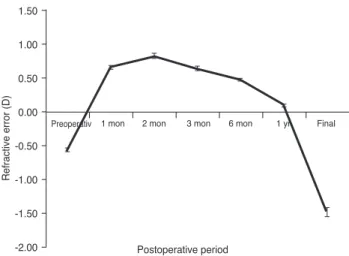
range, -1.21 to +3.50), and the mean final refractive error was -1.48 ± 2.97 D (range, -7.75 to +8.50) in the operated eye. Fig. 3 shows changes in refractive error in these eyes.
The mean rate of refractive change per year was -0.65 D (range, -2.50 to +0.60). For the fellow eyes, the mean final refractive error was -0.85 ± 1.72 D (range, -6.50 to +2.50).
Table 2 shows the characteristics of patients who achieved or did not achieve good visual acuity (final post- operative visual acuity >20 / 30) and the results of a uni- variate analyses. A good visual outcome was significantly associated with better preoperative visual acuity, smaller preoperative refractive error, smaller difference in preop- erative refractive errors between the operated and the fel- low eyes, and absence of strabismus (p = 0.005, 0.005, 0.002, and 0.014, respectively). Preoperative refractive error was excluded from the multiple logistic regression analysis to reduce multicollinearity. The other three variables and
“age at surgery,” which are known to have a decisive effect on surgical outcome, were also included in the analysis.
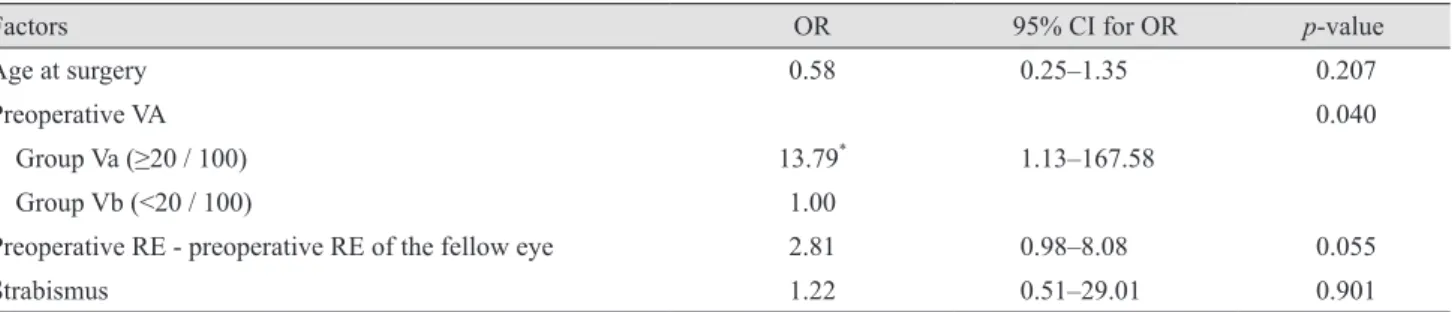
Only preoperative visual acuity remained significant (p = 0.040) (Table 3). Twelve (66.7%) of the 18 patients with a good visual outcome had a preoperative visual acuity of 20 / 100 or better. Patients with a preoperative visual acuity of 20 / 100 or better had a 13.79-fold increased chance of achieving good visual acuity compared to those with a pre- operative visual acuity <20 / 100 (95% confidence interval, 1.13 to 167.58). Fig. 4 illustrates this tendency.
An exudative membrane in the anterior chamber either with or without posterior synechia developed in nine eyes (23.1%) in the immediate postoperative period. This prob- lem resolved rapidly without any further complications with systemic steroid treatment for one or two weeks. No other postoperative complications were recognized.
Discussion
There are plenty of arguments about the best age for congenital cataract surgery, and earlier surgery is general- ly accepted to have a positive correlation with a better vi- sual outcome, especially in eyes with unilateral pediatric cataract [1-6]. The critical period for this type of surgery has been reported to be from 6 to 17 weeks of age in sever- al studies [1-3]. In clinical practice, however, ophthalmolo- gists often encounter older children with unilateral cata- racts for whom the critical period for surgery has already Fig. 2. Changes in visual acuity in all eyes. The mean final post-
operative visual acuity was 0.34 ± 0.29 (0.47 ± 0.54 logarithm of the minimum angle of resolution) in the operated eye. Visual acuity was mostly improved during the first postoperative month.
Error bars indicate standard deviation.
Fig. 3. Changes in refractive error. The mean refractive error was 0.12 ± 2.51 diopters (D) one year postoperatively, and the mean final refractive error was -1.48 ± 2.97 D in the operated eye. The mean rate of refractive change per year was -0.65 D. Error bars indicate standard deviation.
Preoperativ 1 mon 2 mon
Postoperative period
Visual acuity (by Snellen chart)
3 mon 6 mon 1 yr Final 0.45
0.40 0.35 0.30 0.25 0.20 0.15 0.10 0.05 0
Preoperativ 1 mon 2 mon
Postoperative period
Refractive error (D)
3 mon 6 mon 1 yr Final
1.50 1.00 0.50 0.00 -0.50 -1.00 -1.50 -2.00
passed. If it is the children’s first visit to an ophthalmology clinic, it is not easy to determine if the cataract is congeni- tal or developmental. Patients in this study also had no his- tory of ocular trauma or other ophthalmic problems, which made the onset and cause of cataracts unclear. The appear- ance of lens opacities suggested that they were congenital or developmental. Nevertheless, contrary to the aforemen- tioned studies, nearly half of these patients achieved a good postoperative visual acuity of 20 / 30 or better.
Several studies on pediatric cataracts have achieved good postoperative visual acuity even after late surgeries. Zwaan et al. [7] showed visual acuities of 20 / 40 or better in 44%
of eyes, visual acuities of 20 / 50 to 20 / 80 in 27% of eyes, and better visual acuity in children older than four years of age (p = 0.001) despite their inclusion of traumatic and sec- ondary cataracts. The posterior capsular bag IOL of cata-
ract surgery has resulted in good visual acuity (20 / 40 or better in 85.3%) in both unilateral and bilateral develop- mental and traumatic cataract patients over the age of two years [8]. In unilateral cataract cases, visual acuity was 20 / 50 or better in 74% of eyes; a best corrected visual acuity of 20 / 30 or better was achieved in 95% of eyes in bilateral cataract cases [9]. In a retrospective study of 139 eyes with pediatric cataract, the mean visual acuity was better in bi- lateral versus unilateral cases (p < 0.001), and that study found that an older age at surgery was correlated with a better visual acuity outcome in both unilateral and bilateral cases [10]. Early surgery was not a prerequisite for a good postoperative outcome in past studies or in the current study, which might be because the lens opacities started to develop after the critical period of visual development or were not significant during that period. The patients whose Table 2. Univariate analyses
Factor Eyes with a good visual
outcome (VA ≥20 / 30) (n = 18)
Eyes without a good visual outcome (VA <20 / 30)
(n = 21) p-value
Sex 0.621*
Male 8 (42.1) 11 (57.9)
Female 10 (50.0) 10 (50.0)
Age at diagnosis (yr) 6.2 ± 1.7 5.1 ± 2.1 0.087†
Age at surgery (yr) 6.5 ± 1.8 5.5 ± 1.7 0.067†
Preoperative VA 0.005*
Group Va (≥20 / 100) 12 (75.0) 4 (25.0)
Group Vb (<20 / 100) 6 (28.6) 15 (71.4)
Preoperative RE (SE, diopters) 0.69 ± 2.40 -2.09 ± 2.66 0.005†
Preoperative RE - preoperative RE of the fellow eye
(SE, diopters) 0.72 ± 2.21 -2.29 ± 2.82 0.002†
Strabismus 0.014*
(-) 16 (59.3) 11 (40.7)
(+) 2 (16.7) 10 (83.3)
Amblyopia treatment 0.144‡
Spectacles + monocular occlusion 12 (37.5) 20 (62.5)
Spectacles 4 (80.0) 1 (20.0)
Type of cataract 0.106‡
Posterior subcapsular/polar 15 (51.7) 14 (48.3)
Nuclear 3 (60.0) 2 (40.0)
Peripheral 0 (0.0) 5 (100.0)
Axial length (mm) 21.93 ± 0.83 22.60 ± 1.70 0.125†
Values are presented as number (%) or mean ± standard deviation.
VA = visual acuity; RE = refractive error; SE = spherical equivalent.
*Chi-square test; †Student’s t-test; ‡Fisher exact test.
cataracts were already severe during infancy were more likely to be discovered earlier. The strabismus proportion was 30.8% in this study, which was much lower than the rate reported by others [3]. Our findings also suggest that lens opacities were not severe enough to disturb ocular alignment, which could be one of the reasons caregivers only belatedly took their children to the clinic. Moreover, Fig. 2 indicates that the postoperative visual acuity mostly improved during the first postoperative month but made less progress after. This result implies that low preoperative visual acuity resulted from lens opacity rather than ambly- opia, and the depth of amblyopia was not that severe.
The reported predictors of visual outcome in unilateral pediatric cataracts are age at surgery, preexisting nystag-
mus and strabismus, refractive error, compliance with am- blyopia therapy, cataract type, and interocular axial length difference [1-5,11-14]. Preoperative visual acuity was the only factor that affected the surgical outcome for unilater- al pediatric cataractin patients over three years old in this study (p = 0.040); in our study, patients with preoperative visual acuity of 20 / 100 or better had a 13.79-fold in- creased chance of achieving good visual acuity compared to those with preoperative visual acuity <20 / 100. That is, in the presence of good preoperative visual acuity, the vi- sual outcome might be satisfactory even after the known critical period for congenital cataract surgery. Strabismus was not an affecting factor in our multivariate analysis.
However, strabismus was found preoperatively in only 2 children out of 18 who achieved a good postoperative visu- al outcome; 4 children with esodeviation demonstrated a postoperative visual acuity <20 / 30. Weisberg et al. [11]
and Lambert et al. [12] showed that preoperative strabis- mus was a clinical indicator of impending dense amblyo- pia and indicated a compromised visual prognosis in uni- lateral cataract. Our experience with pediatric cataract surgery was in agreement with these findings. Therefore, the results of a multivariate analysis indicated that preop- erative visual acuity affects surgical outcome even after excluding the effect of strabismus.
Cataract type showed no significant effect on postopera- tive visual outcome, which was contrary to the report of Parks et al. [5]. The extent of lens opacity and the degree of obscuring visual axis appeared to be more influential than the type of cataract. Amblyopia treatment did not in- fluence visual outcome in this study, but the study was limited in two points from drawing this conclusion. First, the immediate postoperative visual acuities of those who Table 3. Results of multiple logistic regression analysis of factors that predicted a good visual outcome
Factors OR 95% CI for OR p-value
Age at surgery 0.58 0.25–1.35 0.207
Preoperative VA 0.040
Group Va (≥20 / 100) 13.79* 1.13–167.58
Group Vb (<20 / 100) 1.00
Preoperative RE - preoperative RE of the fellow eye 2.81 0.98–8.08 0.055
Strabismus 1.22 0.51–29.01 0.901
OR = odds ratio of a multiple logistic regression;
CI = confidence interval; VA = visual acuity; RE = refractive error.
*p < 0.05.
Fig. 4. A comparison of changes in visual acuity (VA) according to the preoperative VA. Patients with a preoperative VA of 20 / 100 or better had an increased chance of achieving a good visual outcome compared to those with a preoperative VA less than 20 / 100. Error bars indicate standard deviation.
Preoperativ 1 mon 2 mon
Postoperative period Eyes with preoperative VA ≥ 20 / 100 (n = 16) Eyes with preoperative VA < 20 / 100 (n = 21)
VA (by Snellen chart)
3 mon 6 mon 1 yr Final
0.8 0.7 0.6 0.5 0.4 0.3 0.2 0.1 0
did not need monocular occlusion or spectacles were al- ready satisfactory. Second, many children had trouble maintaining occlusion therapy, especially school-aged chil- dren; therefore, the accurate measurement of compliance with occlusion therapy was not possible in this retrospec- tive study. Unfortunately, interocular axial length differ- ence data were not available in this study because the axial length of the fellow eye was not measured in many cases.
Late surgery for pediatric cataract has the advantage of being performed with simultaneous IOL implantation. Poste- rior chamber IOL implantation has been reported to provide a better visual outcome than lensectomy followed by contact lens use in unilateral pediatric cataract [10,15,16]. Moreover, pseudophakic eyes show a lesser rate of myopic shift and a lower incidence of glaucoma than aphakic eyes [17-19]. The second advantage is that older children undergo less myopic shift resulting from changes in axial length [9,20]. In other words, the target refraction can be determined close to the emmetropia, which aids in amblyopia treatment. Finally, vi- sual acuity could be checked more accurately. Thirty-seven of 39 children had their visual acuity checked using either a Snellen chart or a tumbling E chart preoperatively, and the other two were also cooperative after the surgery.
We are unaware of any previous reports that focused on only unilateral pediatric cataract of no known cause and that was past the critical period for cataract surgery. Our study found that almost half of the patients achieved a good postoperative visual outcome even after this critical period, and the most important predicting factor for surgi- cal outcome was preoperative visual acuity. The result may be helpful in treating and counseling patients with unilat- eral pediatric cataract after three years of age.
Conflict of Interest
No potential conflict of interest relevant to this article was reported.
References
1. Drummond GT, Scott WE, Keech RV. Management of mon- ocular congenital cataracts. Arch Ophthalmol 1989;107:45-51.
2. Birch EE, Stager DR. The critical period for surgical treat- ment of dense congenital unilateral cataract. Invest Oph-
thalmol Vis Sci 1996;37:1532-8.
3. Lundvall A, Kugelberg U. Outcome after treatment of con- genital unilateral cataract. Acta Ophthalmol Scand 2002;80:588-92.
4. Allen RJ, Speedwell L, Russell-Eggitt I. Long-term visual outcome after extraction of unilateral congenital cataracts.
Eye (Lond) 2010;24:1263-7.
5. Parks MM, Johnson DA, Reed GW. Long-term visual re- sults and complications in children with aphakia. A func- tion of cataract type. Ophthalmology 1993;100:826-40.
6. You C, Wu X, Zhang Y, et al. Visual impairment and delay in presentation for surgery in chinese pediatric patients with cataract. Ophthalmology 2011;118:17-23.
7. Zwaan J, Mullaney PB, Awad A, et al. Pediatric intraocular lens implantation. Surgical results and complications in more than 300 patients. Ophthalmology 1998;105:112-8.
8. Crouch ER Jr, Pressman SH, Crouch ER. Posterior chamber intraocular lenses: long-term results in pediatric cataract patients. J Pediatr Ophthalmol Strabismus 1995;32:210-8.
9. Crouch ER, Crouch ER Jr, Pressman SH. Prospective anal- ysis of pediatric pseudophakia: myopic shift and postoper- ative outcomes. J AAPOS 2002;6:277-82.
10. Ledoux DM, Trivedi RH, Wilson ME Jr, Payne JF. Pediat- ric cataract extraction with intraocular lens implantation:
visual acuity outcome when measured at age four years and older. J AAPOS 2007;11:218-24.
11. Weisberg OL, Sprunger DT, Plager DA, et al. Strabismus in pediatric pseudophakia. Ophthalmology 2005;112:1625-8.
12. Lambert SR, Lynn M, Drews-Botsch C, et al. A compari- son of grating visual acuity, strabismus, and reoperation outcomes among children with aphakia and pseudophakia after unilateral cataract surgery during the first six months of life. J AAPOS 2001;5:70-5.
13. Gochnauer AC, Trivedi RH, Hill EG, Wilson ME. Interoc- ular axial length difference as a predictor of postoperative visual acuity after unilateral pediatric cataract extraction with primary IOL implantation. J AAPOS 2010;14:20-4.
14. Lal G, Trivedi RH, Wilson ME Jr, et al. Interocular axial length difference in eyes with pediatric cataracts. J AAPOS 2005;9:358-62.
15. Congdon NG, Ruiz S, Suzuki M, Herrera V. Determinants of pediatric cataract program outcomes and follow-up in a large series in Mexico. J Cataract Refract Surg 2007;33:1775- 80.
16. Greenwald MJ, Glaser SR. Visual outcomes after surgery for unilateral cataract in children more than two years old:
posterior chamber intraocular lens implantation versus contact lens correction of aphakia. J AAPOS 1998;2:168-76.
17. McClatchey SK, Dahan E, Maselli E, et al. A comparison of the rate of refractive growth in pediatric aphakic and pseudophakic eyes. Ophthalmology 2000;107:118-22.
18. Superstein R, Archer SM, Del Monte MA. Minimal myo- pic shift in pseudophakic versus aphakic pediatric cataract
patients. J AAPOS 2002;6:271-6.
19. Asrani S, Freedman S, Hasselblad V, et al. Does primary intraocular lens implantation prevent "aphakic" glaucoma in children? J AAPOS 2000;4:33-9.
20. Trivedi RH, Wilson ME. Biometry data from caucasian and african-american cataractous pediatric eyes. Invest Ophthalmol Vis Sci 2007;48:4671-8.