Biological Enhancement of Healing with Kartogenin Injection at the Tendon-to-Bone Interface in a Rat Rotator Cuff Tear Model
8
0
0
전체 글
(2) DH Kim, et al. Effect of KGN on the Tendon-Bone Interface in a Rat Rotator Cuff Tear Model. much research regarding the effect of KGN in tendon-bone healing. Introduction. of rotator cuff tears. Therefore, the purpose of this study was Rotator cuff tears are an increasing problem as the incidence. to investigate the effects of KGN on the tendon-bone interface. increases in the aging population and affect approximately 30%. in a rat rotator cuff tear model. The hypothesis was that KGN. 1. of people older than 60 years of age . Although surgical treatment. would show increased fibrocartilage formation, superior collagen. is one of the most common orthopedic operations, the failure. fiber organization, and increased biomechanical properties.. 2,3. rate of rotator cuff repair ranges from 11% to 90% . The tendon-to-bone interface is the weakest link in rotator cuff repair,. Methods. and many studies have demonstrated a high rate of incomplete 4. healing at the tendon and bone interface . Previous studies have focused on mechanical reinforcement of the tendon-to-bone. 1. Study approval. insertion site and improvement of healing by administering various. All animal procedures were approved by the Institutional Animal. growth factors, including fibroblast growth factor and. Commission at Kyungpook National University (No. KNU. 5,6. platelet-derived growth factor . There have also been studies. 2018-0083-1).. 7,8. to apply stem cell therapy . These techniques demonstrated some effect on the improvement of rotator cuff healing. Thus, in this study, we evaluated the different types of biochemical adjuvants. 2. Preparation of KGN KGN (5 mg; Sigma-Aldrich, St. Louis, MO, USA) was prepared by dissolving 5 mg of KGN in 15 μL of dimethyl sulfoxide. that may affect the healing of the rotator cuff. Kartogenin (KGN) (C20H15NO3; molecular weight, 317.3 g/mol). to make a 1,000 mM KGN stock solution, which was then diluted. is an inducer that selectively differentiates human mesenchymal. in phosphate-buffered saline (1×) to obtain a 1-mM KGN working. 9. stem cells into cartilage cells . KGN increases chondrocyte. solution. The final concentration was diluted to 500 μM, a. proliferation and induces cartilage differentiation in stem cells. concentration that has been used in the previous studies .. by increasing the levels of aggrecan, collagen II, and Sox-9, which are cartilage formation indicators10. In addition, KGN not only. 13. 3. In vivo animal experiments and surgical procedure. effect. KGN is a very stable small molecule, allowing it to be. Twenty male Sprague Dawley rats (weight, 300– 350 g; 12 weeks old) were divided into two groups. Ten rats were randomly. stored and transported at room temperature10. Even though a small. assigned to each group; group 1 (repair only, n=10) and group. amount of KGN was injected directly into the joints, there was. 2 (KGN single injection, n=10). The rats were anesthetized by. stimulates cartilage regeneration but also has a cartilage protective. 11. intramuscular injection of zolazepam (15 mg/kg, Zoletil; Virbac. no side effect in the experimental rats . Since its discovery, KGN has received attention as a new 12. S.A., Carroscedex, France) and xylazine hydrochloride (5 mg/kg,. cartilage-forming drug for intra-articular treatment . Although. Rompun; Bayer HealthCare, Leverkusen, Germany). Both. developed with a focus on osteoarthritis, it is currently being. shoulders of the rat were shaved and sterilized with iodophor. studied to promote the regeneration of other musculoskeletal. for aseptic conditions. A 3-cm longitudinal incision was made. disorders. Previous studies have shown that when KGN was. along the scapular spine. The deltoid muscle was split and the. injected in the Achilles tendon-bone junction of experimentally. supraspinatus tendon exposed at the greater tuberosity. The. injured mice, the wound of the tendon-bone junction portion was. supraspinatus tendon was isolated and transected the end of the. 9. regenerated by tissue formation . Another study reported that the. tendon insertion site. Decortication of the foot print was performed. collagen composition of mouse dermis was stimulated to produce. with burr. Two bone tunnels were made using a drill at the greater. 13. more collagen composition by KGN . Such effects are expected. tuberosity of the humeral head. The supraspinatus tendon was. to have a positive effect on the restoration process of the rotator. repaired with a single row through the bone tunnel with 3.0. cuff based on wound healing. However, there has not yet been. Ethibond (Ethicon, Cincinnati, OH, USA). The control group. 218. 대한스포츠의학회지.
(3) 김동현 외. 쥐 회전근개 파열 모델의 건 부착부에서 카토제닌 주사의 효과. (group 1) was subjected exclusively to repair. The experimental. junction of the tendon using a sandpaper-attached clamp and. group (group 2; 500 μM of KGN) was injected in the bone-. liquid nitrogen to prevent slippage. The supraspinatus tendon was. to-tendon interface (Fig. 1). Rats in both groups were sacrificed. fixed to this system along its anatomic direction to allow tensile. at 8 weeks after surgery. The right shoulder of each rat was. loading with the tendon-to-bone interface forming a right angle.. used for biomechanical analysis, and the left was used for. The tensile load-to-failure data were automatically collected with. histological analysis.. a personal computer-based data acquisition system.. 4. Biomechanical evaluation. 5. Histological evaluation. At 8 weeks after surgery, each rat was euthanized with CO2. All specimens from both groups were fixed in 10 mL of sterile. gas inhalation. The humeral head and proximal humeral-attached. 10% formalin. After dehydration, they were embedded in paraffin. supraspinatus tendons were separated from both shoulders of each. and cut into 4-μm sections. The sections were stained with. rat. We evaluated the biomechanical strength of the bone-to-tendon. hematoxylin and eosin (H&E), Masson trichrome, picrosirius red,. interface using a Universal Testing Machine (OTT-03; Oriental. and toluidine blue staining.. TM, Siheung, Korea). The specimen was measured to failure. We assessed the healing quality of the tendon-to-bone repair. at a rate of 10 mm/sec with a 20-N load cell using a custom. site using various aspects of tendon tissue. The score items included:. clamping system. The custom fixture clamping system for tensile. (1) continuity of collagen fiber, (2) orientation of collagen fiber,. test of the supraspinatus tendon consisted of two separate fixtures,. (3) maturation of the tendon-to-bone interface, (4) collagen fiber. namely a humeral head fixation unit, which rigidly fixed the. density, (5) vascularity, and (6) cellularity. Each item was evaluated. humeral head and permitted the supraspinatus tendon and muscle. using a 4-point scoring system. For each item, the histological. attached to the humeral head to come out through a hole, and. findings were classified semiquantitatively into four grades (grades. a cryogenic tendon fixation unit, which secured the myotendinous. 0– 3). Grade 0 corresponded with the poorest appearance of the. Fig. 1. The animal model and surgical procedures. (A, B) The deltoid muscle split and the supraspinatus tendon was exposed. (C) The supraspinatus tendon was isolated and transected at the end of the tendon insertion site. (D, E) Two bone tunnels were made at the greater tuberosity of the humeral head and single-row repair. The control group received repair only. (F) In the experimental group, kartogenin (500 μM) was injected into the bone-to-tendon interface. 제38권 제4호 2020. 219.
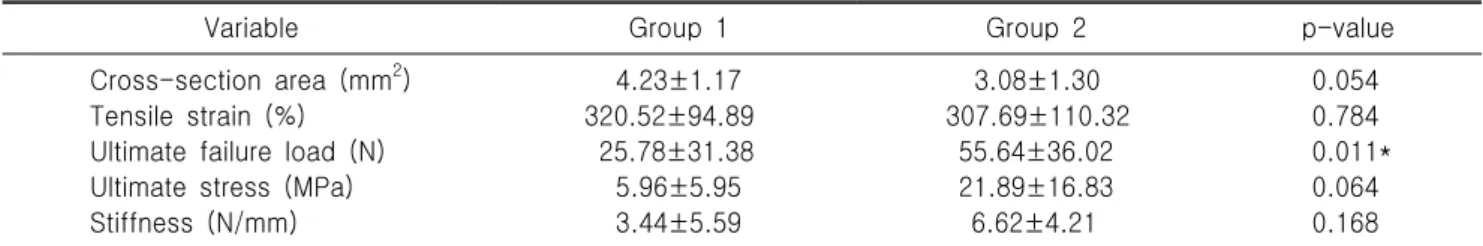
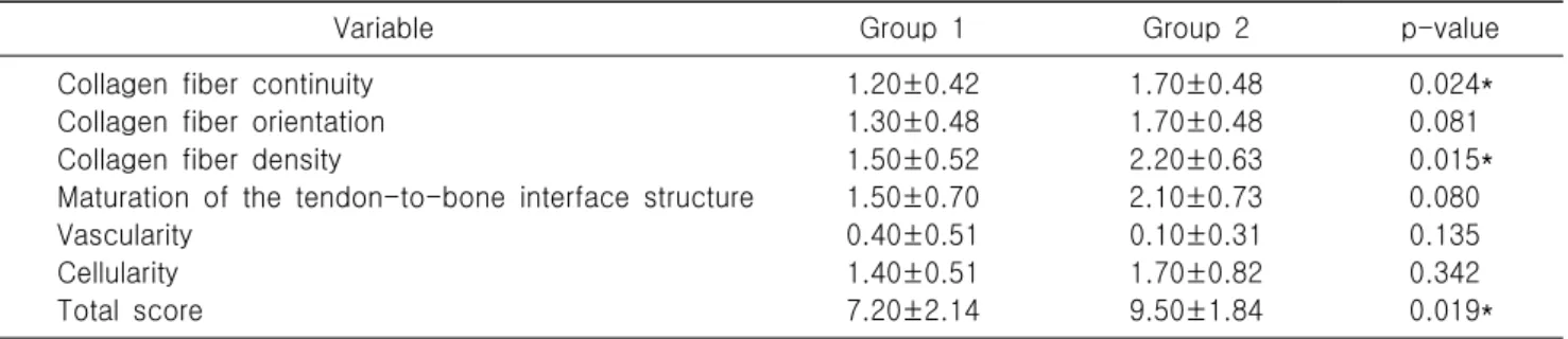
(4) DH Kim, et al. Effect of KGN on the Tendon-Bone Interface in a Rat Rotator Cuff Tear Model. ruptured tendon, grade 1 indicated a poorer; grade 2 indicated. Results. a better grade, and grade 3 indicated a marked regeneration. Overall, the total score of a slide could range from 0 (ruptured tendon). 1. Gross inspection and biomechanical evaluation. 14. to 18 (most marked regeneration) . To eliminate observer error, all tissue slides were evaluated by a pathologist with at least. Gross inspection showed no retear of the supraspinatus. 10 years of experience in a randomized, blinded fashion. All. tendon-to-bone sites in both groups (p=0.531 at 8 weeks) (Table. tissue slides were examined three times in the same position. 1). However, in the biomechanical analysis, the prevalence of. and area of rotator cuff tissue by microscope (Leica DMIL LED;. midsubstance tear was significantly higher in group 1 (p=0.01. Leica, Wetzlar, Germany) and image system (LAS V4.8; Leica). at 8 weeks). Group 1 exhibited a greater tensile strain (group. at ×50 magnification.. 1: 320.52±94.89%, group 2: 307.69±110.32%; p=0.784) and 2. cross-section area (group 1: 4.23±1.17 mm , group 2: 3.08±1.30. 6. Statistical analysis. mm2; p=0.054) compared with group 1. However, there were. All statistical analyses were performed using SPSS ver. 12.0. no significant differences between the groups. Group 2 exhibited. software (SPSS Inc., Chicago, IL, USA). A p-value less than. a greater ultimate failure load (group 1: 25.78±31.38 N, group. 0.05 indicated statistical significance. The Kruskal-Wallis test. 2: 55.64±36.02 N; p=0.011), ultimate stress (group 1: 5.96±5.95. and post-hoc Mann-Whitney U-test were performed to evaluate. N, group 2: 21.89±16.83 N; p=0.064), and stiffness (group 1:. the biomechanical and histological differences based on H&E. 3.44±5.59 N/mm, group 2: 6.62±4.21 N/mm; p=0.168). However,. staining between groups. Data are presented as the mean and. the ultimate failure load showed a significant difference (Table. standard deviation. Interclass correlation coefficients were used. 2).. to assess intra- and interobserver reliability for histological analysis of the fat-to-muscle ratio.. 2. Histological evaluation In the results of H&E staining (magnification, ×50), Masson’s trichrome staining, and picrosirius red staining (Fig. 2), group 2 exhibited a significantly greater total score at 8 weeks (group. Table 1. Findings on gross inspection Variable. Group 1 (n=10). Group 2 (n=10). Retear Bone-to-tendon failure Midsubstance failure. 0 3 7. 0 9 1. 1: 7.20±2.14, group 2: 9.50±1.84; p=0.019) compared with group p-value. 1. Collagen fiber continuity (group 1: 1.20±0.42, group 2:. 0.531. 1.70±0.48; p=0.024) and collagen fiber density (group 1:. 0.01*. 1.50±0.52, group 2: 2.20±0.63; p=0.080) were significantly different in both groups. However, collagen fiber orientation (group. Group 1: supraspinatus repair only, group 2: supraspinatus repair with kartogenin single injection. *Statistically significant.. 1: 1.30±0.48, group 2: 1.70±0.48; p=0.081), maturation of the tendon-to-bone interface structure (group 1: 1.30±0.48, group 2:. Table 2. Comparison of the biomechanical characteristics of both groups Variable. Group 1 2. Cross-section area (mm ) Tensile strain (%) Ultimate failure load (N) Ultimate stress (MPa) Stiffness (N/mm). 4.23±1.17 320.52±94.89 25.78±31.38 5.96±5.95 3.44±5.59. Group 2. p-value. 3.08±1.30 307.69±110.32 55.64±36.02 21.89±16.83 6.62±4.21. 0.054 0.784 0.011* 0.064 0.168. Values are presented as mean±standard deviation. Group 1: supraspinatus repair only, group 2: supraspinatus repair with kartogenin single injection. *Statistically significant. 220. 대한스포츠의학회지.
(5) 김동현 외. 쥐 회전근개 파열 모델의 건 부착부에서 카토제닌 주사의 효과. Fig. 2. Histological evaluation at 8 weeks after repair. Group 1 received only supraspinatus repair; group 2 received supraspinatus repair with a single injection of kartogenin. (A) H&E staining, ×50. (B) Masson trichrome staining, ×50. (C) Picrosirius red staining, ×50. Continuity, orientation, and density of collagen fiber showed a higher average grading in group 2 compared with group 1. Group 2 showed better maturation and density of the tendon-to-bone interface structure (arrows) than group 1. Table 3. Scoring of findings on histological analysis Variable Collagen fiber continuity Collagen fiber orientation Collagen fiber density Maturation of the tendon-to-bone interface structure Vascularity Cellularity Total score. Group 1. Group 2. p-value. 1.20±0.42 1.30±0.48 1.50±0.52 1.50±0.70 0.40±0.51 1.40±0.51 7.20±2.14. 1.70±0.48 1.70±0.48 2.20±0.63 2.10±0.73 0.10±0.31 1.70±0.82 9.50±1.84. 0.024* 0.081 0.015* 0.080 0.135 0.342 0.019*. Values are presented as mean±standard deviation. Group 1: supraspinatus repair only, group 2: supraspinatus repair with kartogenin single injection. *Statistically significant.. 1.70±0.48; p=0.080), vascularity (group 1: 0.40±0.51, group 2:. between groups 1 and 2 (Table 3). The metachromasia area in. 0.10±0.31; p=0.135), and cellularity (group 1: 1.40±0.51, group. toluidine blue staining was measured to confirm fibrocartilage. 2: 1.70±0.82; p=0.342) demonstrated no significant differences. at the tendon-to-bone junction. In group 2, the metachromasia. 제38권 제4호 2020. 221.
(6) DH Kim, et al. Effect of KGN on the Tendon-Bone Interface in a Rat Rotator Cuff Tear Model. Fig. 3. Histological evaluation at 8 weeks after repair. (A) Group 1 received only supraspinatus repair; (B) group 2 received supraspinatus repair with a single injection of kartogenin. The arrows indicate the tendon-to-bone interface. This area is the supraspinatus enthesis, and fibrocartilaginous transition site and was stained with Toluidine blue (×50). The metachromasia were more intense in the supraspinatus enthesis of group 2 compared to the control group.. appeared stronger and higher compared with group 1 (Fig. 3).. 17. rotator cuff tear model. Wang et al. evaluated the effect of KGN in augmenting healing of the repaired enthesis after rotator cuff repair in a murine model. They reported that superior collagen. Discussion. fiber organization and higher ultimate failure loads were seen The purpose of this study was to investigate the effect of. in the KGN group at 4 weeks. Huang et al.18 compared the effects. KGN on tendon-bone healing in a rat rotator cuff tear model.. of repair only (control), microfracture+repair, and microfracture+. In this study, the KGN group showed more improved biomechanical. repair augmentation with a KGN-loaded gelatin methacryloyl. properties and a histologically better healing quality in the. (GelMA) hydrogel scaffold (combined) in tendon-to-bone healing. 15. bone-to-tendon interface than the control group. Johnson et al.. in a rabbit rotator cuff tear model. The authors reported that. reported that KGN, a small synthetic heterocyclic compound,. the KGN-loaded GelMA hydrogel scaffolds group exhibited greater. was found to promote robust chondrogenic differentiation of. ultimate load to failure and stiffness than the other groups at. primary human mesenchymal stem cells. After that, when KGN. 8 and 12 weeks after repair. In histological evaluation, the tendon. was injected intra-articularly into a mouse model of osteoarthritis,. maturation score of the KGN-loaded GelMA hydrogel scaffolds. regeneration of the cartilage and prevention of joint degeneration. group was significantly higher than that of the other groups at. 11,16. was observed. 9. . In 2014, Zhang et al. reported that when injected. 8 and 12 weeks after repair.. into intact rat patellar tendons and injured rat Achilles in vivo,. Our results were similar to previous studies. The tendons of. KGN induced cartilage-like tissue formation in the tendon-to-bone. the KGN group showed a greater mean ultimate load to failure. 9,13. have studied the use of KGN in. than the control group, and KGN treatment appeared to enhance. combination with platelet-rich plasma to facilitate fibrocartilage. the formation and organization of collagen fibrils at the healing. formation at the interface between tendon and bone of rat Achilles. enthesis. This suggests that KGN has significant effects on collagen. tendons. In their studies, KGN treatment was found to enhance. formation and organization at the interface between tendon and. production of collagens I and II and increase expression of Sox-9. bone, even in a rotator cuff tear model. In addition, the two. and scleraxis, which is consistent with the formation of. previous studies used scaffolds to minimize the diffusion of KGN. junctions. Zhang et al.. 13. fibrocartilage-like tissue . In addition, KGN treatment resulted. into surrounding tissues and maximize the retainment of KGN.. in more organized collagen fibers and chondrocytes at the tissue. However, in our study, only a single injection of KGN was applied. interface, and KGN-treated tendon-to-bone constructs demon-. in KGN group. Compared with the control group, there was. strated greater mean ultimate strengths compared with the control. significant biomechanical and histological improvement in KGN. 9,13. group . Two previous studies reported on the effects of KGN in a 222. 대한스포츠의학회지. group. Clinically single injection of the drug is the simplest method that can be further performed for healing after rotator cuff repair..
(7) 김동현 외. 쥐 회전근개 파열 모델의 건 부착부에서 카토제닌 주사의 효과. It is considered important information on the usefulness of KGN. degenerative rotator cuff tears observed in most clinical patients.. to show a significant difference in effect through the simplest. In conclusion, a single-dose injection of KGN reinforces the. method. Therefore, we suggest that our research can provide basic. biomechanical and histological properties at the tendon-to-bone. information in other studies based on KGN.. interface in a rat rotator cuff tear model. Our study confirmed. In our study, the metachromasia area in toluidine blue staining. that KGN improves tissue regeneration and mechanical strength. was measured to confirm fibrocartilage at the tendon-to-bone. of rotator cuff tear. In previous studies, KGN has been widely. junction. In the KGN group, the metachromasia appeared stronger. evaluated as a remedy for cartilage regeneration, but the results. and higher than in the control group. This suggests that KGN,. of the current study confirm the tendon regeneration effect in. which induces cartilage differentiation, promotes the production. a rotator cuff tear model.. 18. of fibrocartilage. Huang et al. also reported better fibrocartilage formation in the KGN group at 4, 8, and 12 weeks in toluidine. Conflict of Interest. blue staining compared with the control and microfracture+repair 17. groups. However, Wang et al. reported that the mean percentage of area of fibrocartilage at the tendon-to-bone interface in Alcian. No potential conflict of interest relevant to this article was reported.. blue staining was higher in the control group compared with the KGN group at 4 weeks. Regarding this difference, it seems. ORCID. that the histological evaluation system of fibrocartilage formation is semiquantitative and difficult to quantify. In the rotator cuff. Dong Hyun Kim https://orcid.org/0000-0001-9078-5953. tear model, a significant increase in fibrocartilage formation can. Seung Gi Min. https://orcid.org/0000-0003-4343-1022. be observed at least 8 weeks after KGN injection.. Hun‐Min Kim. https://orcid.org/0000-0003-4447-6230. It is believed that our study can be a foundation for future. Jin‐Hyun Choi. https://orcid.org/0000-0002-5548-1230. research on the use of KGN in rotator cuff tears. In previous. Hyun Joo Lee. https://orcid.org/0000-0003-2837-3434. cartilage defect models, various modalities such as chitosan nano-. Kyeong Hyeon Park https://orcid.org/0000-0001-7215-6176. 16. and microparticles , photo-cross-linked scaffold with KGN-. Jong Pil Yoon. https://orcid.org/0000-0001-6446-6254. 19. encapsulated nanoparticles , and polyethylene glycol-modified 20. poly (amidoamine) dendrimer have been used to enhance the. Author Contributions. effects of KGN. Future research will require evaluation of other formulations that can improve the effects of KGN in a rotator cuff tear model. The current study had several limitations. First, in the. Conceptualization: HMK. Data curation: SGM. Methodology: HJL. Visualization: JHC, KHP. Writing– original draft: DHK. Writing– review & editing: JPY.. experimental group, only a single dose of KGN was applied at the concentration suggested in a previous experiment. Therefore,. References. it is not possible to know the dose-dependent effect of KGN, and information on the optimal concentration of it is not known. Second, because the grading system applied for histological evaluation was semiquantitative, there is a disadvantage that it is not objectively reliable. Third, the 8-week evaluation period may not be long enough for the final analysis, as there may be further change after 8 weeks. Further study with a longer evaluation period may be needed. Fourth, our tendon damage method is more like an acute rotator cuff tear than the chronic. 1. Yamaguchi K, Ditsios K, Middleton WD, Hildebolt CF, Galatz LM, Teefey SA. The demographic and morphological features of rotator cuff disease: a comparison of asymptomatic and symptomatic shoulders. J Bone Joint Surg Am 2006;88:1699-704. 2. Wang VM, Wang FC, McNickle AG, et al. Medial versus lateral supraspinatus tendon properties: implications for doublerow rotator cuff repair. Am J Sports Med 2010;38:2456-63. 3. Le BT, Wu XL, Lam PH, Murrell GA. Factors predicting. 제38권 제4호 2020. 223.
(8) DH Kim, et al. Effect of KGN on the Tendon-Bone Interface in a Rat Rotator Cuff Tear Model. 4.. 5.. 6.. 7.. 8.. 9. 10. 11.. 12.. rotator cuff retears: an analysis of 1000 consecutive rotator cuff repairs. Am J Sports Med 2014;42:1134-42. Castricini R, Longo UG, De Benedetto M, et al. Platelet-rich plasma augmentation for arthroscopic rotator cuff repair: a randomized controlled trial. Am J Sports Med 2011;39:25865. Wang LL, Yin XF, Chu XC, Zhang YB, Gong XN. Platelet-derived growth factor subunit B is required for tendon-bone healing using bone marrow-derived mesenchymal stem cells after rotator cuff repair in rats. J Cell Biochem 2018;119:8897-908. Yonemitsu R, Tokunaga T, Shukunami C, et al. Fibroblast growth factor 2 enhances tendon-to-bone healing in a rat rotator cuff repair of chronic tears. Am J Sports Med 2019; 47:1701-12. Liu Q, Yu Y, Reisdorf RL, et al. Engineered tendonfibrocartilage-bone composite and bone marrow-derived mesenchymal stem cell sheet augmentation promotes rotator cuff healing in a non-weight-bearing canine model. Biomaterials 2019;192:189-98. Wang C, Hu Q, Song W, Yu W, He Y. Adipose stem cellderived exosomes decrease fatty infiltration and enhance rotator cuff healing in a rabbit model of chronic tears. Am J Sports Med 2020;48:1456-64. Zhang J, Wang JH. Kartogenin induces cartilage-like tissue formation in tendon-bone junction. Bone Res 2014;2:14008. Im GI. Application of kartogenin for musculoskeletal regeneration. J Biomed Mater Res A 2018;106:1141-8. Mohan G, Magnitsky S, Melkus G, et al. Kartogenin treatment prevented joint degeneration in a rodent model of osteoarthritis: a pilot study. J Orthop Res 2016;34:1780-9. Xu X, Shi D, Shen Y, et al. Full-thickness cartilage defects. 224. 대한스포츠의학회지. 13.. 14.. 15. 16.. 17.. 18.. 19.. 20.. are repaired via a microfracture technique and intraarticular injection of the small-molecule compound kartogenin. Arthritis Res Ther 2015;17:20. Zhang J, Yuan T, Zheng N, Zhou Y, Hogan MV, Wang JH. The combined use of kartogenin and platelet-rich plasma promotes fibrocartilage formation in the wounded rat Achilles tendon entheses. Bone Joint Res 2017;6:231-44. Kim DH, Min SG, Yoon JP, et al. Mechanical augmentation with absorbable alginate sheet enhances healing of the rotator cuff. Orthopedics 2019;42:e104-10. Johnson K, Zhu S, Tremblay MS, et al. A stem cell-based approach to cartilage repair. Science 2012;336:717-21. Kang ML, Ko JY, Kim JE, Im GI. Intra-articular delivery of kartogenin-conjugated chitosan nano/microparticles for cartilage regeneration. Biomaterials 2014;35:9984-94. Wang D, Tan H, Lebaschi AH, et al. Kartogenin enhances collagen organization and mechanical strength of the repaired enthesis in a murine model of rotator cuff repair. Arthroscopy 2018;34:2579-87. Huang C, Zhang X, Luo H, et al. Effect of kartogenin-loaded gelatin methacryloyl hydrogel scaffold with bone marrow stimulation for enthesis healing in rotator cuff repair. J Shoulder Elbow Surg 2020 Jul 7 [Epub]. https://doi.org/10.1016/ j.jse.2020.06.013. Shi D, Xu X, Ye Y, et al. Photo-cross-linked scaffold with kartogenin-encapsulated nanoparticles for cartilage regeneration. ACS Nano 2016;10:1292-9. Hu Q, Ding B, Yan X, et al. Polyethylene glycol modified PAMAM dendrimer delivery of kartogenin to induce chondrogenic differentiation of mesenchymal stem cells. Nanomedicine 2017;13:2189-98..
(9)
수치


관련 문서
This report detailed the cases of 3 patients who experienced shoulder pain caused by rotator cuff tear with SASD bursitis despite conservative treatment and their
A number of animal studies 6,22-24) not only support the use of BMAC to augment tendon repair by improv- ing enthesis regeneration but also highlight the function of BMAC to
For biomechanical tests, the tibia with affixed tendon was posted in a MTS machine for measurement of maximal pull-out load (interface strength). At 4 and 8 weeks the
광범위 회전근 개 파열(massive rotator cuff tear)이란 일반적으로 2개 이상의 건의 전층 파열 또는 파열된 단면 부위의 최대 직경이 5 cm 이상인 경우로 정의되고 있다.
In this in vivo experiment on rat calvarial bone defect, histological analyses showed that Danshen and Ge Gan ex- tractions increased bone formation activity when used with
Purpose: The causes of rotator cuff tendon tear were excessive overuse, aging process and impingement syndrome and a lot of research of these factors have been performed. But
03) Gonzalez D and Lopez RA: Concurrent rotator cuff tear and brachial plexus palsy associated with anterior dislocation of the shoulder. A report of two cases. J Bone Joint
