Herbal medicines have been used for centuries to treat a variety of diseases and conditions including promotion of fractured bone healing and bone diseases such as osteoporo- sis7. Puerariae radix, also known as Ge Gen, comes from the root of Pueraria lobata; this commonly used traditional medi- cine has been demonstrated to decrease loss in bone density8 as well as treat fever9, liver diseases10, and cardiovascular disease11. Danshen or Radix salvia miltiorrhiza is another common traditional medicine that has been used to treat car- diovascular disease, improve perfusion of ischemic myocar- dium, and enhance blood circulation12-14. Several studies have examined the effectiveness of Danshen in promoting healing in bone fracture15,16.
Like aspirin (from leaves of the willow tree, nonsteroidal antiinflammatory drug), Taxol (from the cortex of Taxus brevifolia, chemotherapy drug), and Stillen (from leaves of Artemisia argyi, antiulcerant), several studies using herbal medicine have been and are still being performed. The aim of this study was to evaluate the osteogenic capacity of Ge Gen and Danshen extracts and to establish a screening system of
I. Introduction
Bone defects are caused by tumors, cysts, infections, trau- ma, and aging. They play a significant role in mastication, as well as in esthetics. Slow bone regeneration in such defects can pose therapeutic problems. Various methods are currently used to obtain adequate defect closure including autogenous bone grafting, allogenic bone grafting, and application of growth factors, polymeric membranes, and enamel matrix derivatives1-6.
Il-Kyu Kim
Oral and Maxillofacial Surgery, Department of Dentistry, Inha University Hospital, Inha University School of Medicine, 27 Inhang-ro, Jung-gu, Incheon 22332, Korea
TEL: +82-32-890-2470 FAX: +82-32-890-2475 E-mail: [email protected]
ORCID: http://orcid.org/0000-0003-3930-766X
This is an open-access article distributed under the terms of the Creative Commons Attribution Non-Commercial License (http://creativecommons.org/
licenses/by-nc/4.0/), which permits unrestricted non-commercial use, distribution, and reproduction in any medium, provided the original work is properly cited.
CC
Effect of herbal extracts on bone regeneration in a rat calvaria defect model and screening system
Dong-Hwan Lee1, Il-Kyu Kim1, Hyun-Young Cho1, Ji-Hoon Seo1, Jun-Min Jang1, Jin Kim2
1Oral and Maxillofacial Surgery, Department of Dentistry, Inha University School of Medicine, Incheon,
2Oral Cancer Research Institute and Department of Oral Pathology, Yonsei University College of Dentistry, Seoul, Korea
Abstract(J Korean Assoc Oral Maxillofac Surg 2018;44:79-85)
Objectives: The aim of this study was to evaluate the effects of herbal extracts on bone regeneration. Two known samples were screened.
Materials and Methods: We previously established a rat calvaria defect model using a combination of collagen scaffold and herbal extracts. An 8 mm diameter trephine bur with a low-speed dental hand piece was used to create a circular calvaria defect. The experimental group was divided into 4 classifications: control, collagen matrix, Danshen with collagen, and Ge Gan with collagen. Animals in each group were sacrificed at 4, 6, 8, and 10 weeks after surgery, and bone regeneration ability was evaluated by histological examination.
Results: Results revealed that both Danshen and Ge Gan extracts increased bone formation activity when used with collagen matrix. All groups showed almost the same histological findings until 6 weeks. However, after 6 weeks, bone formation activity proceeded differently in each group. In the experimental groups, new bone formation activity was found continuously up to 10 weeks. In the Danshen and Ge Gan groups, grafted materials were still present until 10 weeks after treatment, as evidenced by foreign body reactions showing multinucleated giant cells in chronic inflammatory vascular connective tissue.
Conclusion: Histological analyses showed that Danshen and Ge Gan extractions increased bone formation activity when used in conjunction with collagen matrix.
Key words: Bone regeneration, Herb extract, Danshen, Ge Gan, Rat calvaria defect
[paper submitted 2017. 11. 23 / revised 2018. 1. 3 / accepted 2018. 1. 3]
Copyright © 2018 The Korean Association of Oral and Maxillofacial Surgeons. All rights reserved.
The defects were grafted with different materials. In the Danshen and Ge Gan groups, the defects were filled with a collagen matrix containing Danshen or Ge Gan extract. In the collagen group, the defects were filled with collagen matrix containing saline; in the control group, the defects were left untreated. The graft materials were prepared 15 minutes be- fore grafting.
All experimental areas were closed with 4.0 black surgical silks. Postoperatively, all rats received an intramuscular injec- tion of 25,000 IU/kg penicillin G once daily for 3 days.
4. Histological preparation
Animals were sacrificed by CO2 inhalation at 4, 6, 8, and 10 weeks after surgery. Defects and surrounding tissues were removed for histological preparation and immediately fixed by immersion in 10% neutral buffered formalin.
The specimens was dehydrated in 70% ethanol and then embedded in methacrylate-based resin. Polymerization of the embedding solution occurred in a photopolymerization unit (EXAKT Apparatebau, Norderstedt, Germany) with exposure to daylight for 2 hours and to ultraviolet light for 10 hours.
Each polymerized block was affixed to the vacuum head of the EXAKT macrocutter, and sections were sliced to a thick- ness of approximately 100 µm. Sections were ground and polished using the EXAKT micro grinder to a thickness of 15 µm, mounted on microscope slides, stained with hematoxy- lin and eosin, and coverslipped. Bone healing patterns were observed under a light microscope (BX50; Olympus, Tokyo, Japan).
III. Results
In total, 4 study groups underwent microscopic evaluation based on the injected biomaterial. Each group was tested at four time intervals, and none of them showed complete fill- ing of the defect with new bone.
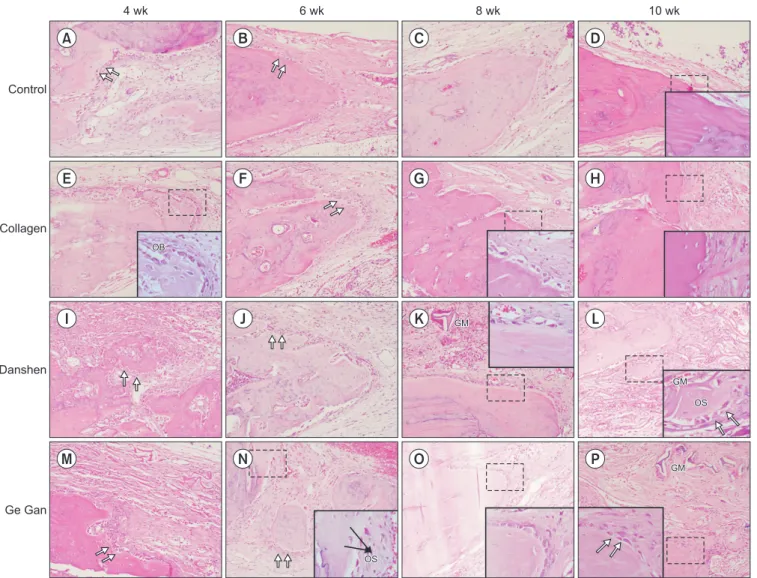
In all groups, the 4 and 6 week time points showed almost the same histological findings. Activated osteoblasts rim- ming newly formed woven bone (Fig. 1; double arrows) were observed in the bone repair areas. Osteocytes were clearly distinguishable at the inner parts of newly formed bone, indi- cating direct bone repair. The presence of granulation tissue was characterized by numerous newly formed small blood vessels, an intense inflammatory cell infiltrate, and immature connective tissue with large amounts of fibroblasts and im- mature collagen fiber bundles in the center of the bone defect herbal medicines.
II. Materials and Methods
1. Experimental materials and animals
Forty-eight Sprague-Dawley male rats, 8 weeks old, weighing 250 g were selected for the study. Rat size increases rapidly during this stage, providing a favorable model for studying normal bone healing. Before the study, the animals were housed in an environmentally controlled animal facility for 1 week for acclimatization.
An 8 mm diameter trephine bur with a low-speed dental hand piece was used to create circular calvaria defects. Col- lagen membranes (Rapi-Plug; Dalim Tissen, Seoul, Korea) were used to fill the defects in the experimental group.
Animal selection and management, surgical protocol, and preparation followed routines approved by the Institutional Animal Care and Use Committee of Inha University (INHA 160802-426).
2. Preparation of herb extraction
Crude Danshen and Ge Gan were extracted with 70%
methanol reflux for 3 hours at 70°C. The liquid/solid ratio was 1:10. The extracts were lyophilized and sealed in steril- ized tubes. Before starting the experiment, 4 mL distilled water was added to 4 g of each lyophilized powder. The col- lagen matrix was soaked for 15 minutes before grafting in the Danshen and Ge Gan groups.
3. Surgical procedure
Surgery was conducted on all rats under sterile conditions.
General anesthesia was induced by intramuscular injection of 10 mg/kg xylazine HCl and 40 mg/kg ketamine HCl and was maintained by injection of the same mixture in half doses.
Rat skulls were shaved and sterilized using 70% ethanol. An injection of 2% lidocaine HCl 1.8% containing 1:100,000 epinephrine was administrated locally at the operation site to reduce bleeding.
The bone surface of the calvaria was exposed by a 3 cm longitudinal midline incision along the sagittal suture. In each parietal bone, a standardized round defect was created using an 8 mm diameter trephine bur in a low-speed hand piece. To prevent overheating of the bone, the operation site was irri- gated continuously with sterile saline solution.
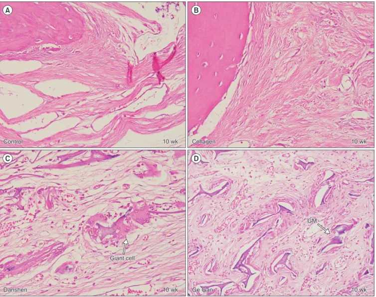
for each group. For example, in the collagen group, only one section showed graft material at 10 weeks; most of them totally resorbed in 6 weeks. At 10 weeks, the collagen group showed a defect area filled with fibroblastic dense colla- gen tissue without inflammation. However, in the Danshen and Ge Gan groups, grafted materials were present until 10 weeks, as evidenced by a foreign body reaction showing multinucleated giant cells in chronic inflammatory vascular connective tissue.(Fig. 2)
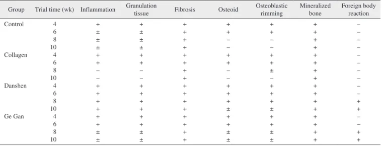
These results were divided into seven classifications and are summarized in Table 1.
IV. Discussion
The demand for use of alloplastic materials for bone repair is increasing since there are limitations in grafting autogenous area.
Interestingly, bone formation activity was attenuated at dif- ferent time points in each group. In the control group, bone formation was reduced at 6 weeks. Fibroblast and collagen fiber volume density significantly increased in the first 6 weeks, followed by a reduction in the subsequent periods (8 and 10 weeks). In the collagen group, activated osteoblasts rimming newly formed woven bone were seen at 6 weeks, but from 8 weeks on, peripheral osteoid and osteoblastic rim- ming were markedly reduced. By 10 weeks, a mineralized bone margin was found without osteoblastic rimming. In contrast, in the Danshen and Ge Gan groups, osteoid forma- tion with osteoblasts rimming was found continuously in the trabecular bone surface at the edges and the center of the de- fect at 8 and 10 weeks, respectively.(Fig. 1)
Grafted biomaterials were resorbed at different time points
Control
Collagen
Danshen
Ge Gan
4 wk 6 wk 8 wk 10 wk
OB OB
OS OS GM GM GM
GM
GM GM
OS OS
A B C D
E F G H
I J K L
M N O P
Fig. 1. Representative H&E sections depicting the new bone formation at 4, 6, 8, and 10 weeks for each groups (H&E staining, A-P: ×200, inset: ×1,000). (OB: osteoblasts [double arrows], GM: grafted material, OS: osteoid)
Dong-Hwan Lee et al: Effect of herbal extracts on bone regeneration in a rat calvaria defect model and screening system. J Korean Assoc Oral Maxillofac Surg 2018
and microcirculation is crucial19; this process is known as an- giogenesis. The formation of new vasculature transports oxy- gen and nutrients to bone tissues that can differentiate into os- teoblasts, chondroclasts, and osteoclasts20. Many experiments have shown that endothelial cells provide a microvasculature during bone remodeling, and this angiogenesis is necessary for bone formation21,22. Hence, the processes of osteogenesis and angiogenesis are closely correlated. Danshen and Ge Gan have both been reported to have angiogenesis and osteogen- esis activity23.
Danshen, or Radix salvia miltiorrhiza, is a common tra- ditional medicine that has been used to treat cardiovascular disease, improve perfusion of ischemic myocardium, and enhance blood circulation. Several studies have examined the bone. Autogenous bone grafts require additional surgery and
are associated with donor site morbidity, including infection, pain, and hematoma17. To develop more effective bone graft materials with enhanced osteogenic properties, alloplastic materials are combined with osteoinductive substances.
Many in vitro and in vivo studies have shown the positive effects of herbs on bone formation by the promotion of osteo- blastic proliferation and inhibition of osteoclastic formation18. However, questions regarding, mechanism of action, main compounds, and experimental systems remain. Thus, it is worth evaluating herbal extracts to analyze their osteogenic enhancement properties.
Bone is a highly vascularized tissue. To maintain homeo- stasis and regeneration, the development of microvasculature
10 wk
10 wk
10 wk
10 wk Control
Control CollagenCollagen
Danshen
Danshen Ge GanGe Gan
GM GM
Giant cell Giant cell
A B
C D
Fig. 2. Representative H&E section depicting healing process in defect area at 10-week trial (H&E staining, A-D: ×400). A, B. Fibroblastic dense collagen tissue without inflammation in control and collagen group. C, D. Grafted materials (GMs) are surrounded by foreign body reaction showing multinucleated giant cells in chronic inflammatory vascular connective tissue in Danshen and Ge Gan groups, respectively.
Dong-Hwan Lee et al: Effect of herbal extracts on bone regeneration in a rat calvaria defect model and screening system. J Korean Assoc Oral Maxillofac Surg 2018
Gan groups, bone formation was found at 10 weeks. This comparison showed that Danshen and Ge Gan extractions increased bone formation activity when used with a collagen matrix. The mechanism of this effect is likely angiogenesis.
However, in the Danshen and Ge Gan groups, grafted materi- als were still present at 10 weeks, as evidenced by foreign body reaction showing multinucleated giant cells in chronic inflammatory vascular connective tissue. Thus, angiogenesis mechanisms cannot rule out the inflammation effect, and fur- ther study is needed to understand the mechanism of inflam- matory vascular connective tissue.
Pryor et al.28 evaluated the validity of radiographic evalu- ations of bone formation in a critical-size rat calvaria oste- otomy defect model. Bilateral, calvaria osteotomy defects in rats treated with a platelet-rich plasma preparation or control treatments were evaluated by radiographic and histometric measures following a 4- or 8-week healing interval. They re- ported low accuracy of radiographic evaluations employed in identifying and characterizing bone filling in the rat calvaria osteotomy defects. Nyan et al.29 treated rat calvaria defects using calcium sulfate and simvastatin. They observed the smallest amount of bone formation at 2 and 4 weeks but re- markable bone formation at 8 weeks. According to their find- ings, bone formation in the experimental group seemed to be delayed as a result of soft tissue inflammation, but graft ma- terials appeared to promote bone formation in spite of the in- flammatory response. Yoon et al.30 implanted adipose-derived stem cells (ADSCs) with polymer constructs in rat calvaria defect. They reported that, at 12 weeks post-implantation, effectiveness of Danshen in promoting osteogenesis. Chin
et al.24 reported an in vivo study showing that Danshen en- hanced bone remodeling by regulating the gene expression of alkaline phosphatase (ALP), osteocalcin (OCN), osteoprote- gerin (OPG), and receptor activator of nuclear factor kappa-Β ligand (RANKL). An in vivo study by Wong and Rabie16 found that Danshen significantly promoted new bone forma- tion when an extract in collagen matrix was placed in bone defects. Lay et al.25 showed that Danshen extract and salvi- anolic acid B (a component of Danshen) enhanced angiogen- esis in a murine SVEN 1 ras (SVR) endothelial cell line by up-regulating vascular endothelial growth factor (VEGF) and VEGF-R2 gene expression.
Ge Gan or Puerariae radix comes from the root of Pueraria lobata and has been shown to have effects on decreasing loss in bone density. Wang et al.8 examined the role of Ge Gan in bone metabolism by feeding OVX mice a diet containing different doses of the herb. They suggest that Ge Gan may prevent osteoporosis in post-menopausal women. Huh et al.26 found that Ge Gan increased the mRNA expression of VEGF and dose-dependently increased ALP activity related to os- teoblastic activity. Zhang et al.27 found that Ge Gan was able to induce angiogenesis in an ischemic myocardial infarction model.
In this study, we observed several interesting results. Bone formation activity was attenuated at different time points in each group. In the control group, bone formation was reduced at 6 weeks. In the collagen group, osteoid and osteoblastic rimming were found until 8 weeks. In the Danshen and Ge
Table 1. Histological findings at 4, 6, 8, and 10 weeks for each groups Group Trial time (wk) Inflammation Granulation
tissue Fibrosis Osteoid Osteoblastic
rimming
Mineralized bone
Foreign body reaction Control
Collagen
Danshen
Ge Gan
4 6 8 10
4 6 8 10 4 6 8 10 4 6 8 10
+
±
±
± + + – – + + + + + +
±
±
+
±
±
± + + – – + + + + + +
±
±
+ + + + + + + + + + + + + + + +
+ + – – + + – – + + +
± + +
±
±
+ + – – + +
± – + + +
± + +
±
±
+ + + + + + + + + + + + + + + +
– – – – – – – – – – + + – – + + (+: positive, –: negative)
Dong-Hwan Lee et al: Effect of herbal extracts on bone regeneration in a rat calvaria defect model and screening system. J Korean Assoc Oral Maxillofac Surg 2018
reported.
References
1. Park JC, Kim YH, Choi HS, Oh JS, Shin SH, Kim YD. The rate and stability of mandibular block bone graft in recent 5 years.
Maxillofac Plast Reconstr Surg 2017;39:21.
2. Burg KJ, Porter S, Kellam JF. Biomaterial developments for bone tissue engineering. Biomaterials 2000;21:2347-59.
3. Gomes KU, Carlini JL, Biron C, Rapoport A, Dedivitis RA. Use of allogeneic bone graft in maxillary reconstruction for installation of dental implants. J Oral Maxillofac Surg 2008;66:2335-8.
4. Peng W, Kim IK, Cho HY, Seo JH, Lee DH, Jang JM, et al. The healing effect of platelet-rich plasma on xenograft in peri-implant bone defects in rabbits. Maxillofac Plast Reconstr Surg 2016;38:16.
5. Lekovic V, Camargo PM, Weinlaender M, Nedic M, Aleksic Z, Kenney EB. A comparison between enamel matrix proteins used alone or in combination with bovine porous bone mineral in the treatment of intrabony periodontal defects in humans. J Periodontol 2000;71:1110-6.
6. Warnke PH, Springer IN, Wiltfang J, Acil Y, Eufinger H, Wehm- öller M, et al. Growth and transplantation of a custom vascularised bone graft in a man. Lancet 2004;364:766-70.
7. Wang ZQ, Li JL, Sun YL, Yao M, Gao J, Yang Z, et al. Chinese herbal medicine for osteoporosis: a systematic review of random- ized controlled trails. Evid Based Complement Alternat Med 2013;2013:356260.
8. Wang X, Wu J, Chiba H, Umegaki K, Yamada K, Ishimi Y. Puerar- iae radix prevents bone loss in ovariectomized mice. J Bone Miner Metab 2003;21:268-75.
9. Chueh FS, Chang CP, Chio CC, Lin MT. Puerarin acts through brain serotonergic mechanisms to induce thermal effects. J Phar- macol Sci 2004;96:420-7.
10. Overstreet DH, Lee YW, Rezvani AH, Pei YH, Criswell HE, Janowsky DS. Suppression of alcohol intake after administration of the Chinese herbal medicine, NPI-028, and its derivatives. Alcohol Clin Exp Res 1996;20:221-7.
11. Wang LY, Zhao AP, Chai XS. Effects of puerarin on cat vascular smooth muscle in vitro. Zhongguo Yao Li Xue Bao 1994;15:180-2.
12. Chang PN, Mao JC, Huang SH, Ning L, Wang ZJ, On T, et al.
Analysis of cardioprotective effects using purified Salvia miltior- rhiza extract on isolated rat hearts. J Pharmacol Sci 2006;101:245- 13. Cheng TO. Cardiovascular effects of Danshen. Int J Cardiol 9.
2007;121:9-22.
14. Zhou L, Zuo Z, Chow MS. Danshen: an overview of its chemistry, pharmacology, pharmacokinetics, and clinical use. J Clin Pharma- col 2005;45:1345-59.
15. Liu YR, Qu SX, Maitz MF, Tan R, Weng J. The effect of the major components of Salvia Miltiorrhiza Bunge on bone marrow cells. J Ethnopharmacol 2007;111:573-83.
16. Wong RW, Rabie AB. Effect of Salvia miltiorrhiza extract on bone formation. J Biomed Mater Res A 2008;85:506-12.
17. Younger EM, Chapman MW. Morbidity at bone graft donor sites. J Orthop Trauma 1989;3:192-5.
18. Wong RW, Rabie AB. Traditional Chinese medicines and bone formation--a review. J Oral Maxillofac Surg 2006;64:828-37.
19. Schmid J, Wallkamm B, Hämmerle CH, Gogolewski S, Lang NP. The significance of angiogenesis in guided bone regenera- tion. A case report of a rabbit experiment. Clin Oral Implants Res 1997;8:244-8.
20. Dai J, Rabie AB. VEGF: an essential mediator of both angiogen- esis and endochondral ossification. J Dent Res 2007;86:937-50.
21. Rabie AB, Shum L, Chayanupatkul A. VEGF and bone formation in the glenoid fossa during forward mandibular positioning. Am J Orthod Dentofacial Orthop 2002;122:202-9.
all of the cell-loaded defects were partly filled with newly formed bone. Partial solid osseous bridging of the defect was also noted. In this study, all groups showed the same level of activated osteoblasts and rimmed newly formed woven bone until 6 weeks. After 6 weeks, bone formation activity was different in each group. In the experimental groups, bone for- mation activity was found continuously up to 10 weeks. Our results suggest that it is appropriate to evaluate the time of new bone formation by histologic observation 8 to 10 weeks after surgery.
V. Conclusion
In this in vivo experiment on rat calvarial bone defect, histological analyses showed that Danshen and Ge Gan ex- tractions increased bone formation activity when used with collagen matrix. The mechanism of this effect is likely angio- genesis. However, it is possible that this behavior is simply an inflammation effect.
ORCID
Dong-Hwan Lee, http://orcid.org/0000-0002-1091-2543 Il-Kyu Kim, http://orcid.org/0000-0003-3930-766X Hyun-Young Cho, http://orcid.org/0000-0003-3055-0591 Ji-Hoon Seo, http://orcid.org/0000-0003-3618-6380 Jun-Min Jang, http://orcid.org/0000-0002-2484-2718 Jin Kim, http://orcid.org/0000-0003-2357-2290
Authors’ Contributions
Conception and design: D.H.L. and I.K.K. Performed the experiments: D.H.L., H.Y.C., J.H.S., and J.M.J. Analysis and interpretation of data: D.H.L., I.K.K., and J.K. Writing, review, and/or revision of the manuscript: D.H.L. and I.K.K.
All authors read and approved the final manuscript.
Ethics Approval and Consent to Participate
Animal selection and management, surgical protocol, and preparation followed routines approved by the Institutional Animal Care and Use Committee of Inha University (INHA 160802-426).
Conflict of Interest
No potential conflict of interest relevant to this article was
22. Augustin G, Antabak A, Davila S. The periosteum. Part 1: anatomy, histology and molecular biology. Injury 2007;38:1115-30.
23. Yang Y, Chin A, Zhang L, Lu J, Wong RW. The role of traditional Chinese medicines in osteogenesis and angiogenesis. Phytother Res 2014;28:1-8.
24. Chin A, Yang Y, Chai L, Wong RW, Rabie AB. Effects of medicinal herb Salvia miltiorrhiza on osteoblastic cells in vitro. J Orthop Res 2011;29:1059-63.
25. Lay IS, Chiu JH, Shiao MS, Lui WY, Wu CW. Crude extract of Salvia miltiorrhiza and salvianolic acid B enhance in vitro angiogenesis in murine SVR endothelial cell line. Planta Med 2003;69:26-32.
26. Huh JE, Yang HR, Park DS, Choi DY, Baek YH, Cho EM, et al.
Puerariae radix promotes differentiation and mineralization in hu- man osteoblast-like SaOS-2 cells. J Ethnopharmacol 2006;104:345-
27. Zhang S, Chen S, Shen Y, Yang D, Liu X, Sun-Chi AC, et al. Pu-50.
erarin induces angiogenesis in myocardium of rat with myocardial infarction. Biol Pharm Bull 2006;29:945-50.
28. Pryor ME, Susin C, Wikesjö UM. Validity of radiographic evalua- tions of bone formation in a rat calvaria osteotomy defect model. J Clin Periodontol 2006;33:455-60.
29. Nyan M, Sato D, Oda M, Machida T, Kobayashi H, Nakamura T, et al. Bone formation with the combination of simvastatin and cal- cium sulfate in critical-sized rat calvarial defect. J Pharmacol Sci 2007;104:384-6.
30. Yoon E, Dhar S, Chun DE, Gharibjanian NA, Evans GR. In vivo osteogenic potential of human adipose-derived stem cells/poly lactide-co-glycolic acid constructs for bone regeneration in a rat critical-sized calvarial defect model. Tissue Eng 2007;13:619-27.