Standardized Approaches to Syncope Evaluation for Reducing Hospital Admissions and Costs in Overcrowded
Emergency Departments
Tae Gun Shin,
1June Soo Kim,
2Hyoung Gon Song,
3Ik Joon Jo,
1Min Seob Sim,
1and Seung-Jung Park
21Department of Emergency Medicine, 2Division of Cardiology, Department of Medicine, Cardiac and Vascular Center, Samsung Medical Center, Sungkyunkwan University School of Medicine, Seoul;
3Executive Board Member of Public Relations, Korean Medical Association, Seoul, Korea.
Received: July 3, 2012 Revised: October 26, 2012 Accepted: October 29, 2012
Co-corresponding authors: Dr. June Soo Kim, Division of Cardiology, Department of Medicine, Cardiac and Vascular Center, Samsung Medical Center, Sungkyunkwan University School of Medicine, 81 Irwon-ro, Gangnam-gu, Seoul 135-710, Korea.
Tel: 82-2-3410-3414, Fax: 82-2-3410-3417 E-mail: juneskim@skku.edu and Dr. Hyoung Gon Song,
Executive Board Member of Public Relations, Korean Medical Association,
302-75 Ichon 1-dong, Yongsan-gu, Seoul 140-721, Korea.
Tel: 82-2-794-2424, Fax: 82-2-792-1296 E-mail: cprkings@gmail.com
∙ The authors have no financial conflicts of interest.
© Copyright:
Yonsei University College of Medicine 2013 This is an Open Access article distributed under the terms of the Creative Commons Attribution Non- Commercial License (http://creativecommons.org/
licenses/by-nc/3.0) which permits unrestricted non- commercial use, distribution, and reproduction in any medium, provided the original work is properly cited.
Purpose: The evaluation of syncope is often disorganized and ineffective. The ob- jective of this study was to examine whether implementation of a standardized emergency department (ED) protocol improves the quality of syncope evaluation.
Materials and Methods: This study was a prospective, non-randomized study conducted at a 1900-bed, tertiary teaching hospital in South Korea. We compared two specific periods, including a 12-month observation period (control group, Jan- uary-December 2009) and a 10-month intervention period after the implementa- tion of standardized approaches, comprising risk stratification, hospital order sets and establishment of a syncope observational unit (intervention group, March-De- cember 2010). Primary end points were hospital admission rates and medical costs related to syncope evaluation. Results: A total of 244 patients were enrolled in this study (116 patients in the control group and 128 patients in the intervention group).
The admission rate decreased by 8.3% in the intervention group (adjusted odds ra- tio 0.31, 95% confidence interval 0.13-0.70, p=0.005). There was a cost reduction of about 30% during the intervention period [369000 Korean won (KRW), inter- quartile range (IQR) 240000-602000 KRW], compared with the control period (542000 KRW, IQR 316000-1185000 KRW). The length of stay in the ED was also reduced in the intervention group (median: 4.6 hours vs. 3.4 hours). Conclu- sion: Standardized approaches to syncope evaluation reduced hospital admissions, medical costs and length of stay in the overcrowded emergency department of a tertiary teaching hospital in South Korea.
Key Words: Syncope, diagnosis, education, costs and cost analysis
INTRODUCTION
Syncope is a common clinical problem in the emergency department (ED), ac- counting for 3-5% of all ED visits and 1-6% of all hospital admissions.1-5 Moreover, it has a substantial financial impact on health care and imposes a significant socio- economic burden.6,7 The total annual costs for syncope-related hospital admissions
consent, previous enrollment in other studies, and non-syn- copal episodes such as lightheadedness, vertigo, hypogly- cemia, seizure or stroke.
Syncope evaluation in the observation period
Patients who visited the ED with syncope were evaluated without standardized guidelines or educational programs on proper syncope evaluation. There were no ED protocols for syncope or syncope observational unit. Clinical decisions were made largely by the ED resident or attending physi- cian for each case.
Syncope evaluation in the intervention period
Between January and February 2010, we conducted an edu- cational program for physicians-in-training and ED faculty, including a 1-hour lecture. The educational program cov- ered an initial evaluation for syncope, risk stratification, a standardized ED protocol, important electrocardiogram (ECG) findings and several clinical cases. The educational program and the ED protocol were largely based on the 2009 European Society of Cardiology (ESC) guidelines.2 Risk stratification was conducted by initial history taking and evaluation in the ED. We defined “high risk” according to the ESC guidelines, summarized as follows: 1) severe structural heart disease or coronary heart disease, (1) heart failure, (2) low left ventricular ejection fraction, or (3) pre- vious myocardial infarction; 2) clinical or ECG features suggesting arrhythmic syncope, (1) syncope during exer- tion or supine, (2) palpitations at the time of syncope, (3) family history of sudden cardiac death, (4) non-sustained ventricular tachycardia, (5) bifascicular-block or intraven- tricular conduction abnormalities with QRS duration ≥120 ms, (6) inadequate sinus bradycardia (<50 bpm) or sinoatri- al block in the absence of negative chronotropic medica- tions or physical training, (7) pre-excited QRS complex, (8) prolonged or short QT interval, (9) Brugada pattern, (10) ECG findings suggestive of arrhythmogenic right ventricu- lar cardiomyopathy; or 3) important comorbidities (severe anemia, electrolyte disturbance). “Low risk” was defined as follows: 1) age <50 years, 2) no previous history of cardio- vascular disease, 3) symptoms consistent with reflex-medi- ated or vasovagal syncope, 4) normal cardiovascular exam- ination, and 5) normal ECG findings.18 If patients were neither low nor high risk, they were categorized as “inter- mediate risk”.
When patients visited the ED due to syncope, the ED physician obtained their clinical history and performed a are estimated to be $2.4 billion in the United States.8
The evaluation of syncope is often not standardized and ineffective.9,10 Despite the recent publication of guidelines regarding appropriate and structured evaluation of synco- pe,2,11,12 these guidelines have not been widely disseminated and are sometimes difficult to apply in clinical practice.9,13 Even hospital-based education may be insufficient to en- sure optimal evaluation. As a result, inappropriate diagnos- tic tests and unnecessary hospitalizations are frequent in the processes of diagnosing and managing syncope.14
Recently, several studies reported that the implementa- tion of standardized care pathways significantly improved diagnostic yields and reduced the rate of hospital admis- sions as well as overall medical costs related to syncope evaluation.13,15-19 However, to our knowledge, few have re- ported on improvements in the quality of syncope evalua- tion as a result of the implementation of standardized ap- proaches in hospitals of different settings with limited medical resources in Asia.19
The aim of this study was to examine the effectiveness of a simple, standardized ED protocol comprising an educa- tional program, risk stratification, hospital order sets, and establishment of a syncope observational unit at a tertiary- care hospital in South Korea.
MATERIALS AND METHODS
Study design and setting
This study was a prospective, nonrandomized study con- ducted at Samsung Medical Center, a 1900-bed, tertiary- care teaching hospital, with an annual ED census of 67000 visits, in Seoul, South Korea. We compared two specific pe- riods: a 12-month observation period before the implemen- tation of a standardized ED protocol (control group, Janu- ary-December 2009) and a 10-month intervention period after protocol implementation (intervention group, March- December 2010). The study was approved by the hospital’s institutional review board. Informed consent was obtained from all study participants or their legal designates.
Selection of participants
Consecutive patients aged 18 years or older presenting to the ED with syncope were recruited to participate in the study.
Syncope was defined as a sudden, transient loss of con- sciousness with spontaneous recovery due to transient glob- al cerebral hypoperfusion.2,18 Exclusion criteria were lack of
the protocol for evaluation of syncope.
End points and data collection
We evaluated two primary end points: hospital admission rates and medical costs. When patients were hospitalized after follow-up visits at the OPD, these hospitalizations were also included. Costs were calculated during diagnostic evaluation according to hospital accounting reports. Indi- rect costs such as loss of earnings or costs paid by patients at other hospitals were not evaluated. Costs are expressed in Korean won (KRW), the currency of South Korea (ap- proximately 1000 won=1 United States dollar).
The secondary end points were ED length of stay (LOS), hospital LOS, rate of certain diagnosis, all-cause mortality and recurrent syncope during six-month follow-up. Certain diagnosis was recognized when the initial evaluation lead to a diagnosis based on symptoms, signs, ECG findings or when a suspected diagnosis was confirmed by direct testing.18
We prospectively collected data on each patient’s demo- graphic factors, comorbidities, family history, previous syn- copal episodes, presenting symptoms, risk stratification, syncope-related trauma, as well as diagnostic tests and pro- cedures. The ED occupancy rate was also calculated in terms of enrolled patient visits.20 Diagnoses were established based on previously described criteria:2,21 neurally mediated, orthostatic hypotension, cardiac arrhythmia, structural cardi- ac, cerebrovascular, and unknown causes of syncope. Bony fractures, cerebral concussions and lacerations caused by syncopal episodes were defined as significant trauma. At the 6-month follow-up, recurrences of syncope and survival data were obtained through telephone interviews by a trained nurse coordinator.
Power estimates and statistics
We expected to recruit 100 patients for each period accord- ing to previous data. If we assumed that a 20% difference in the hospital admission rate (30% vs. 10%) would be con- sidered significant, the statistical power would be 92.5% (al- pha error, 0.05). For a 15% difference in the admission rate, the estimated statistical power was 74.4 percent.
Continuous variables were expressed as the mean±stan- dard deviation or the median (interquartile range, IQR).
These variables were compared using Student’s t-test or Wilcoxon’s rank-sum test, while categorical variables were compared by chi-square test or Fisher’s exact test. A multi- ple logistic regression analysis was used to estimate the adjusted odds ratio for hospitalization. A multiple linear re- physical examination. After the ECG was performed, the
ED physician stratified subjects into specific risk groups.
Hospital order sets were provided according to risk. All or- der sets included regular vital sign checking, postural blood pressure evaluation, and order for initial evaluation at the ED. The order set for the low-risk group included a com- plete blood count and serum electrolytes (with or without blood chemistries depending on the physician’s decision).
For the intermediate and high-risk groups, blood chemistries were routinely assessed (glucose, blood urea nitrogen, creat- inine, liver function tests). Continuous ECG monitoring, cardiac enzymes, and cardiologic consultation were added for the high-risk group. The sets were modified or tests were added depending on the patient’s history, condition, or if fur- ther tests were deemed necessary by the physician.
Low-risk patients were discharged without further inves- tigation in the ED and referred to the outpatient department (OPD) for additional evaluation, if needed. Outpatient fol- low-up was available for both the control and the interven- tion groups. We also recommended early follow-up and in- vestigation for intermediate-risk patients who were stable and did not have any significant findings in the ED. All pa- tients of high risk and patients of intermediate risk with sig- nificant findings in the ED were placed in a syncope obser- vational unit in the ED, which consisted of two beds with continuous monitoring equipment for prompt and intensive monitoring of patients of high or intermediate risk. Unfor- tunately, the observational unit shared beds with a previous- ly existing observational unit for coronary syndrome pa- tients because of a severe overcrowding problem in the ED.
However, during the observation period, no monitoring section was allowed to be used exclusively, and the ED bed- management protocol for patients with syncope, including decisions about which patients to monitor, was dependent on the physicians in charge without formal risk stratifica- tion. Additional tests including echocardiography, head-up tilt test, Holter monitoring, treadmill test, brain imaging, electroencephalography and carotid Doppler were per- formed at the ED or the OPD, according to the clinical de- cision of the emergency physicians or cardiologists based on the current ESC guidelines.2 Coronary angiography, electrophysiological study and implantable loop recorder evaluations were performed after hospitalization, if appro- priate. The on-duty cardiologist made decisions regarding whether or not to admit individual patients.
There were no differences in medico-sociological condi- tions or hospital policies between the two periods, except
vention group). Finally, 244 patients were enrolled in this study (116 patients in the control group and 128 patients in the intervention group).
There were no significant differences in baseline charac- teristics between the two groups with the exception of age.
The mean age of the intervention group was older than that of the control group (52.3±17.2 vs. 46.4±18.4, p=0.01).
High-risk patients were slightly more frequent in the inter- vention group, but this difference was not statistically signif- icant (18.7% vs. 10.3%, p=0.16). Neurally mediated synco- pe was the most common diagnosis in both groups (65.5%
vs. 62.9%, p=0.35). The ED occupancy rates were very high for both periods (Table 1).
Fifty-five patients (47.4%) in the control group and 75 patients (58.5%) in the intervention group visited the OPD gression analysis was also performed to predict indepen-
dent factors associated with total medical costs. In those multivariate models, we adjusted variables including age, gender, risk strata and syncope-related trauma. Stata ver- sion 12.0 (Stata Corp LP, College Station, TX, USA) was used to perform the statistical analyses, and two-tailed p- values <0.05 were considered to be significant.
RESULTS
Characteristics of the study subjects
A total of 281 eligible patients were identified. Among them, 37 patients who did not consent to participate were exclud- ed (15 patients in the control group, 22 patients in the inter-
Table 1. Baseline Clinical Characteristics of the Control and Intervention Groups
All (n=244) Control group (n=116) Intervention group (n=128) p value
Age (yrs) 49.5±18.2 46.4±18.4 52.3±17.2 0.010
Age >65 yrs 55 (22.5) 26 (22.4) 29 (22.6) 0.96
Male gender 122 (50.0) 58 (50.0) 64 (50.0) 1.00
Comorbidities
Hypertension 64 (26.2) 28 (24.1) 36 (28.1) 0.49
Diabetes 24 (9.8) 11 (9.4) 13 (10.1) 0.86
Structural heart disease 15 (6.1) 8 (6.9) 7 (5.4) 0.64
Coronary disease 13 (5.3) 6 (5.1) 7 (5.4) 0.91
Arrhythmia* 15 (6.1) 9 (7.7) 6 (4.6) 0.31
Cerebrovascular disease 15 (6.1) 6 (5.1) 9 (7.0) 0.54
Chronic lung disease 3 (1.2) 0 (0) 3 (2.3) 0.24
Malignancy 7 (2.8) 4 (3.4) 3 (2.3) 0.60
Previous syncope 123 (50.4) 57 (49.1) 66 (51.5) 0.70
Prodromal symptom 187 (76.6) 93 (80.2) 94 (73.4) 0.21
Significant trauma 80 (32.8) 41 (35.3) 39 (30.5) 0.42
Risk stratification 0.16
High 36 (14.7) 12 (10.3) 24 (18.7)
Low 83 (34.0) 43 (37.0) 40 (31.2)
Intermediate 125 (51.2) 61 (52.5) 64 (50.0)
Final diagnosis 0.35
Neurally mediated 157 (64.3) 73 (62.9) 84 (65.5)
Orthostatic hypotension 24 (9.8) 9 (7.7) 15 (11.7)
Cardiopulmonary 13 (5.3) 8 (6.2) 5 (4.3)
Arrhythmia† 9 (3.6) 4 (3.4) 5 (3.9)
Cerebrovascular 2 (0.8) 2 (1.7) 0 (0)
Others 2 (0.8) 2 (1.7) 0 (0)
Unexplained 37 (15.1) 21 (18.1) 16 (12.5)
ED occupancy rate (%) 157 (134-186) 154 (135-185) 160 (133-188) 0.85 ED, emergency department.
Data are shown as mean±SD, median (interquartile range) or n (%).
*In the control group, 6 patients had atrial fibrillation, 1 had second-degree atrioventricular block and 2 patients had sinus bradycardia. In the intervention group, 4 patients had atrial fibrillation and 2 patients had sinus bradycardia.
†In the control group, 1 patient had atrial fibrillation, 2 patients had high-degree atrioventricular block, and 1 patient had tachy-bradycardia syndrome. In the intervention group, 3 patients had atrial fibrillation, 1 patient had high-degree atrioventricular block and 1 patient had sick-sinus syndrome.
cies of other diagnostic tests were not significantly different between the two groups (Table 2).
Primary endpoint-hospital admission rate
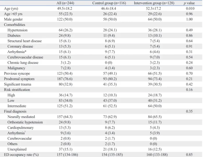
The admission rate decreased by 8.3% in the intervention group [unadjusted odds ratio (OR) 0.55, 95% confidence interval (CI) 0.28-1.09, p=0.08]. In particular, the admission rate was reduced by 13.4% for low or intermediate-risk pa- tients (unadjusted OR 0.22, 95% CI 0.08-0.63, p=0.005) (Fig. 1). In the multivariate logistic regression test, mem- bership in the intervention group was associated with a re- duction in hospitalization after adjustment for potential confounding factors (OR 0.31, 95% CI 0.13-0.70, p=
0.005) (Table 3).
Primary endpoint-medical cost
There was a substantial cost reduction of about 30% in the intervention period (369000 KRW, IQR 240000-602000 KRW), compared with the control period (542000 KRW, IQR 316000-1185000 KRW). This difference was especial- ly prominent when we compared the costs paid by low- or intermediate-risk patients, the costs at the ED, the costs paid by patients revisiting the OPD and the costs for diag- nostic tests (Table 4). In the multivariate linear regression analysis of medical costs, membership in the intervention group was associated with decreased costs (coefficient after visiting the ED. The total numbers of OPD visits dur-
ing the evaluation of syncope were 147 and 127, respec- tively. In the intervention group, the syncope unit treated a total of 37 patients (28.9%).
Diagnostic tests for the evaluation of syncope
Postural blood pressure measurement was more frequently performed in the intervention group than the control group (97.6% vs. 87.9%, p=0.003). However, the use of cardiac enzymes and electroencephalography were lower in the in- tervention group than the control group, respectively (42.9%
vs. 56.0%, p=0.041; 2.3% vs. 8.6%, p=0.029). The frequen- Table 2. Frequencies of Diagnostic Tests in Syncope Evaluation
Control group (n=116) Intervention group (n=128) p value
Postural BP check 102 (87.9) 125 (97.6) 0.003
Electrocardiogram 116 (100) 128 (100) 1.00
Laboratory tests* 116 (100) 128 (100) 1.00
Cardiac enzymes 65 (56.0) 55 (42.9) 0.041
Echocardiography 54 (46.5) 54 (42.1) 0.49
Head-up tilt test 45 (38.7) 52 (40.6) 0.77
Carotid sinus massage 0 (0) 3 (2.3) 0.24
Holter recording 22 (18.9) 37 (28.9) 0.07
Electrophysiological study 3 (2.59) 2 (1.5) 0.67
Coronary angiography 6 (5.1) 6 (4.6) 0.54
Treadmill test 16 (13.7) 26 (20.3) 0.23
Brain CT scan 71 (61.2) 76 (59.3) 0.77
Brain MRI scan 13 (11.2) 8 (6.2) 0.16
Electroencephalography 10 (8.6) 3 (2.3) 0.029
Carotid Doppler 1 (0.9) 3 (2.3) 0.62
Implantable loop recorder 0 (0) 2 (1.5) 0.27
BP, blood pressure; CT, computed tomography; MRI, magnetic resonance imaging.
Data are shown as n (%).
*Additional analysis showed that there was a significant reduction in the use of less informative tests for syncope evaluation, such as basic blood chemis- try, d-dimer, C-reactive protein, creatine kinase, etc.
Fig. 1. Comparisons of hospital admission rates between the control and in- tervention groups.
0 10 20 30
Hospital admission rate (%)
Overall group Low & intermediate subgroup Control group Intervention group
p=0.08 21.5
18.2 13.2
4.8 p=0.005
they were deaths due to malignancies. Recurrent syncopal events were reported for 3 patients (one in the control group and two in the intervention group). Because very few ad- verse events were reported, we were unable to compare the two groups regarding this measure, although we did evalu- ate the outcomes of all patients through OPD visits or tele- phone interviews (Table 6).
DISCUSSION
Although syncope is a commonly presenting symptom, the -712000 KRW, 95% CI -1341000 to -83000 KRW, p=
0.027) (Table 5).
Secondary endpoints
The ED LOS was significantly reduced in the intervention group, although the hospital LOS was not different between the two groups (median: 4.6 hours vs. 3.4 hours). There was an increasing trend in the rate of certain diagnosis in the in- tervention group, but it was not statistically significant.
During the six-month follow-up, three deaths were re- ported (1 patient in the control group and 2 patients in the intervention group). These were not unexpected events, as Table 3. Univariate and Multivariate Analysis for Hospital Admissions
Variable Univariate Multivariate
Odds ratio (95% CI) p value Odds ratio (95% CI) p value
Age, per 10 yrs 1.29 (1.06-1.56) 0.009 1.02 (0.99-1.04) 0.13
Male gender 1.79 (0.90-3.54) 0.09 1.48 (0.69-3.19) 0.30
Risk (reference: low risk group)
Intermediate risk 2.15 (0.81-5.69) 0.12 1.18 (0.34-4.07) 0.78
High risk 12.8 (4.46-36.92) <0.001 10.5 (2.99-36.9) <0.001
Significant trauma 0.75 (0.36-1.56) 0.45 0.73 (0.32-1.65) 0.46
Intervention group 0.55 (0.28-1.09) 0.08 0.31 (0.13-0.70) 0.005
CI, confidence interval.
Table 4. Medical Costs during Syncope Evaluation
Control group (n=116) Intervention group (n=128) p value Medical costs during syncope evaluation (1000 KRW)
Cost per patient (all) 542 (316-1185) 369 (240-602) <0.001
Cost per patient (patients who were followed at the
outpatient department) 530 (361-819) 382 (259-548) 0.002
Cost per patient (hospitalized) 2464 (1377-7703) 2285 (1630-7469) 0.90
Cost per patient (low risk) 381 (288-616) 237 (126-386) <0.001
Cost per patient (intermediate risk) 604 (329-1208) 380 (266-668) 0.003
Cost per patient (high risk) 1277 (723-2518) 637 (440-3374) 0.38
Cost per patient at the emergency department 429 (275-895) 313 (210-490) <0.001
Cost for diagnostic tests 354 (213-553) 302 (106-517) 0.019
KRW, Korean won.
Data are shown as median (interquartile range) or n (%). The exchange rate used is roughly 1000 KRW for 1 United States dollar.
Table 5. Univariate and Multivariate Linear Regression Analysis of Total Cost Per Patient
Variable Univariate Multivariate
Coefficient (95% CI) p value Coefficient (95% CI) p value
Age, per 10 yrs 363 (182 to 543) <0.001 348 (161 to 580) 0.003
Male gender 268 (-401 to 939) 0.43 -112 (-758 to 533) 0.73
Risk (reference: low risk group)
Intermediate risk 709 (10 to 1409) 0.047 -115 (-1029 to 798) 0.80
High risk 2791 (1805 to 3777) <0.001 2202 (1098 to 3306) <0.001
Significant trauma 303 (-406 to 1012) 0.40 365 (-296 to 1027) 0.27
Intervention group -456 (-112 to 212) 0.18 -712 (-1341 to -83) 0.027
CI, confidence interval.
Unit: 1000 Korean won.
fact, increase costs.2,15,18,25-27 However, risk stratification may be useful if clinical evaluation is appropriately per- formed, depending on risk. Shen, et al.18 showed that fo- cused evaluation of intermediate-risk patients, based on a specific syncope unit, significantly improved diagnostic yield and reduced hospital admission.
The creation of formal syncope units may increase diag- nostic yield and prevent unnecessary admissions and test- ing.15,17,18,28 In our study, we were unable to fully evaluate the effectiveness of a dedicated syncope unit, because our unit was not exclusively used for syncope patients, and most tests and specialist assessments could be performed in other areas of the ED if clinically indicated.
One change that was made as a result of our study was the modification of the ED bed management policy. There was no established protocol for bed assignment of syncope patients during the control period, despite ED overcrowd- ing. We hypothesized that the implementation of such a protocol would enable more rapid and safe monitoring and evaluation of high-risk or intermediate-risk patients. How- ever, we anticipate that such protocols might have the nega- tive effects of increasing ED LOS and costs in overcrowd- ed EDs. Further investigations concerning the overall effects of dedicated syncope units in the ED are needed.
Unfortunately, there was no significant reduction in major adverse events. This is one of our study’s main limitations.
We, however, focused on the efficacy of care provision in acute settings. In addition, to the best of our knowledge, most previous studies showed similar outcomes: that there were significant improvements in admission rates, medical costs and diagnostic yields without long-term data or with no significant differences in long-term outcomes.13,17,18,22
There are several other limitations to this study. First, this study was a nonrandomized, single center study. Also, we compared the two patient groups on a historical basis, and our sample size was too small for sufficient statistical pow- er to assess all end points. Additionally, we could not evalu- evaluation of syncope in the ED is often challenging and
time-consuming, and may incur unnecessary health care costs and delays in the diagnostic process.6,7,22 In our study, admission rates, medical costs and ED LOS were signifi- cantly reduced after the implementation of systematic ap- proaches for syncope evaluation. We observed outcome im- provements, particularly in low or intermediate-risk patients.
These results have important clinical implications, as inap- propriate admission, medical costs, and ED length of stay in the low and intermediate-risk patients were significantly reduced. Our results provide evidence for the importance of standardized evaluation and care for syncope in the ED.
Education alone may be insufficient to modify physician behavior, as local hospital education programs often do not lead to notable changes in the evaluation of syncope.9,23 We hypothesized that a simple, organized approach would help to overcome this barrier. The educational program and pro- tocol used in this study focused on initial risk stratification and evaluation in the ED. We also recommended that ED physicians use a hospital order set according to patient risk.
The effectiveness of a standardized order set in the evalu- ation of syncope has not yet been demonstrated. Such use may ensure that necessary procedures are performed and may allow for new practice changes.24 Regarding diagnos- tic tests, we hypothesized that a standardized order set would have the positive effect of reducing the use of less specific tests for diagnosis and encourage the measurement of pos- tural blood pressure during initial investigations. Our stan- dardized order sets, however, were limited in the initial eval- uation in the ED and should be optimized according to current evidence. Further study of a well-organized hospital order set and an extended decision-making system for addi- tional testing are needed.
Although several methods for risk stratification have dem- onstrated prognostic value, there is no clear evidence that any method directly improves diagnostic accuracy or re- duces costs; moreover, risk stratification methods may, in Table 6. Secondary Outcomes
Control group (n=116) Intervention group (n=128) p value Length of stay
ED length of stay (hrs) 4.6 (3.3-8.5) 3.4 (2.3-5.8) <0.001
Hospital length of stay, admitted patients (days) 4 (2-8) 6 (3.5-7.3) 0.55
Certain diagnosis 70 (60.3) 88 (68.7) 0.17
All-cause mortality 2 (1.7) 1 (0.8) 0.50
Recurrent syncope 1 (0.8) 2 (1.7) 0.50
KRW, Korean won; ED, emergency department.
Data are shown as median (interquartile range) or n (%). The exchange rate used is roughly 1000 KRW for 1 United States dollar.
sis and management of syncope (version 2009). Eur Heart J 2009;30:2631-71.
3. Kenny RA, O’Shea D, Walker HF. Impact of a dedicated syncope and falls facility for older adults on emergency beds. Age Ageing 2002;31:272-5.
4. Soteriades ES, Evans JC, Larson MG, Chen MH, Chen L, Benja- min EJ, et al. Incidence and prognosis of syncope. N Engl J Med 2002;347:878-85.
5. Blanc JJ, L’Her C, Touiza A, Garo B, L’Her E, Mansourati J. Pro- spective evaluation and outcome of patients admitted for syncope over a 1 year period. Eur Heart J 2002;23:815-20.
6. Chen LY, Benditt DG, Shen WK. Management of syncope in adults: an update. Mayo Clin Proc 2008;83:1280-93.
7. Malasana G, Brignole M, Daccarett M, Sherwood R, Hamdan MH. The prevalence and cost of the faint and fall problem in the state of Utah. Pacing Clin Electrophysiol 2011;34:278-83.
8. Sun BC, Emond JA, Camargo CA Jr. Direct medical costs of syn- cope-related hospitalizations in the United States. Am J Cardiol 2005;95:668-71.
9. Sclafani JJ, My J, Zacher LL, Eckart RE. Intensive education on evidence-based evaluation of syncope increases sudden death risk stratification but fails to reduce use of neuroimaging. Arch Intern Med 2010;170:1150-4.
10. Ammirati F, Colivicchi F, Santini M. Diagnosing syncope in clini- cal practice. Implementation of a simplified diagnostic algorithm in a multicentre prospective trial-the OESIL 2 study (Osservatorio Epidemiologico della Sincope nel Lazio). Eur Heart J 2000;21:
935-40.
11. Strickberger SA, Benson DW, Biaggioni I, Callans DJ, Cohen MI, Ellenbogen KA, et al. AHA/ACCF Scientific Statement on the evaluation of syncope: from the American Heart Association Councils on Clinical Cardiology, Cardiovascular Nursing, Cardio- vascular Disease in the Young, and Stroke, and the Quality of Care and Outcomes Research Interdisciplinary Working Group; and the American College of Cardiology Foundation: in collaboration with the Heart Rhythm Society: endorsed by the American Auto- nomic Society. Circulation 2006;113:316-27.
12. Huff JS, Decker WW, Quinn JV, Perron AD, Napoli AM, Peeters S, et al. Clinical policy: critical issues in the evaluation and man- agement of adult patients presenting to the emergency department with syncope. Ann Emerg Med 2007;49:431-44.
13. Brignole M, Ungar A, Bartoletti A, Ponassi I, Lagi A, Mussi C, et al. Standardized-care pathway vs. usual management of syncope patients presenting as emergencies at general hospitals. Europace 2006;8:644-50.
14. Brignole M, Malasana G, Sherwood RP, Daccarett M, Jetter TL, Hamdan MH. Evaluation of patients with “faint” in an American teaching hospital: a dire need for a standardized approach. Pacing Clin Electrophysiol 2011;34:284-90.
15. Sheldon RS, Morillo CA, Krahn AD, O’Neill B, Thiruganasam- bandamoorthy V, Parkash R, et al. Standardized approaches to the investigation of syncope: Canadian Cardiovascular Society posi- tion paper. Can J Cardiol 2011;27:246-53.
16. Elesber AA, Decker WW, Smars PA, Hodge DO, Shen WK;
American College of Emergency Physicians. Impact of the applica- tion of the American College of Emergency Physicians recommen- dations for the admission of patients with syncope on a retrospec- tively studied population presenting to the emergency department.
Am Heart J 2005;149:826-31.
17. Ammirati F, Colaceci R, Cesario A, Strano S, Della Scala A, Col-
ate whether or not our standardization procedure had any effect on morbidity and mortality. Therefore, our findings should be cautiously interpreted if considering similar im- plementation in other institutions. Second, we did not modi- fy the diagnostic process outside the ED because our proto- col was focused on ED evaluation. Hence, use of additional diagnostic tests was not strictly controlled. Further studies must be conducted after the adoption of a hospital-wide standardized protocol at our institution. Third, the approach- es we used in our study might not be optimal and did not completely reflect the current guidelines, as we modified the guidelines to suit our hospital. For instance, basic blood tests and chest radiographs were conducted solely to pre- vent malpractice lawsuits, even when not clinically indicat- ed. Fourth, we are unable to exclude the possibility that our findings resulted from a Hawthorne effect during the study period. Finally, we observed a difference in age between the two groups that might represent random variation or be associated with selection bias. However, we may have drawn the same conclusions after adjusting for potential confound- ers including age.
In conclusion, the implementation of standardized ap- proaches to evaluate syncope reduced hospital admissions, medical costs and LOS in the overcrowded ED of a tertia- ry-care teaching hospital in South Korea.
ACKNOWLEDGEMENTS
We would like to thank research nurses, Su Jin Bae and Hee Jung Park, in the clinical trial center of the Samsung Medi- cal Center ED, as well as paramedics in the ED, including Hyun Yong Wi, for their assistance in data collection. We appreciate the provision of hospital accounting data by Jongsoo Seo, Manager, Department of Patient Affairs.
This study was supported by the Samsung Medical Cen- ter Clinical Research Development Program grant #CRS- 109-35-2.
REFERENCES
1. Morichetti A, Astorino G. [Epidemiological and clinical findings in 697 syncope events]. Minerva Med 1998;89:211-20.
2. Task Force for the Diagnosis and Management of Syncope; Euro- pean Society of Cardiology (ESC); European Heart Rhythm As- sociation (EHRA); Heart Failure Association (HFA); Heart Rhythm Society (HRS), Moya A, et al. Guidelines for the diagno-
evaluation of an educational programme for physicians involved in the management of syncope. Europace 2005;7:400-6.
24. Thiel SW, Asghar MF, Micek ST, Reichley RM, Doherty JA, Kollef MH. Hospital-wide impact of a standardized order set for the management of bacteremic severe sepsis. Crit Care Med 2009;
37:819-24.
25. Gabayan GZ, Derose SF, Asch SM, Chiu VY, Glenn SC, Man- gione CM, et al. Predictors of short-term (seven-day) cardiac out- comes after emergency department visit for syncope. Am J Cardi- ol 2010;105:82-6.
26. Serrano LA, Hess EP, Bellolio MF, Murad MH, Montori VM, Er- win PJ, et al. Accuracy and quality of clinical decision rules for syncope in the emergency department: a systematic review and meta-analysis. Ann Emerg Med 2010;56:362-73.e1.
27. Reed MJ, Newby DE, Coull AJ, Prescott RJ, Jacques KG, Gray AJ. The ROSE (risk stratification of syncope in the emergency de- partment) study. J Am Coll Cardiol 2010;55:713-21.
28. Brignole M, Ungar A, Casagranda I, Gulizia M, Lunati M, Ammi- rati F, et al. Prospective multicentre systematic guideline-based management of patients referred to the Syncope Units of general hospitals. Europace 2010;12:109-18.
angelo I, et al. Management of syncope: clinical and economic impact of a Syncope Unit. Europace 2008;10:471-6.
18. Shen WK, Decker WW, Smars PA, Goyal DG, Walker AE, Hodge DO, et al. Syncope Evaluation in the Emergency Department Study (SEEDS): a multidisciplinary approach to syncope manage- ment. Circulation 2004;110:3636-45.
19. Li YW, Chen L, Du JB, Yang YY, Jin HF. Cost-effectiveness of diagnostic approaches to vasovagal syncope. Chin Med J (Engl) 2010;123:2635-9.
20. McCarthy ML, Aronsky D, Jones ID, Miner JR, Band RA, Baren JM, et al. The emergency department occupancy rate: a simple measure of emergency department crowding? Ann Emerg Med 2008;51:15-24, 24.e1-2.
21. Brignole M, Alboni P, Benditt DG, Bergfeldt L, Blanc JJ, Thom- sen PE, et al. Guidelines on management (diagnosis and treat- ment) of syncope-update 2004. Executive Summary. Eur Heart J 2004;25:2054-72.
22. Fedorowski A, Burri P, Juul-Möller S, Melander O. A dedicated investigation unit improves management of syncopal attacks (Syncope Study of Unselected Population in Malmo--SYSTEMA I). Europace 2010;12:1322-8.
23. Blanc JJ, L’her C, Gosselin G, Cornily JC, Fatemi M. Prospective