INTRODUCTION
Given concerns regarding late drug eluting stent (DES) throm-
bosis, imaging modalities have attempted to identify risk fac- tors for the occurrence of stent thrombosis.1-4 Of these, late stent malapposition or incomplete coverage of stent struts af- ter DES implantation have been suggested as major potential risks for late adverse clinical outcomes, such as late stent thrombosis.1-5 Additionally, several clinical and procedural features after DES implantation have been postulated as po- tential risk factors for late stent malapposition and incomplete coverage of stent struts. Optical coherence tomography (OCT) has been used to evaluate these risk factors due to its superior resolution over other imaging modalities.4,6,7 Nevertheless, lit- tle data exists regarding the impact of plaque characteristics or composition of de novo lesions on vascular healing responses, particularly late stent malapposition or incomplete strut cov-
Impact of Coronary Plaque Characteristics
on Late Stent Malapposition after Drug-Eluting Stent Implantation
Sung-Jin Hong1*, Byeong-Keuk Kim2,3*, Dong-Ho Shin2,3, Jung-Sun Kim2,3, Young-Guk Ko2,3, Donghoon Choi2,3, Yangsoo Jang2,3,4, and Myeong-Ki Hong2,3,4
1Division of Cardiology, Department of Internal Medicine, Sanggye Paik Hospital, Inje University College of Medicine, Seoul;
2Division of Cardiology, Severance Cardiovascular Hospital, Yonsei University College of Medicine, Seoul;
3Cardiovascular Institute, Yonsei University College of Medicine, Seoul;
4Severance Biomedical Science Institute, Yonsei University College of Medicine, Seoul, Korea.
Purpose: To evaluate the impact of pre-procedural coronary plaque composition assessed by virtual histology intravascular ultra- sound (VH-IVUS) on late stent malapposition assessed by optical coherence tomography (OCT) following drug-eluting stent (DES) implantation.
Materials and Methods: The study population consisted of 121 patients (121 lesions) who underwent both pre-procedural VH- IVUS and follow-up OCT after DES implantation. The association between pre-procedural plaque composition [necrotic core (NC), dense calcium (DC), fibrotic (FT), and fibro-fatty (FF) volumes] assessed by VH-IVUS and late stent malapposition (percent malapposed struts) or strut coverage (percent uncovered struts) assessed by follow-up OCT was evaluated.
Results: Pre-procedural absolute total NC, DC, FT, and FF plaque volumes were 22.9±19.0, 7.9±9.6, 63.8±33.8, and 16.5±12.4 mm3, respectively. At 6.3±3.1 months post-intervention, percent malapposed and uncovered struts were 0.8±2.5% and 15.3±16.7%, re- spectively. Pre-procedural absolute total NC and DC plaque volumes were positively correlated with percent malapposed struts (r=0.44, p<0.001 and r=0.45, p<0.001, respectively), while pre-procedural absolute total FT plaque volume was weakly associated with percent malapposed struts (r=0.220, p=0.015). Pre-procedural absolute total DC plaque volume was the only independent predictor of late stent malapposition on multivariate analysis (β=1.12, p=0.002). There were no significant correlations between pre-intervention plaque composition and percent uncovered struts.
Conclusion: Pre-procedural plaque composition was associated with late stent malapposition but not strut coverage after DES implantation. Larger pre-procedural absolute total DC plaque volumes were associated with greater late stent malapposition.
Key Words: Drug-eluting stent, intravascular ultrasound, optical coherence tomography Yonsei Med J 2015 Nov;56(6):1538-1544
http://dx.doi.org/10.3349/ymj.2015.56.6.1538 pISSN: 0513-5796 · eISSN: 1976-2437
Received: September 3, 2014 Revised: November 18, 2014 Accepted: December 23, 2014
Corresponding author: Dr. Myeong-Ki Hong, Division of Cardiology, Severance Cardiovascular Hospital, Yonsei University College of Medicine, 50-1 Yonsei-ro, Seodaemun-gu, Seoul 03722, Korea.
Tel: 82-2-2228-8458, Fax: 82-2-393-2041, E-mail: [email protected]
*Sung-Jin Hong and Byeong-Keuk Kim contributed equally to this work.
•The authors have no financial conflicts of interest.
© Copyright: Yonsei University College of Medicine 2015
This is an Open Access article distributed under the terms of the Creative Com- mons Attribution Non-Commercial License (http://creativecommons.org/ licenses/
by-nc/3.0) which permits unrestricted non-commercial use, distribution, and repro- duction in any medium, provided the original work is properly cited.
erage, after DES implantation.8-10
We hypothesized that quantitative assessment of plaque composition could predict late stent malapposition or incom- plete strut coverage after DES implantation. We sought to prove this hypothesis by evaluating the association between pre-procedural coronary plaque composition on virtual his- tology intravascular ultrasound (VH-IVUS) and strut malap- position or coverage on follow-up OCT after DES implanta- tion.
MATERIALS AND METHODS
Between July 2010 and December 2011, we identified 121 eli- gible patients (121 lesions) who underwent both pre-proce- dural VH-IVUS examination on de novo lesions and follow-up OCT after DES implantation from an OCT registry database at our institute. Patients or lesions with the following character- istics were excluded from this study: 1) untreated significant left main coronary artery disease, bifurcation treated with two-stent techniques, in-stent restenosis, graft stenosis, or overlapping DES; 2) ST-elevation myocardial infarction; 3) hemodynamically unstable status or ejection fraction <30%; 4) renal insufficiency with baseline creatinine ≥2.0 mg/dL; and 5) poor intravascular ultrasound (IVUS) or OCT image quali- ty. This study protocol was approved by the Institutional Re- view Board of Yonsei University College of Medicine. All pa- tients provided written informed consent.
DESs were selected by operators at the time of implantation and included sirolimus-eluting stents (Cypher, Cordis, Miami, FL, USA), zotarolimus-eluting stents (Resolute or Integrity, Medtronic, Santa Rosa, CA, USA), everolimus-eluting stents (Xience, Abbott Vascular, Santa Clara, CA, USA), or biolimus A9-eluting stents (Nobori, Terumo Corporation, Tokyo, Japan or Biomatrix, Biosensors International, Singapore). Each DES was implanted with conventional techniques. Unfractionated heparin was administered as an initial bolus of 100 IU/kg, with additional boluses administered during the procedure to achieve an activated clotting time of 250 to 300 seconds. Dual antiplatelet therapy (aspirin and clopidogrel) was prescribed at least until the follow-up OCT was performed. Quantitative coronary angiography analyses were performed before and after stent implantation and at follow-up using an off-line quantitative coronary angiographic system (CASS system, Pie Medical Instruments, Maastricht, the Netherlands) in an inde- pendent core laboratory (Cardiovascular Research Center, Seoul, Korea). Reference vessel diameters and minimal luminal di- ameters were measured with a guiding catheter for magnifica- tion-calibration from diastolic frames in a single, matched view that showed the smallest minimal luminal diameter.
The VH-IVUS examination was performed in the target le- sion before pre-dilation using a 20-MHz 2.9 Fr, phased-array IVUS catheter (Eagle Eye, Volcano Therapeutics, Rancho Cor-
dova, CA, USA). After intracoronary administration of nitro- glycerin (200 μg), the IVUS catheter was placed distal to the target lesion and then pulled back using a motorized pullback system at 0.5 cm/s. During pullback, gray-scale IVUS was re- corded, and raw radiofrequency data were captured at the top of the R wave for reconstruction of a color-coded map by a VH- IVUS data recorder (Volcano Therapeutics). Gray-scale quanti- tative IVUS analyses of the external elastic membrane, lumen, and plaque and media (external elastic membrane-lumen) were performed according to the Clinical Expert Consensus Document on IVUS.11 VH-IVUS data were analyzed for the en- tire length of the target lesion covered by DES with 1-mm in- tervals for volumetric analysis. On the pre-procedural IVUS images, the diseased segment was selected and the references were defined as the most normal-appearing segments within 5 mm proximal and distal to the lesion shoulders. Also, the re- gion of interest, matched frames of the stented segment on follow-up OCT images, was identified based on anatomic landmarks, such as side branches or calcification, aided by an- giographic images revealing the IVUS catheter position. VH- IVUS analysis coded tissue as red (necrotic core, NC), white (dense calcium, DC), green (fibrotic, FT), or yellow-green (fi- bro-fatty, FF).12,13 The findings of VH-IVUS analyses were de- scribed as absolute volumes and percentages (relative amounts) of plaque volumes.
OCT was performed using two OCT systems (Model M2 Imaging System and C7-XR Imaging System, LightLab Imag- ing, Inc., St. Jude Medical, St. Paul, MN, USA).6,9 All of the OCT images were analyzed at a core laboratory (Cardiovascular Research Center, Seoul, Korea) by analysts who were blinded to patient and procedural information. Cross-sectional OCT images were analyzed at 1-mm intervals. Stent and luminal cross-sectional areas (CSA) were measured, and neointimal hyperplasia (NIH) CSA was calculated as the stent CSA minus the luminal CSA.6 Total stent, lumen, and NIH volumes within stented segments were calculated. NIH thickness, which was the distance between the endoluminal surface of the neointi- ma and the strut, was measured inside each strut with a line perpendicular to the neointima and strut.14 An uncovered strut was defined as having a NIH thickness of 0 μm.7,14,15 Stent malapposition was defined as the presence of any malap- posed struts.15-17 A malapposed strut was defined as a strut that was detached from the vessel wall as follows: Cypher,
≥160 μm; Resolute or Integrity, ≥110 μm; Xience, ≥100 μm;
and Nobori or Biomatrix, ≥130 μm.15-19 The percent of malap- posed or uncovered struts in each stented lesion was calculat- ed as: (number of malapposed or uncovered struts/total number of struts in all cross-sections of the lesion)×100.7
Continuous data are presented as mean±standard devia- tion, and categorical data are presented as number (%). Pear- son’s two-way test was used to assess the relationships be- tween two quantitative variables. Each pre-procedural plaque component (NC, DC, FT, and FF) was classified into quartiles.
Analysis of variance was used to compare the percentage of malapposed or uncovered struts according to each plaque component quartile. Multivariate analysis was performed to determine the independent predictors of late stent malappo- sition. Variables that reached a p<0.1 in univariate analysis were included in a multivariate forward stepwise regression analysis. Statistical analysis was performed with SPSS soft- ware, version 18.0 (SPSS Inc., Chicago, IL, USA), and a p-value
<0.05 was considered statistically significant.
RESULTS
Clinical, procedural, and pre-procedural VH-IVUS character- istics according to the presence of late stent malapposition are
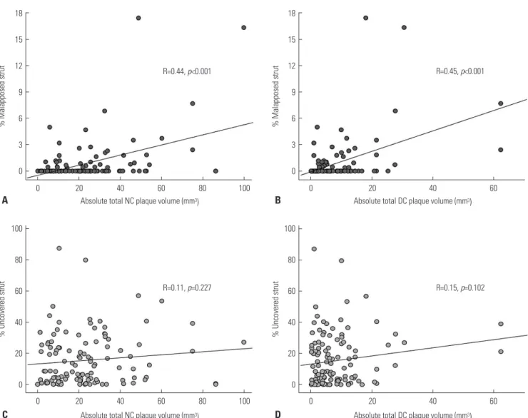
summarized in Table 1 and 2. Follow-up OCT characteristics are summarized in Table 3. Pre-procedural absolute total NC and DC plaque volumes were positively correlated with the percentage of malapposed struts (r=0.44, p<0.001, Fig. 1A and r=0.45, p<0.001, Fig. 1B, respectively), and pre-procedural ab- solute total FT plaque volume was weakly associated with the percentage of malapposed struts (r=0.220, p=0.015). When each relative total plaque volume was classified into quartiles according to the plaque components, the highest NC plaque volume quartile and the highest DC plaque volume quartile had a significantly higher percentage of malapposed struts (p=0.013 for relative NC plaque volume, p=0.020 for relative DC plaque volume) (Fig. 2A). The highest FT plaque volume quartile had a lower percentage of malapposed struts (p=0.040).
The lesions with stent malapposition on follow-up OCT had a
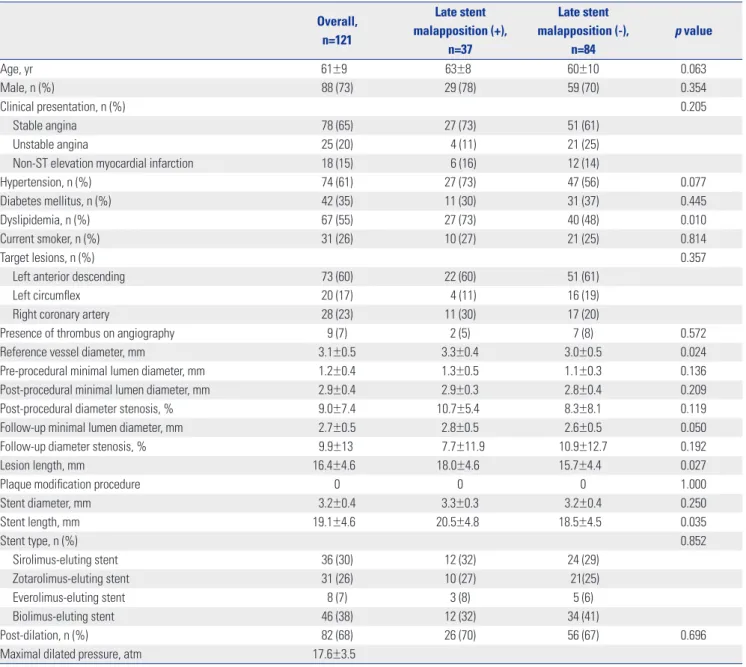
Table 1. Baseline Clinical and Procedural Characteristics
Overall, n=121
Late stent malapposition (+),
n=37
Late stent malapposition (-),
n=84
p value
Age, yr 61±9 63±8 60±10 0.063
Male, n (%) 88 (73) 29 (78) 59 (70) 0.354
Clinical presentation, n (%) 0.205
Stable angina 78 (65) 27 (73) 51 (61)
Unstable angina 25 (20) 4 (11) 21 (25)
Non-ST elevation myocardial infarction 18 (15) 6 (16) 12 (14)
Hypertension, n (%) 74 (61) 27 (73) 47 (56) 0.077
Diabetes mellitus, n (%) 42 (35) 11 (30) 31 (37) 0.445
Dyslipidemia, n (%) 67 (55) 27 (73) 40 (48) 0.010
Current smoker, n (%) 31 (26) 10 (27) 21 (25) 0.814
Target lesions, n (%) 0.357
Left anterior descending 73 (60) 22 (60) 51 (61)
Left circumflex 20 (17) 4 (11) 16 (19)
Right coronary artery 28 (23) 11 (30) 17 (20)
Presence of thrombus on angiography 9 (7) 2 (5) 7 (8) 0.572
Reference vessel diameter, mm 3.1±0.5 3.3±0.4 3.0±0.5 0.024
Pre-procedural minimal lumen diameter, mm 1.2±0.4 1.3±0.5 1.1±0.3 0.136
Post-procedural minimal lumen diameter, mm 2.9±0.4 2.9±0.3 2.8±0.4 0.209
Post-procedural diameter stenosis, % 9.0±7.4 10.7±5.4 8.3±8.1 0.119
Follow-up minimal lumen diameter, mm 2.7±0.5 2.8±0.5 2.6±0.5 0.050
Follow-up diameter stenosis, % 9.9±13 7.7±11.9 10.9±12.7 0.192
Lesion length, mm 16.4±4.6 18.0±4.6 15.7±4.4 0.027
Plaque modification procedure 0 0 0 1.000
Stent diameter, mm 3.2±0.4 3.3±0.3 3.2±0.4 0.250
Stent length, mm 19.1±4.6 20.5±4.8 18.5±4.5 0.035
Stent type, n (%) 0.852
Sirolimus-eluting stent 36 (30) 12 (32) 24 (29)
Zotarolimus-eluting stent 31 (26) 10 (27) 21(25)
Everolimus-eluting stent 8 (7) 3 (8) 5 (6)
Biolimus-eluting stent 46 (38) 12 (32) 34 (41)
Post-dilation, n (%) 82 (68) 26 (70) 56 (67) 0.696
Maximal dilated pressure, atm 17.6±3.5
significantly larger initial absolute total NC plaque volume (30.7±22.3 mm3 vs. 19.5±16.4 mm3, p=0.008) and absolute to- tal DC plaque volume (12.3±14.5 mm3 vs. 5.9±5.4 mm3, p=
0.012) (Table 2). Similarly, the lesions with stent malapposi- tion had a significantly larger relative total NC plaque volume (22.1±8.7% vs. 17.4±8.9%, p=0.009) and a larger relative total DC plaque volume (8.6±5.9% vs. 5.9±4.5%, p=0.008) (Table 2).
On the pre-procedural IVUS images, among the 37 lesions with late stent malapposition, superficial calcium was observed in 24 lesions (65%) and deep calcium was observed in eight le- sions (21%). The remaining five lesions (14%) did not have cal- cium. Also, the mean calcium length of the 31 calcified lesions was 8.9±8.3 mm, and the proportion of calcium length with re- gard to total lesion length was 44±30%.
As for the percentage of uncovered struts, there was no sig- nificant relationship with absolute total NC plaque volume (r=0.11, p=0.227) (Fig. 1C) or absolute total DC plaque volume (r=0.15, p=0.102) (Fig. 1D). Similarly, there were no significant differences in the percentage of uncovered struts among the rel- ative total volume quartiles of each plaque component (Fig. 2B).
On multivariate analyses, pre-procedural absolute total DC plaque volume was the only independent predictor of late stent malapposition (β=1.12, p=0.002).
DISCUSSION
The main findings of the present study were as follows: 1) pre- procedural quantitative assessment of plaque composition by
VH-IVUS may predict the degree of late stent malapposition detected by OCT; 2) greater pre-procedural DC plaque bur- den was associated with greater late stent malapposition and was the only independent predictor for late stent malapposi- tion; and 3) pre-procedural plaque composition was not asso- ciated with strut coverage on follow-up OCT after DES im- plantation.
Until now, there has been little data regarding the associa- tion between pre-procedural plaque composition evaluated by VH-IVUS and strut coverage on follow-up OCT after DES implantation. Even though plaque characteristics seem to strongly affect vascular healing responses after DES implanta- tion, no data exists regarding the impact of plaque character- istics on late stent malapposition or coverage of struts, which may be considered potential risk factors for the occurrence of late stent thrombosis.1-5 In addition, many previous OCT stud- ies evaluating vascular responses after DES implantation have commonly been limited by a lack of information regarding pre-procedural plaque characteristics of de novo lesions.16,17 Given this situation, we sought to investigate the impact of plaque characteristics of de novo lesions on vascular respons- es following DES implantation using pre-procedural VH-IVUS and follow-up OCT.
Mechanisms of late persistent malapposition have been proposed. Malapposition might be mediated partly by calci- fied lesions that do not allow for homogeneous stent expan- sion and could result in the lack of contact of stent struts with the vessel walls.1 Also, as for plaque characteristics affecting the occurrence of stent malapposition, a recent OCT study re- Table 2. Pre-Procedural VH-IVUS
Total, n=121
Late stent malapposition (+),
n=37
Late stent malapposition (-),
n=84
p value Pre-procedural gray-scale IVUS characteristics
Target lesion CSA with minimum lumen area
EEM CSA, mm2 13.0±4.1 14.2±4.1 12.4±4.1 0.028
Lumen CSA, mm2 2.7±0.9 3.2±1.2 2.5±0.6 0.003
P&M CSA, mm2 10.2±4.0 11.0±3.8 9.9±4.0 0.139
Plaque burden, % 77.3±9.0 76.5±9.2 77.6±8.9 0.529
Pre-procedural VH-IVUS characteristics Absolute total plaque volume, mm3
Necrotic core 22.9±19.0 30.7±22.3 19.5±16.4 0.008
Dense calcium 7.9±9.6 12.3±14.5 5.9±5.4 0.012
Fibrotic 63.8±33.8 68.1±28.7 61.9±35.8 0.358
Fibrofatty 16.5±12.4 16.5±13.1 16.6±12.1 0.973
Relative total plaque volume, %
Necrotic core 18.9±9.1 22.1±8.7 17.4±8.9 0.009
Dense calcium 6.7±5.1 8.6±5.9 5.9±4.5 0.008
Fibrotic 59.3±9.8 56.4±9.8 60.5±9.6 0.031
Fibrofatty 15.1±10.3 12.7±7.8 16.2±11.1 0.081
IVUS, intravascular ultrasound; EEM, external elastic membrane; CSA, cross-sectional area; P&M, plaque and media; VH, virtual histology.
Values were presented as mean±standard deviation.
Fig. 1. Correlation between absolute total plaque component volumes and percentage of malapposed (A and B) or uncovered struts (C and D) on fol- low-up OCT. Absolute total NC plaque volume and absolute total DC plaque volume were positively correlated with percentage of malapposed struts but not with percentage of uncovered struts. OCT, optical coherence tomography; NC, necrotic core; DC, dense calcium.
18 15 12 9 6 3 0
% Malapposed strut
0 20
Absolute total NC plaque volume (mm3) R=0.44, p<0.001
40 60 80 100
A 100
80 60 40 20 0
% Uncovered strut
0 20
Absolute total NC plaque volume (mm3) R=0.11, p=0.227
40 60 80 100
C
18 15 12 9 6 3 0
% Malapposed strut
0 20
Absolute total DC plaque volume (mm3) R=0.45, p<0.001
40 60
B 100
80 60 40 20 0
% Uncovered strut
0 20
Absolute total DC plaque volume (mm3) R=0.15, p=0.102
40 60
D
76 54 32 10
Q1 Q2
Relative NC plaque volume quartilesQ3 p=0.013
Q4
% Malapposed strut
40 30 20 10 0
Q1 Q2
Relative NC plaque volume quartilesQ3 p=0.581
Q4
% Uncovered strut
76 54 32 10
Q1 Q2
Relative DC plaque volume quartiles p=0.020
Q3 Q4
% Malapposed strut
40 30 20 10 0
Q1 Q2
Relative DC plaque volume quartiles p=0.941
Q3 Q4
% Uncovered strut
76 54 32 10
Q1 Q2
Relative FT plaque volume quartiles p=0.040
Q3 Q4
% Malapposed strut
40 30 20 10 0
Q1 Q2
Relative FT plaque volume quartiles p=0.955
Q3 Q4
% Uncovered strut
76 54 32 10
Q1 Q2
Relative FF plaque volume quartiles p=0.547
Q3 Q4
% Malapposed strut
40 30 20 10 0
Q1 Q2
Relative FF plaque volume quartiles p=0.312
Q3 Q4
% Uncovered strut
Fig. 2. Percentage of malapposed (A) and uncovered struts (B) according to the relative total plaque volume quartiles of each plaque component. The highest relative NC plaque volume quartiles and the highest relative DC plaque volume quartiles had a significantly higher percentage of malapposed struts. NC, necrotic core; DC, dense calcium; FT, fibrotic; FF, fibrofatty.
A
B
ported that the presence of calcium was one independent predictor of late persistent stent malapposition after DES im- plantation.6 Therefore, although we could not classify late stent malapposition into late-acquired and persistent malap- position in the present study, larger DC volumes may be im- portant for late persistent stent malapposition. Other IVUS studies have suggested that late-acquired stent malapposition occurs mainly due to positive remodeling and plaque/throm- bus resolution.20,21 In the present study, we found that abso- lute total NC plaque volume is also positively related to the degree of late stent malapposition in univariate analysis. Thus, larger NC volumes may be important for late-acquired stent malapposition. Similar to our study, a previous study reported that a post-stented NC component was associated with the development of late stent malapposition on follow-up IVUS after DES implantation, particularly in patients with acute myocardial infarction and diabetes mellitus.22 However, the present study is different from the previous study in that we assessed the plaque characteristics pre-intervention, not post- intervention. Furthermore, we assessed vascular healing re- sponses using OCT, not IVUS, which might weight the impor- tance of this study. Our study showed that pre-procedural plaque composition may play an essential role in formation of late stent malapposition after DES implantation.
Late stent thrombosis after DES implantation is a serious concern because of delayed vascular healing and inflamma- tory reaction. Uncovered struts on follow-up OCT have been proposed as potential risk factors for late stent thrombosis af- ter DES implantation.4,5 A previous study showed that cover- Table 3. Pre-Procedural VH-IVUS and Matched Follow-Up OCT Charac- teristics
Follow-up OCT characteristics Total, n=121 Time interval to follow-up OCT, months 6.3±3.1 Total mean number of cross sections, n 17.2±5.0 Total mean number of analyzable struts, n 189±58
Mean stent CSA, mm2 7.5±2.0
Mean lumen CSA, mm2 7.0±2.0
Mean NIH CSA, mm2 0.5±0.4
Percentage of NIH CSA, % 6.5±5.3
Median NIH thickness, μm 54.5 (35.1–96.3)*
Percent malapposed struts, % 0.8±2.5
Percent uncovered struts, % 15.3±16.7
Percentage of both of malapposed and uncovered
struts, % 0.5±1.8
Cross sections with any uncovered strut, % 31.1±37.6 Cross sections with a ratio of uncovered to total
strut >0.3, % 15.4±26.9
Cross sections with any malapposed strut, % 3.0±9.5
Stent malapposition, n (%) 37 (31)
OCT, optical coherence tomography; CSA, cross-sectional area; NIH, neointi- mal hyperplasia; VH-IVUS, virtual histology intravascular ultrasound.
*Was presented as median (interquartile range).
age of malapposed stent segments is delayed, compared to well-apposed segments, and that the larger the acute malap- position, the greater the likelihood of persistent malapposi- tion and delayed healing at follow-up.4 Thus, we hypothesized that plaque composition, particularly a DC or NC component, may result in delayed strut coverage. However, in the present study, there were no significant associations between the spe- cific plaque components and strut coverage after DES im- plantation. Considering the results of our previous OCT study, DES type rather than plaque characteristics on VH-IVUS may be the more powerful factor in determining strut coverage.7
This study has several limitations. First, it was not clear whether the malappositions at follow-up were residual or newly acquired because post-procedural intravascular imag- ing studies were not preformed immediately after DES im- plantation. Second, this study was based on registry data, and the sample size was quite small. Finally, different types of DES were used.
In conclusion, pre-procedural assessment of plaque com- position by VH-IVUS may predict the formation of late stent malapposition detected by OCT at follow-up. Larger pre-pro- cedural DC plaque volumes were associated with late stent malapposition. However, pre-procedural plaque composition was not associated with decreased strut coverage at follow-up after DES implantation.
ACKNOWLEDGEMENTS
This study was supported by a grant from the Korea Health- care Technology R&D Project, Ministry for Health, Welfare &
Family Affairs, Republic of Korea (No. A085012 and A102064);
a grant from the Korea Health 21 R&D Project, Ministry of Health & Welfare, Republic of Korea (No. A085136); and the Cardiovascular Research Center, Seoul, Korea.
REFERENCES
1. Cook S, Wenaweser P, Togni M, Billinger M, Morger C, Seiler C, et al. Incomplete stent apposition and very late stent thrombosis af- ter drug-eluting stent implantation. Circulation 2007;115:2426- 34.
2. Siqueira DA, Abizaid AA, Costa Jde R, Feres F, Mattos LA, Staico R, et al. Late incomplete apposition after drug-eluting stent implan- tation: incidence and potential for adverse clinical outcomes. Eur Heart J 2007;28:1304-9.
3. Hassan AK, Bergheanu SC, Stijnen T, van der Hoeven BL, Snoep JD, Plevier JW, et al. Late stent malapposition risk is higher after drug-eluting stent compared with bare-metal stent implantation and associates with late stent thrombosis. Eur Heart J 2010;31:
1172-80.
4. Guagliumi G, Sirbu V, Musumeci G, Gerber R, Biondi-Zoccai G, Ikejima H, et al. Examination of the in vivo mechanisms of late drug-eluting stent thrombosis: findings from optical coherence tomography and intravascular ultrasound imaging. JACC Cardio- vasc Interv 2012;5:12-20.
5. Won H, Shin DH, Kim BK, Mintz GS, Kim JS, Ko YG, et al. Optical
coherence tomography derived cut-off value of uncovered stent struts to predict adverse clinical outcomes after drug-eluting stent implantation. Int J Cardiovasc Imaging 2013;29:1255-63.
6. Im E, Kim BK, Ko YG, Shin DH, Kim JS, Choi D, et al. Incidences, predictors, and clinical outcomes of acute and late stent malap- position detected by optical coherence tomography after drug- eluting stent implantation. Circ Cardiovasc Interv 2014;7:88-96.
7. Kim BK, Kim JS, Oh C, Ko YG, Choi D, Jang Y, et al. Major determi- nants for the uncovered stent struts on optical coherence tomog- raphy after drug-eluting stent implantation. Int J Cardiovasc Im- aging 2012;28:705-14.
8. Guo N, Maehara A, Mintz GS, He Y, Xu K, Wu X, et al. Incidence, mechanisms, predictors, and clinical impact of acute and late stent malapposition after primary intervention in patients with acute myocardial infarction: an intravascular ultrasound sub- study of the Harmonizing Outcomes with Revascularization and Stents in Acute Myocardial Infarction (HORIZONS-AMI) trial.
Circulation 2010;122:1077-84.
9. Gutiérrez-Chico JL, Wykrzykowska J, Nüesch E, van Geuns RJ, Koch KT, Koolen JJ, et al. Vascular tissue reaction to acute malap- position in human coronary arteries: sequential assessment with optical coherence tomography. Circ Cardiovasc Interv 2012;5:20- 9, S1-8.
10. Ozaki Y, Okumura M, Ismail TF, Naruse H, Hattori K, Kan S, et al.
The fate of incomplete stent apposition with drug-eluting stents:
an optical coherence tomography-based natural history study.
Eur Heart J 2010;31:1470-6.
11. Mintz GS, Nissen SE, Anderson WD, Bailey SR, Erbel R, Fitzgerald PJ, et al. American College of Cardiology Clinical Expert Consen- sus Document on Standards for Acquisition, Measurement and Reporting of Intravascular Ultrasound Studies (IVUS). A report of the American College of Cardiology Task Force on Clinical Expert Consensus Documents. J Am Coll Cardiol 2001;37:1478-92.
12. Nair A, Kuban BD, Tuzcu EM, Schoenhagen P, Nissen SE, Vince DG. Coronary plaque classification with intravascular ultrasound radiofrequency data analysis. Circulation 2002;106:2200-6.
13. Nasu K, Tsuchikane E, Katoh O, Vince DG, Virmani R, Surmely JF, et al. Accuracy of in vivo coronary plaque morphology assess- ment: a validation study of in vivo virtual histology compared
with in vitro histopathology. J Am Coll Cardiol 2006;47:2405-12.
14. Takano M, Inami S, Jang IK, Yamamoto M, Murakami D, Seimiya K, et al. Evaluation by optical coherence tomography of neointi- mal coverage of sirolimus-eluting stent three months after im- plantation. Am J Cardiol 2007;99:1033-8.
15. Barlis P, Dimopoulos K, Tanigawa J, Dzielicka E, Ferrante G, Del Furia F, et al. Quantitative analysis of intracoronary optical coher- ence tomography measurements of stent strut apposition and tis- sue coverage. Int J Cardiol 2010;141:151-6.
16. Kim BK, Hong MK, Shin DH, Kim JS, Ko YG, Choi D, et al. Optical coherence tomography analysis of strut coverage in biolimus- and sirolimus-eluting stents: 3-month and 12-month serial fol- low-up. Int J Cardiol 2013;168:4617-23.
17. Kim BK, Ha J, Mintz GS, Kim JS, Shin DH, Ko YG, et al. Ran- domised comparison of strut coverage between Nobori biolimus- eluting and sirolimus-eluting stents: an optical coherence tomog- raphy analysis. EuroIntervention 2014;9:1389-97.
18. Tanigawa J, Barlis P, Dimopoulos K, Dalby M, Moore P, Di Mario C. The influence of strut thickness and cell design on immediate apposition of drug-eluting stents assessed by optical coherence tomography. Int J Cardiol 2009;134:180-8.
19. Davlouros PA, Mavronasiou E, Xanthopoulou I, Karantalis V, Tsig- kas G, Hahalis G, et al. An optical coherence tomography study of two new generation stents with biodegradable polymer carrier, eluting paclitaxel vs. biolimus-A9. Int J Cardiol 2012;157:341-6.
20. Hong MK, Mintz GS, Lee CW, Park DW, Park KM, Lee BK, et al.
Late stent malapposition after drug-eluting stent implantation:
an intravascular ultrasound analysis with long-term follow-up.
Circulation 2006;113:414-9.
21. Ako J, Morino Y, Honda Y, Hassan A, Sonoda S, Yock PG, et al.
Late incomplete stent apposition after sirolimus-eluting stent im- plantation: a serial intravascular ultrasound analysis. J Am Coll Cardiol 2005;46:1002-5.
22. Hong YJ, Jeong MH, Choi YH, Song JA, Jang SY, Yoo JH, et al. Rela- tion between poststenting peristent plaque components and late stent malapposition after drug-eluting stent implantation: virtual histology-intravascular ultrasound analysis. Int J Cardiol 2013;
167:1882-7.