ISSN 1225-6552, eISSN 2287-7630 http://dx.doi.org/10.7853/kjvs.2014.37.3.213
< Case Report >
Veterinary Service
Available online at http://kjves.org
*Corresponding author: Choi-Kyu Park, Tel. +82-53-950-5973, Fax. +82-53-950-5973, E-mail. [email protected]
대본청 앵무( Psittacula eupatria )로부터 PCR에 의한 avian polyomavirus 최초 검출
김희정
1ㆍ이선락
2ㆍ박최규
1*
경북대학교 수의과대학 & 수의전염병제어센터1, 경주 버드 파크2
First detection of avian polyomavirus by PCR from Alexandrine Parakeet (Psittacula eupatria) in Korea
Hee-Jung Kim
1, Sun-Rock Lee
2, Choi-Kyu Park
1*
1College of Veterinary Medicine & Animal Disease Intervention Center, Kyungpook National University, Daegu 702-701, Korea, 2Gyungju Bird Park, Gyungju 780-280, Korea
(Received 2 July 2014; revised 20 August 2014; accepted 5 September 2014)
Abstract
In early April 2014, a month-old Alexandrine Paraqeet (Psittacula eupatria) that was raised in a domes- tic aviary located in Gyungju-si, Korea was suddenly died and submitted to Animal Disease Intervention Center, Kyungpook National University in order to diagnose the causative agent. In post-mortem exami- nation, the bird had abnormally developed feathers on the neck and abdomen region and subcutaneous hemorrhages on the neck and cheek adjacent to the beak. At necropsy, the bird had hemorrhage on the muscle of the femoral region, ascites, multi-focal hemorrhages on the epicardium, and diffuse hemor- rhages on the sub-serosa of proventriculus and gizzard, suggesting typical avian polyomavirus (APV) infection. The partial large tumor (T) antigen gene of APV was detected by PCR from tissues of the heart, lung, liver, kidney, proventriculus and feathers of the APV-suspected birds. However, other patho- genic virus-specific nucleic acid common with psittacine birds such as avian bornavirus, psittacine beak and feather disease virus and psittacid herpesvirus were not detected from the mixed tissue samples of the bird, indicating this case is due to single infection of APV. Nucleotide sequence analysis of the partially amplified large T antigen DNA was confirmed to have 99∼100% homology with that of the previously reported APV strains. This case report describes the first detection of APV in Alexandrine Paraqeet in Korea.
Key words : Avian polyomavirus, Alexandrine paraqeet, Polymerase chain reaction (PCR)
서 론
조류 폴리오마바이러스(avian polyoma virus; APV) 는 Polyomaviridae에 속하는 DNA 바이러스로 1981년 미국과 캐나다의 조류원에서 고폐사율을 보이는 잉꼬 (Melopsittacus undulatus)로부터 처음 확인되었기 때문 에 budgerigar fledgling disease (BFD) virus 또는 BFD
polyomavirus로 명명되었다(Bernier 등, 1981; Bozeman 등, 1981; Davis 등, 1981). 그러나 이후 앵무새를 포함 한 다양한 조류 종에서 APV 감염이 추가로 확인됨에 따라 현재는 APV로 통칭하고 있다(Johne과 Müller, 2003, Hou 등, 2005; Phalen, 2007). BFD에 이환된 잉 꼬는 간염, 복수 및 심낭수 등의 특징적인 임상소견 과 함께 100%에 가까운 높은 치사율을 보이는 것이 특징이며, 생존한 잉꼬는 만성적인 깃털 이상을 나타 낸다(Kaleta 등, 1984; Krautwald 등, 1989). APV의 감
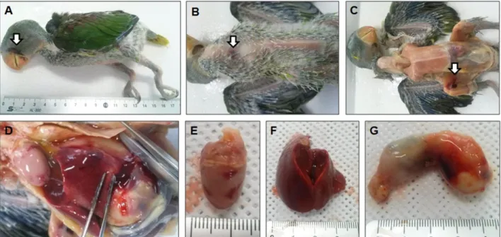
Fig. 1. Post-mortem examination of Alexndrine Parakeet (Psittacula eupatria) suspected with avian polyomavirus infection. (A and B) Abnormalities and lack of feathers on the neck and abdomen, and subcutaneous hemorrhage on the neck and cheek adjacent to the beak (arrow) (C) hemorrhage on the muscle of the femoral region (arrow) (D) ascites in the thoracic and abdominal cavities (E) multi-focal hemorrhages on the epi- cardium (F) enlargement of liver (G) diffuse hemorrhages on the sub-serosa of proventriculus and gizzard are shown.
등, 2005; Ogawa 등, 2006; Rahaus와 Wolff, 2005;
Zhuang 등, 2012), 우리나라에서는 아직까지 APV 감 염 사례에 대해 보고된 바가 없다. 이번의 연구에서 는 국내 조류원에서 급사하여 의뢰된 대본청 앵무 (Psittacula eupatria) 폐사체 시료로부터 최초로 APV 를 검출하였기에 그 증례를 보고하는 바이다.
증 례
경주시에 소재한 조류원에서 사육 중인 1개월령의 어린 앵무새 1수가 급사하여 2014년 4월 초순에 폐사 체가 경북대학교 수의전염병제어센터에 진단 의뢰되 었다. 의뢰된 앵무새는 대본청 앵무로 해당 조류원에 서 자체 부화하여 포육 중이었으며, 시판 이유식
복하여 내부 장기를 관찰한 결과, 흉수 및 복수가 다 소 저류되어 있었으며, 심외막의 다발성 출혈반점, 간장의 종대, 근위와 선위에 걸친 미만성 충․출혈 소 견이 관찰되었다(Fig. 1E∼G).
상기 임상증상(급사 및 피하출혈)과 부검소견(흉․복 수, 간장 종대 및 실질장기의 출혈)을 고려할 때 APV 의 감염이 의심되어 APV 유전자 검출을 위한 PCR을 실시하였다. 즉, 의뢰된 앵무새의 심장, 폐장, 간장, 신장 및 깃털을 채취하여 조직분쇄기(Bertin technol- ogy, Rockville, USA)를 이용하여 분쇄한 다음, 8,000 rpm에서 10분간 원심분리하여 상층액 200 l를 수거 하였고, 시판 핵산 추출 키트(Inclone biotech, Korea) 를 이용하여 핵산을 추출하였다. 추출한 핵산을 이용 하여 Johne과 Müller (1998)의 방법에 따라 PCR을 실시 한 다음, 1.2% agarose gel 전기영동을 실시하여 APV
Fig. 3. Allignment of nucleotide sequence of the amplified partial large T antigen gene of avian polyomavirus (APV) in this case and seven strains of APV reported by Katoh et al. (2009). The boxes indicate primer sequences for PCR.
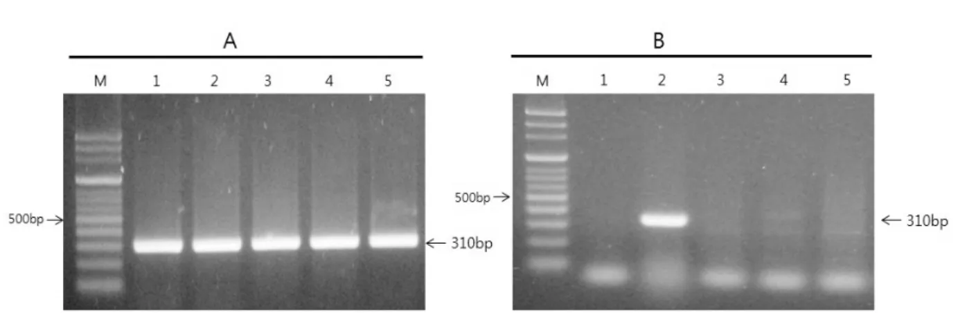
Fig. 2. Amplification of partial large T-antigen gene of avian polyomavirus (APV). (A) Amplified large T antigen gene of APV from tissues and feather of APV-infected Alexandrine Paraqeet. Lane M; 100 bp DNA ladder, Lane 1; heart, Lane 2; lung, Lane 3; liver, Lane 4; kidney, Lane 5;
feather. (B) Detection of 4 psittacine viral pathogens by PCR or RT-PCR. Lane M; 100 bp DNA ladder, Lane 1-5 are the results of amplification of avian bornavirus, APV, psittacine beak and feather disease virus and psittacine herpesvirus-specific gene and negative control, respectively.
과, 공시된 앵무새의 전 장기에서 310 bp의 특이 유 전자가 증폭되어 해당 개체가 APV에 감염되었음을 확인할 수 있었다(Fig. 2A). 또한, APV 이외 앵무새에 흔히 감염되는 바이러스성 질병의 감염 여부를 확인 하기 위하여 의뢰 앵무새의 장기로부터 추출한 핵산 을 이용하여 이전 연구자의 방법에 따라 avian borna- virus (ABV, Guo 등, 2012), psittacine beak and feather disease virus (PBFDV, Ypelaar 등, 1999), psittacid her- pesvirus (PHV, Tomaszewski 등, 2001)의 검출을 위하 여 PCR 및 RT-PCR을 실시하였다. 시험에 사용한 pri- mer는 ABV 검출을 위하여 Guo 등(2012)이 보고한 ABVM-F1 5'-GGTAATTGTTCCTGGATGGC-3', ABVM- R15'-ACACCAATGTTCCGAAGACG-3', PBFDV 검출을 위하여 Ypelaar 등(1999)이 보고한 M-F1 5'-AACCC- TACAGACGGCGAG-3', M-R1 5'-GTCACAGTCCTCC- TTGTACC-3', PHV 검출을 위하여 Tomaszewski 등 (2001)이 보고한 5'-CATGAACGGCATGCTGCCGT-3', 5'-GACTGCCACGGAGTATTGC-3'를 이용하여 PCR 및 RT-PCR을 수행하였으며, 동일한 방법으로 전기영동 을 실시하여 각각의 증폭산물을 확인한 결과, APV를 제외한 3종의 다른 바이러스는 검출되지 않아 이번 의 증례는 APV 단독감염에 의한 것으로 확인되었다 (Fig. 2B). 증폭된 APV 유전자 단편에 대한 핵산 염기 서열을 분석하고, DNASTAⓇLasergene (DNASTAR Inc.
USA)를 이용하여 Katoh 등(2009)이 보고한 APV 분리주 들의 핵산 염기서열과 비교분석한 결과, 99∼100% 일 치함이 확인되었다(Fig. 3).
고 찰
1981년 북미대륙에서 처음 APV가 확인된 이후 유럽 및 아시아 각국에서도 다양한 조류 종에서 APV 감염이 확인되었으나(Bert 등, 2005; Ogawa 등, 2006;
Rahaus와 Wolff, 2005; Zhuang 등, 2012), 현재까지 한
나타내는 것이 특징이다(Kaleta 등, 1984; Klautward 등, 1989; Phalen, 2007). 비 잉꼬류의 앵무새 종에서도 유사한 병증을 유발하지만 질병에 대한 감수성과 병 증의 정도는 감염된 조류 종과 감염숙주의 연령에 따 라 다양하며, 특히 포육 중에 있는 마카우(Macows), 코뉴어(Conures), 목도리 앵무(Ring-necked Paraqeets), 카이큐(Caiques) 및 뉴기니아 앵무(Eclectus Parrots) 종 에서 치명적인 병증을 유발하는 것으로 알려져 있다 (Enders 등, 1997; Phalen, 2007). 이번의 증례에서 APV 감염이 확인된 대본청 앵무는 1개월령의 포육 중인 어린 앵무새로서 조류원 관리 수의사가 급사 이전 피 하출혈을 확인하였다고 품고하였으며, 외관상 깃털의 비정상적인 발달과 피하출혈이 관찰되었고, 부검소견 으로 실질장기(심장, 선위)의 출혈 병변 등이 관찰되 었다(Fig. 1). 이는 이전의 연구자들이 보고한 APV 감 염 앵무새에서 나타난 임상증상 및 부검소견과 일치 하였다(Bernier 등, 1981; Davis 등 1981; Krautwald 등, 1989).
APV 감염증은 특징적인 임상증상과 부검소견에 근거하여 가진단할 수는 있지만 psittacine beak and feather disease virus와 같은 다른 병원체들도 유사한 병변을 유발하기 때문에 PCR과 같은 유전자진단법 으로 APV 바이러스 핵산을 검출하여 확진해야 한다 (Phalen 등, 1991; Johne과 Müller 1998). PCR 진단용 primer로는 APV의 유전자 증에서 가장 안정하여 모 든 APV를 검출할 수 있는 large T antigen 유전자 부 위에서 주로 선발하여 이용하고 있다(Johne과 Müller, 1998; Ogawa 등, 2006). 증례에서도 Johne과 Müller (1998)가 사용한 primer를 이용하여 PCR을 실시한 결 과, 공시한 모든 장기에서 310 bp의 large T antigen의 유전자를 증폭할 수 있었다(Fig. 2). 특정 장기에 친화 성을 가지는 포유류의 polyomavirus들과는 달리 APV 는 감염 개체의 여러 장기에서 증식하면서 세포를 파 괴하기 때문에 급성의 전신성 질병을 유발하게 된다 (Hou 등, 2005; Johne 등, 2000, Phalen 등, 1993). 따라
서 이번의 증례의 APV 이환 앵무새의 실질장기에서 공히 APV 유전자가 증폭된 것은 APV 감염의 이러한 특성을 반영한 결과라고 생각된다. 추가적으로 앵무 새 종에서 흔히 감염되는 바이러스성 질병 3종에 대한 유전자 검사를 실시한 결과(Fig. 2B), APV 이외 다른 바이러스의 유전자가 검출되지 않아 이번의 증례는 APV 단독감염에 의한 것으로 확인되었다.
PCR로 증폭된 APV large T antigen DNA의 핵산 염기 서열을 분석하여 이전에 보고된 APV의 해당 유전자 염 기서열과 비교한 결과, Katoh 등(2009)이 보고된 APV 유 전자염기서열들과 99∼100% 일치하였다(Fig. 3). APV는 바이러스가 분리된 지역 및 분리된 조류 종에 따라 해당 분리주간에 유사성이 높아서 독특한 1개 유전 형 또는 1개 혈청형으로 분류될 수 있다고 보고하였 다(Johne과 Müller, 1998; Phalen 등, 1993). 이러한 보 고를 고려할 때, 현재까지 국내에서는 APV에 대한 예찰이나 보고가 이루어지지 않았으나 국외로부터 반입되거나 국내에서 발생했을 가능성이 크게 존재 하므로, 한국에도 조류 종별로 특정 유전형의 APV가 감염되고 있을 가능성이 높을 것으로 추정된다. 그러 나 아직 한국에는 APV 유전자 염기서열에 대한 정보 가 거의 없기 때문에 현재로서는 분석이 곤란하며, 향후 국내의 다양한 조류 종에서의 APV 감염의 추가 확인과 유전자 정보 확보 노력이 필요하다.
APV는 감염 앵무새의 깃털 먼지, 호흡기 및 뇨 분 비물에 의해 수평감염되며, 알을 통한 수직감염도 가 능한 것으로 보고되고 있다(Bernier 등, 1984; Phalen, 2007). 또한, 특별한 임상증상을 보이지 않는 성조에 서도 다양한 비율로 APV가 검출되고 있어 불현성 감 염 성조와의 직․간접 접촉에 의한 수평전파 가능성도 흔히 발생할 것으로 추정된다(Bert 등, 2005; Rahaus 와 Wolff, 2005; Ogawa 등, 2006). 이번 증례의 대본청 앵무는 포육중인 어린 일령이기 때문에 어미새로부 터 수직감염되었을 가능성이 높다고 판단되나 해당 조류원에서 앵무새를 포함한 다양한 조류가 같이 사 육되고 있기 때문에 불특정의 불현성 감염 성조로부 터 수평감염되었을 가능성도 배제할 수 없다. 따라서 APV의 추가 발생을 예방하기 위해서는 해당 조류원에 서 사육중인 조류들에 대한 추가적인 조사와 함께 APV의 수직 및 수평감염을 막기 위한 체계적인 방역 대책 수립이 필요하다. 또한 이번 증례를 통하여 APV 의 국내 발생이 촤초로 확인되었기 때문에 국내 앵무 새에 대한 정확한 감염상황 파악을 위한 확대 조사가 필요하며, APV 피해 예방을 위한 바이러스 특성 조
사와 예방약 개발 등 체계적인 연구가 이루어져야 할 것으로 생각된다.
결 론
2014년 4월초에 경주 소재 조류원에서 사육 중이 던 1개월령 대본청 1수가 급사하여 경북대학교 수의 전염병제어센터에 질병 진단을 위하여 폐사체가 의 뢰되었다. 외관상 목과 복부의 비정상적인 깃털 발달 과 함께 목과 부리에 인접한 뺨 부위에 피하출혈이 관찰되었고, 부검을 실시한 결과, 대퇴부 근육의 출 혈, 복수, 심외막의 다발성 점상출혈, 선위 및 근위 장막하의 미만성 출혈 소견 등 전형적인 조류폴리오 마바이러스(avian polyomavirus; APV) 감염으로 의심 되는 병변이 관찰되었다. 내부 장기(심장, 폐장, 신장, 선위) 및 깃털을 대상으로 PCR을 실시한 결과, APV 의 large T antigen에 해당하는 특이 유전자가 검출되 었다. 반면에 앵무새에 흔히 감염되는 다른 3종의 바 이러스 즉, avian bornavirus, psittacine beak and feather disease virus 및 psittacid herpesvirus의 DNA는 검출되 지 않아 이번의 증례는 APV 단독감염에 의한 것으로 확인되었다. 증폭된 DNA 단편에 대한 유전자염기서 열 분석 결과, 이전에 보고된 APV 분리주들의 핵산 염기서열과 99∼100% 일치하는 것으로 확인되었다.
이상 이번 증례를 통하여 국내에서 급사한 대본청 앵 무새에서 APV가 처음으로 확인되었음을 보고한다.
감사의 글
이 논문은 2013 학년도 경북대학교 학술연구비의 지원을 받아 연구되었음.
참 고 문 헌
Bernier G, Morin M, Marsolais G. 1981. A generalized inclusion body disease in the budgerigar (Melopsittacus undulatus) caused by a papova-like agent. Avian Dis 25: 1083-1092.
Bemier G, Morin M, Marsolais G. 1984. Papovavirus induced feather abnormalities and skin lesions in the budgerigar:
clinical and pathological findings. Can Vet J 25: 307-310.
Bert E, Tomassone L, Peccati C, Navarrete MG, Sola SC. 2005.
Detection of beak and feather disease virus (BFDV) and avian polyomavirus (APV) DNA in psittacine birds in
Gardner SD, Knowles WA, Hand JF, Porter AA. 1989. Characte- rization of a new polyomavirus (Polyomavirus papio- nis-2) isolated from baboon kidney cell cultures. Arch Virol 105: 223-233.
Guo J, Covaleda L, Heatley JJ, Baroch JA, Tizard I, Payan SL.
2012. Widespread avian bornavirus infection in mute swans in the Northeast United States. Vet Med Res Rep 3: 49-52.
Hou J, Jens PJ, Major EO, zur Hausen HJ, Almeida J, van der Noordaa D, Walker D, Lowy D, Bernard U, Butel JS, Cheng D, Frisque RJ, Nagashima K. 2005. Polyo- maviridae. pp. 231-238. In: Fauquet CM, Mayo MA, Maniloff J, Desselberger U, Ball LA(ed.), Virus taxo- nomy. Eighth Report of the ICTV. Elsevier Academic Press, Amsterdam.
Johne R, Jungmann A, Müller H. 2000. Agnoprotein 1a and ag- noprotein 1b of avian polyomavirus are apoptotic inducers. J Gen Virol 81: 1183-1190.
Johne R, Müller H. 1998. Avian polyomavirus in wild birds: ge- nome analysis of isolates from Falconiformes and Psittaciformes. Arch Virol 143: 1501-1512.
Johne R, Müller H. 2003. The genome of goose hemorrhagic pol- yomavirus, a new member of the proposed subgenus Avipolyomavirus. Virology 308: 291-302.
Kaleta EF, Herbst W, Kaup FJ, Jank-Ludwig R, Marschall HJ, Drommer W, Krautwald ME. 1984. Investigations on the viral aetiology of a disease of budgerigars (Melopsitta- cus undulatus) with hepatitis and feather disorders. J Vet Med B 31: 219-224.
Katoh H, Ohya K, Une Y, Yamaguchi T, Fukushi H. 2009. Mole-
206-215. In: Thomas NJ, Hunter DB, Atkinson CT(ed.).
Infectious diseases of wild animal. 1st ed. Blackwell Publishing, Ames, Iowa.
Phalen DN, Wilson VG, Graham DL. 1991. Polymerase chain-re- action assay for avian polyomavirus. J Clin Microbiol 29: 1030-1037.
Phalen DN, Wilson VG, Graham DL. 1993. Organ distribution of avian polyomavirus DNA and virus neutralizing anti- body-titres in healthy adult budgerigars. Am J Vet Res 54: 2040-2047.
Rahaus M, Wolff MH. 2005. A survey to detect subclinical poly- omavirus infections of captive psittacine birds in Ger- many. Vet Microbiol 105: 73-76.
Rossi G, Taccini E, Tarantino C. 2005. Outbreak of avian poly- omavirus infection with high mortality in recently cap- tured Crimson's seedcrackers (Pyrenestes sanguineus). J Wildl Dis 41: 236-240.
Tomaszewski E, Wilson VG, Wigle WL, Phalen DN. 2001.
Detection and heterogeneity of Herpesviruses causing Pacheco’ disease in parrots. J Clin Microbiol 39: 533-538.
Ypelaar IM, Bassami R, Wilcox GE, Raidal SR. 1999. A univer- sal polymerase chain reaction for the detection of psitta- cine beak and feather disease virus. Vet Microbiol 68:
141-148.
Zhuang Q, Chen J, Mushtaq MH, Chen J, Liu S, Hou G, Li J, Huang B, Jiang W. 2012. Prevalence and genetic charac- terization of avian polyomavirus and psittacine beak and feather disease virus isolated from budgerigars in Mainland China. Arch Virol 157: 53-61.