Hot Water Extract of Scutellaria baicalensis Inhibits Migration, Invasion and Tube Formation in a Human Umbilical Vein Endothelial Cell Model and a Rat Aortic Ring Sprouting Model
Eok-Cheon Kim
1, Kiho Bae
1, Han Sung Kim
2, Yeong-Min Yoo
2, Michael Gelinsky
3and Tack-Joong Kim
1*
1Division of Biological Science and Technology, Yonsei-Fraunhofer Medical Device Lab, College of Science and Technology, Yonsei University, Wonju 26493, Korea
2Department of Biomedical Engineering, Yonsei-Fraunhofer Medical Device Lab, College of Health Science, Yonsei University, Wonju 26493, Korea
3Center for Translational Bone, Joint and Soft Tissue Research, Medical Faculty and University Hospital, Technische Universität Dresden, Dresden 01307, Germany
Received November 1, 2015 /Revised December 4, 2015 /Accepted December 13, 2015
Angiogenesis is essential for the pathophysiological processes of embryogenesis, tissue growth, dia- betic retinopathy, psoriasis, wound healing, rheumatoid arthritis, cardiovascular diseases, and tumor growth. Inhibition of angiogenesis represents an attractive therapeutic approach for the treatment of angiogenic diseases such as cancer. However, uncontrolled angiogenesis is also necessary for tumor development and metastasis. Inhibition of vascular endothelial growth factor (VEGF) signaling, a crit- ical factor in the induction of angiogenesis, cause robust and rapid changes in blood vessels of tumors and therefore VEGF constitutes a target for such anti-angiogenic therapy. Recently, since natural com- pounds pose significantly less risk of deleterious side effects than synthetic compounds, a great many natural resources have been assessed for useful substance for anti-angiogenic treatment. Here we eval- uated the anti-angiogenic effects of a hot water extract of Scutellaria baicalensis (SBHWE) using in vitro assays and ex vivo animal experiments. Our results show that SBHWE dose-dependently abrogated vascular endothelial responses by inhibiting VEGF-stimulated migration and invasion as well as tube formation in a human umbilical vein endothelial cell (HUVEC) model, without cytotoxicity, as de- termined by a cell viability assay. Further study revealed that SBHWE prevented VEGF-induced neo- vascularization in a rat aortic ring sprouting model. Taken together, our findings reveal an anti-angio- genic activity of Scutellaria baicalensis and suggest that SBHWE is a novel candidate inhibitor of VEGF-induced angiogenesis.
Key words :
Angiogenesis, Endothelial cell, Scutellaria baicalensis
*Corresponding author
*Tel : +82-33-760-2242, Fax : +82-33-760-2183
*E-mail : [email protected]
This is an Open-Access article distributed under the terms of the Creative Commons Attribution Non-Commercial License (http://creativecommons.org/licenses/by-nc/3.0) which permits unrestricted non-commercial use, distribution, and reproduction in any medium, provided the original work is properly cited.
Journal of Life Science 2016 Vol. 26. No. 1. 91~100 DOI : http://dx.doi.org/10.5352/JLS.2016.26.1.91
Introduction
Angiogenesis, a neovascularization from pre-existing ves- sels, is a key step for tumor growth and metastasis and is a complex process that includes degradation of the ex- tracellular matrix, migration, proliferation, sprouting, elon- gation, and tube formation of endothelial cells, controlled by a delicate balance between angiogenic inducers and an- giogenic inhibitors [6, 7, 10, 41]. Therefore, anti-angiogenic agents that can interfere with the essential steps of cancer
development are a promising strategy for human cancer treatment [2, 33, 43]. Numerous studies in cell culture and in animal models demonstrate that natural products derived from medicinal plants are able to exert chemopreventive and antitumor activities targeting angiogenesis in the tumor en- vironment [32, 40].
Scutellariae baicalensis, a medicinal plant, grows in various
regions of Asia, including Siberia, the far east of Russia,
Mongolia, China, Japan, and Korea, where it has been used
as an ingredient in botanical formulations for thousands of
years with positive results for the treatment of inflammatory
diseases, allergies, hyperlipemia, diabetes, arteriosclerosis,
and bacterial and viral infections [5, 17, 23, 25, 51, 52, 53,
57]. This herb has been included as an important ingredient
in various combination therapies such as Ger-Gen-Chyn-Tang
[15], Soshiho-tang [28], and Shuanghuanglian [61]. S. baicalensis
extracts have also been shown to modulate production of
cytokines related to antiviral activity [3], exert chemo- preventive effects against a variety of cancers [4, 22, 36, 56], induce apoptosis of human monocytic leukemia and osteo- genic sarcoma cells [14], and inhibit metastasis in hep- atocellular carcinoma cells [37]. These beneficial effects are due to its active constituents, which include baicalin, baica- lein, wogonoside, and wogonin [4, 11, 53, 55, 56]. Although numerous biological activities of S. baicalensis have been re- ported, no studies have examined the anti-angiogenic effect of the extract.
Therefore, the purpose of the present study was to inves- tigate the anti-angiogenic activity of SBHWE on VEGF-in- duced angiogenesis through measurement of migration, in- vasion, and differentiation of HUVECs in vitro, and an aortic ring sprouting angiogenesis assay ex vivo.
Materials and Methods
Animals
Seven-week-old Sprague-Dawley rats were purchased from Orient Co. (Sungnam, Korea). The rats were housed in a temperature-controlled room with a 12 hr light and 12 hr dark schedule. They were given food and water ad libitum.
All experiments were conducted in accordance with the
“Guide for the Care and Use of Laboratory Animals” adopt- ed by the United States National Institutes of Health and reviewed and approved by the Ethics Committee, Institutio- nal Animal Care and Use Committee (IACUC, Approval # YWC-131127-1) of Yonsei University (Wonju, Korea).
Plant extract and reagents
The S. baicalensis extract used in the current investigation was provided by the Korea Rural Development Administra- tion. The extract was prepared in distilled deionized water as a stock solution at a concentration of 100 mg/ml, and was stored at -80°C. Before use in the experiments, the stock solution was clarified and diluted with M199 medium at dif- ferent concentrations.
3-(4,5-dimethylthiazol-2-yl)-2,5-diphenyltetrazolium bro- mide (MTT) was purchased from USB Corporation (Cleve- land, OH, USA). Matrigel basement membrane matrix was obtained from BD Biosciences (Bedford, MA, USA). hVEGF
165, recombinant human VEGF
165which has cross reactivity for rat, was purchased from PeproTech (Rocky Hill, NJ, USA).
Antibodies to VEGFR2, phospho-VEGFR2 (Tyr 1175), phos- pho-ERK (Thr 202/Tyr 204), ERK, phospho-p38 MAP kinase
(Thr 180/Tyr 182), and p38 MAP kinase were obtained from Cell Signaling Technology (Beverly, MA, USA). Horseradish peroxidase-conjugated secondary antibody was purchased from Thermo Scientific Inc. (Rockford, IL, USA). Cell culture reagents and most other biochemical reagents were pur- chased from Sigma (St. Louis, MO, USA), unless otherwise specified.
Cell culture
Human umbilical vein endothelial cells (HUVECs) pur- chased from the American Type Culture Collection (ATCC
®, Manassas, VA, USA) were maintained on gelatin-coated plates in M199 medium (GIBCO
®, Grand Island, NY, USA) supplemented with 20% fetal bovine serum (FBS) (GIBCO
®, Gaithersburg, MD, USA), 3 ng/ml basic fibroblast growth factor (KOMABIOTECH, Seoul, Korea), 100 U/ml penicillin, 100 mg/ml streptomycin (GIBCO
®, Grand Island, NY, USA), and 5 U/ml heparin (Sigma) at 37°C in 5% CO
2in air, and used at early passages (passages 6-10) in all experiments.
For in vitro experiments, HUVECs were serum- and growth factor-starved for 6 hr, after which the effects of SBHWE were assessed.
Cell viability assay
An MTT assay, a measure of cell viability, was used to assess the cytotoxicity of SBHWE. Briefly, confluent HUVECs in gelatin-coated 24-well plates were treated with different concentrations (1-100 μg/ml) of SBHWE in triplicate. The treated cells were incubated with the extract for 24 hr, fol- lowed by the addition of 100 μl of MTT solution (5 mg/ml in PBS) to each well. The plates were further incubated at 37°C for 4 hr and subsequently the medium was carefully removed from the plates, and dimethyl sulfoxide (DMSO) was added to solubilize the formazan produced from MTT by viable cells. The optical density (OD) was read using a plate reader (Molecular Devices, Sunnyvale, CA, USA) at 595 nm. The cytotoxicity of SBHWE was calculated as follows:
cell viability (%) = (average absorbance of SBHWE-treated group / average absorbance of control group)×100.
Cellular proliferation assay
HUVECs were seeded at a density of 4×10
4cells per well
in gelatinized 12-well plates containing M199 medium with
20% FBS and incubated for 24 hr. These cells were serum-
and growth factor-starved and treated either with VEGF (20
ng/ml) or VEGF (20 ng/ml) + SBHWE (1-100 μg/ml), and
incubated at 37°C for 24 hr. Thereafter, proliferation of these cells was measured using an MTT assay. OD readings were taken at 595 nm with a spectrophotometer (Molecular Devices) and the absorbance values were normalized to un- treated controls to calculate growth percentages.
Migration assay
To evaluate the effects of SBHWE on VEGF-induced HUVEC migration, endothelial cell migration was assessed using gelatin-coated 3-μm pore Transwell inserts (Costar, Corning, NY, USA) placed in 24-well plates. HUVECs (5×10
4cells/100 μl/insert) were plated in M199 medium (1% FBS) containing various concentrations of SBHWE in the upper chamber of the transwell and M199 medium (1% FBS, 600 μl) containing VEGF (20 ng/ml) was added into the lower chamber as a chemoattractant. After 4 hr, non-migrated cells were removed by cotton swab and migrated cells were stained with H&E and examined under a microscope. The number of migrated cells was quantified by counting the cells under a 40× objective. Each treatment was done in- triplicate and eight images were quantified per each trans- well membrane.
Cell invasion assays
The invasion assay was performed using a Transwell chamber containing a polycarbonate filter with a pore size of 3 μm as described [20]. HUVECs were suspended in the Matrigel-coated upper chambers in M199 medium (1% FBS) containing SBHWE, at a final concentration of 5×10
4cells/
100 μl. The bottom chambers (600 μl) were filled with M199 (1% FBS) containing VEGF (20 ng/ml). After incubation for 24 hr, the noninvasive cells that remained on the upper sur- face of the filter were removed by a cotton swab. Cells that traversed through the Matrigel were attached to the under- surface of the insert filter. The cells were stained with H&E and then eight fields of each membrane were counted under an inverted optical microscope (200×), and the average num- ber of cells in each field was calculated. The data were ex- pressed as percent invasion compared with the control.
Tube formation analysis
This study examined endothelial tube formation using an
in vitro angiogenesis assay. Matrigel Growth Factor Reduced(GFR) (BD Biosciences, Bedford, MA) was used as a sub- strate for the study of angiogenesis. Matrigel GFR solution was thawed overnight at 4°C, and the Matrigel GFR solution
(150 μl) was distributed onto each 24-well plate and allowed to solidify for 30 min at 37°C. Cells were trypsinized, re- suspended in M199 medium (1% FBS) at 2×10
5cells/ml with either VEGF (20 ng/ml) or VEGF (20 ng/ml) + SBHWE, and the cell suspension was seeded onto a 24-well plate pre-coat- ed with Matrigel and allowed to form capillary tubes. The capillaries formed were photographed after 20 hr using an inverted microscope, and the average tube areas were quan- tified using Image-Pro Plus (Media Cybernetics, Bethesda, MD, USA).
Rat aortic ring sprouting assay
The standard rat aortic ring assay was used as a model for our ex vivo angiogenesis study. Freshly sacrificed dorsal aortas from seven-week-old male Sprague Dawley rats (n=3) were rinsed with ice-cold HBSS, separated from fibroadipose tissue, and cut into 1 mm-long pieces using a sterile surgical blade. Each aorta ring was placed in a well of a 48-well plate pre-coated with Matrigel matrix (120 μl), and then covered with another layer of Matrigel matrix (50 μl). After polymer- ization for 30 min, serum-free M199 media was added to each well in the presence or absence of SBHWE with or with- out 20 ng/ml VEGF. On day 7 the rings were photographed at 40× magnification by phase-contrast microscopy and mi- crovessel outgrowth was quantified. Sprouting was meas- ured using the following scale: 0 = no sprouting; 1 = mi- grated cells without sprouting; 2 = isolated sprouting; 3 = sprouting in 25-50% of the arterial ring circumference; 4 = sprouting in 50-75% of the circumference; and 5 = sprouting in 75-100% of the circumference. The assay was scored from 0 to 5 in a double-blinded manner, and each data point was assessed six times.
Statistical analysis
All numerical values are represented as the mean ± stand- ard deviations. Statistical significance was determined using Student's t-test. Each experiment was repeated at least three times to yield comparable results. Values with p<0.05 and
p<0.01 were considered to indicate a statistically significantdifference.
Results
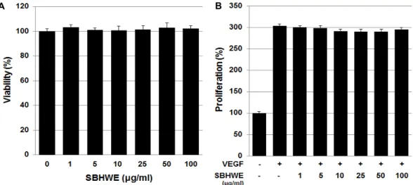
Effect of SBHWE on viability and proliferation of HUVECs.
To rule out the possibility that inhibition of angiogenesis
A B
Fig. 1. Effects of SBHWE on the viability and VEGF-induced proliferation of HUVECs. (A) HUVECs were incubated with various concentrations (1, 5, 10, 25, 50, and 100 μg/ml) of SBHWE. After 24 hr, cytotoxicity was quantified by an MTT assay. (B) HUVECs were pretreated for 30 min with various concentrations (1, 5, 10, 25, 50, and 100 μg/ml) of SBHWE before exposure to VEGF (20 ng/ml). After 24 hr, the number of proliferated cells was determined by an MTT assay. VEGF treatment alone served as a positive control. Each bar represents the average ± SE of three independent experiments.
was due to cytotoxic effects, we analyzed the viability of HUVECs treated with SBHWE extract by MTT assay. As shown in Fig. 1A, following 24 hr of incubation with various concentrations of SBHWE, endothelial cell viabilities were not significantly changed by treatment with this extract. The results indicate that exposure to SBHWE does not induce cytotoxic effects in the conditions of this study.
To characterize the anti-angiogenic activity of SBHWE, we first determined whether SBHWE inhibited growth factor-in- duced endothelial cell proliferation. HUVECs were exposed to various concentrations of SBHWE, then cultured with or without VEGF (20 ng/ml) for 24 hr. HUVEC proliferation was significantly increased in response to VEGF treatment, but it was not suppressed as the concentration of SBHWE increased (Fig. 1B). This result indicates that SBHWE does not affect the growth of HUVECs in response to VEGF at these concentrations.
Effect of SBHWE on motility of HUVECs
Migration is representative of the ability of endothelial cells to form new blood vessels. We next studied the effects of SBHWE on VEGF-induced cell migration using a Trans- well plate. Fig. 2A shows a photomicrograph of endothelial cells migrating across the filter membrane after 4 hr of incubation. In the absence of SBHWE, a large number of HUVECs migrated to the lower side of the filter in the Transwell chamber following stimulation with VEGF (20 ng/ml). This VEGF-induced migration of endothelial cells
was dose-dependently inhibited by SBHWE treatment (Fig.
2B). SBHWE alone had no significant effect on basal migra- tion of these endothelial cells. These results revealed that SBHWE significantly inhibited the motility of HUVECs.
Effect of SBHWE on invasion of HUVECs
Since invasion of vascular endothelial cells into the ex- tracellular matrix is an important step for neovascular for- mation, we used Matrigel-coated polycarbonate filters in Transwell chambers to elucidate the anti-invasive effect of SBHWE on HUVECs. HUVECs plated in the upper chamber, which contained M199 media plus various concentrations of SBHWE, were allowed to invade the lower chamber, which contained VEGF acting as a chemoattractant. Our results showed that SBHWE dose-dependently abolished the VEGF- stimulated invasion of HUVECs across the Matrigel-coated filter (Fig. 3). This result indicates that SBHWE markedly inhibited invasion of HUVECs in response to the pro-angio- genic factor, VEGF.
Effect of SBHWE on tube formation by HUVECs To verify its anti-angiogenic activity, we examined the ef- fects of SBHWE on VEGF-induced tube formation by HUVECs on Matrigel, a well-established angiogenesis assay.
Our study showed that VEGF stimulation of HUVECs seed-
ed on Matrigel promotes differentiation to form capil-
lary-like tubes within 20 hr; however, in the presence of
SBHWE, the width and length of the endothelial tubes in-
A B
Fig. 2. SBHWE inhibits the VEGF-induced migration of HUVECs. HUVECs were treated with VEGF (20 ng/ml) in the presence or absence of SBHWE (1, 5, 10, 25, 50, and 100 μg/ml). Chemotactic migration was assayed after incubation in Transwell plates for 4 hr. (A) Representative migrated cells were photographed. (B) Cells that migrated to the bottom of the filter were counted using optical microscopy. The in vitro angiogenesis assay was performed as described in Materials and Methods.
VEGF treatment alone served as a positive control. Data are expressed as mean ± SE (n = 3). *p<0.05 and **p<0.01 versus VEGF alone.
A B
Fig. 3. SBHWE inhibits VEGF-induced invasion of endothelial cells. Effect of SBHWE on HUVEC invasion measured using Transwell culture plates. HUVECs were treated for 24 hr with VEGF (20 ng/ml) and various concentrations (10, 50, and 100 μg/ml) of SBHWE. (A) Representative invaded cells were photographed. (B) Cells that invaded to the bottom of the filter were counted using optical microscopy. VEGF treatment alone served as a positive control. **p<0.01 versus VEGF alone.
duced by VEGF were reduced dose-dependently. At a con- centration of 100 μg/ml, SBHWE completely abrogated en- dothelial tube formation. To quantify the degree of tube for- mation, photographs were taken under an inverted micro-
scope (Fig. 4A), and the tube area was calculated using im-
age analysis software (Fig. 4B). This result suggests that
SBHWE may have an inhibitory effect on angiogenesis.
A B
Fig. 4. SBHWE inhibits VEGF-induced tube formation by endothelial cells. HUVECs were preincubated for 30 min with SBHWE (10, 50, or 100 μg/ml) and plated on Matrigel-coated plates at a density of 2×10⁵ cells per well. They were incubated in the presence or absence of 20 ng/ml VEGF, and microphotographs were obtained after 20 hr (40×). (A) Representative endothe- lial tubes are shown. (B) The area covered by the tube network was measured using Image-Pro Plus software. The experiments were repeated three times, and values represent mean ± SE values of triplicate determinations. **p<0.01 versus VEGF alone.
A B
Fig. 5. SBHWE inhibits VEGF-induced vessel sprouting ex vivo. Aortas in Matrigel were cultured with or without VEGF (20 ng/ml) in the presence or absence of SBHWE, and analyzed on day 7. (A) Representative aortic rings were photographed. (B) SBHWE blocks VEGF-induced vessel sprouting. The assay was scored from 0 (least positive) to 5(most positive), and the data represent mean ± SE values (n = 6). **p<0.01 versus VEGF alone.
Effect of SBHWE on angiogenesis ex vivo To further characterize its anti-angiogenic activity, we performed the aortic ring sprout formation assay, a widely used ex vivo angiogenesis assay that mimics several stages of angiogenesis, including endothelial cell proliferation, mi- gration, invasion, and tube formation. Treatment with VEGF significantly stimulated vessel sprouting from aortic rings
embedded in Matrigel, compared to the results with me-
dium alone. However, compared with the VEGF alone-treat-
ed group, SBHWE significantly inhibited the growth of
blood vessels in the aortic rings of rats in a concentration-
dependent manner (Fig. 5). These results demonstrate that
SBHWE caused a dramatic decrease in sprout length and
density from the aortic ring. In conclusion, SBHWE exhibited
anti-angiogenic activity both in vitro and ex vivo.
Discussion
Natural products are potential sources of alternative med- icines for chemoprevention in humans [46, 47]. These prod- ucts have various chemical properties and can reduce the development of tumors through multiple mechanisms, which include inhibition of angiogenesis and stimulation of apoptosis [49]. Scutellaria baicalensis has been widely used as a traditional medicine for the treatment of various ail- ments, such as inflammation, cancer, infectious diseases, hy- percholesterolemia, and hypertension in East Asia since an- cient times [8, 21, 44, 60, 62]. Additional studies have demon- strated that baicalin, baicalein, and wogonin, the main bio- active flavonoids derived from S. baicalensis, show antitumor and anti-angiogenic effects on various cancer cells [29-31].
Recent studies have reported that an aqueous extract of S.
baicalensis has a neuroprotective effect on excitotoxic primary
neuronal cell death in cortical cells [54], can ameliorate drug addiction-related behavior through functional regulation of dopamine receptors in rat [58], can prevent hematuria and blood-brain barrier disruption in rats because of its anti-in- flammatory effects [19, 45], has a hepatoprotective effect in mice fed a high-fat diet [27], and promotes a strong chemo- therapeutic effect in lung cancer cells [38]. Although numer- ous studies on the physiological and pharmacological func- tions of S. baicalensis have been conducted, the detailed func- tions and mechanisms thereof have not been elucidated thus far.
Angiogenesis plays a critical role in tumor progression and is a tightly regulated process that involves proliferation, migration, invasion, and organization into capillaries by en- dothelial cells [9, 12]. Currently, one of the most thriving fields in drug development, with a plethora of new drugs on the market, is inhibition of angiogenesis through the spe- cific targeting of VEGF, a potent stimulator of angiogenesis, and of its receptors expressed on the cell surface, which are the major mediators of VEGF-induced proangiogenic signal- ing in endothelial cells, by therapeutic antibodies and natu- ral products [1, 16, 24, 35, 50]. Thus in the present study, we examined the effects of SBHWE on VEGF-induced angio- genesis both in vitro and ex vivo.
Since endothelial cells play major roles in each step of VEGF-mediated angiogenesis, in order to investigate the an- ti-angiogenic effects of SBHWE in HUVECs, we first de-
termined the non-toxic concentration of SBHWE to be used for our experiments by examining the cytotoxicity of various concentrations of SBHWE (1-100 μg/ml). No cytotoxic effects of SBHWE were observed at the given concentrations (Fig.
1A). We then examined the effects of a non-toxic concen- tration of SBHWE on the process of angiogenesis, in the form of endothelial cell proliferation, migration, and invasion in response to VEGF, resulting in the formation of capillary tubes by HUVECs. Our in vitro study demonstrated that SBHWE significantly inhibited the migratory properties of cells toward a chemoattractant, VEGF (Fig. 2), and of the ability of VEGF-stimulated cells to pass through a Matrigel- coated membrane (Fig. 3) and differentiate into capillary-like structures formed of VEGF-induced endothelial cells (Fig. 4), although VEGF-induced proliferation was not affected by SBHWE treatment (Fig. 1B). It is to be noted that SBHWE alone had no effects on proliferation of the endothelial cells.
These results indicate that the inhibitory angiogenic activity of SBHWE is due to suppression of migration, invasion, and tube formation, but not of proliferation. To determine the underlying molecular mechanism of the anti-angiogenic ef- fects of SBHWE, we examined its effects on phosphorylation of key signal transduction pathways. Binding of VEGF to the receptor tyrosine kinase VEGFR2 leads to activation of VEGFR2’s cytoplasmic domain and initiation of intracellular signaling cascades, including activation of extracellular sig- nal-regulated kinase (ERK), phosphoinositide 3-kinase (PI3K)-AKT, c-Jun N-terminal kinase (JNK), and p38 mi- togen-activated protein kinase (p38 MAPK), which are nec- essary for essential angiogenic processes [18, 26, 34, 39, 42, 48, 59]. In this study, we found that the phosphorylation of VEGFR2, ERK1/2, and p38 MAPK in response to VEGF were not downregulated by SBHWE treatment (data not shown). Although we did not investigate the effect of this extract on activation of the PI3K-AKT and JNK pathways by VEGF, at the cellular level SBHWE completely sup- pressed the migration, invasion, and tube formation of VEGF-treated endothelial cells. These results indicate that the inhibitory effect of SBHWE on angiogenesis is not via ERK1/2 and p38 MAPK, and that the anti-angiogenic activ- ity of SBHWE may be due to the blockage of another activa- tion pathway. More experiments are in progress to under- stand the molecular targets and mechanisms underlying the inhibitory effect of SBHWE on VEGF-induced angiogenesis.
To further confirm the anti-angiogenic potential of SBHWE,
we conducted an ex vivo rat aortic ring assay. Neoangiogen-
esis, the growth of new capillaries, in response to VEGF syn- thesized by tumor cells plays a crucial role in tumor growth and metastasis [13]. Similar to the results of the tube for- mation assay, the VEGF-stimulated outgrowth of vessels from the fragmented aorta ring is suppressed as the dose of SBHWE escalates. This implies that SBHWE significantly inhibited the formation of new vessels.
Based on the data provided in the present study, we con- clude that a hot water extract of S. baicalensis can sig- nificantly inhibit VEGF-induced angiogenesis via sup- pression of endothelial cell migration, invasion, and tube for- mation in vitro, and sprout formation from aortic rings ex
vivo. Consequently, S. baicalensis may be a promising chemo-preventive and therapeutic agent for the treatment of angio- genesis-related diseases such as cancer.
Acknowledgements
We are grateful to Dr. Yusu Shin (National Institute Horticultural & Herbal Science, RDF, KOREA) for providing us with SBHWE. This research was supported by the Leading Foreign Research Institute Recruitment Program through the National Research Foundation of Korea (NRF) funded by the Ministry of Science, ICT & Future Planning (2010-00757), and by the Basic Science Research Program through the National Research Foundation of Korea (NRF), funded by the Ministry of Science, ICT & Future Planning (NRF-2014R1A1A2059027).
References
1. Bernatchez, P. N., Soker, S. and Sirois, M. G. 1999.
Vascular endothelial growth factor effect on endothelial cell proliferation, migration, and platelet-activating factor synthesis is Flk-1-dependent. J. Biol. Chem. 274, 31047- 31054.
2. Bhat, T. A. and Singh, R. P. 2008. Tumor angiogenesis - a potential target in cancer chemoprevention. Food Chem.
Toxicol. 46, 1334-1345.
3. Błach-Olszewska, Z., Jatczak, B., Rak, A., Lorenc, M., Gulanowski, B., Drobna, A. and Lamer-Zarawska, E.
2008. Production of cytokines and stimulation of resist- ance to viral infection in human leukocytes by Scutellaria baicalensis flavones. J. Interferon Cytokine Res. 28, 571-581.
4. Bonham, M., Posakony, J., Coleman, I., Montgomery, B., Simon, J. and Nelson, P. S. 2005. Characterization of chem- ical constituents in Scutellaria baicalensis with antiandrogenic and growth-inhibitory activities toward prostate carcinoma.
Clin. Cancer Res. 11, 3905-3914.
5. Bruno, M., Piozzi, F. and Rosselli, S. 2002. Natural and hemi- synthetic neoclerodane diterpenoids from scutellaria and their antifeedant activity. Nat. Prod. Rep. 19, 357-378.
6. Bussolino, F., Mantovani, A. and Persico, G. 1997. Molecular mechanisms of blood vessel formation. Trends Biochem. Sci.
22, 251-256.
7. Carmeliet, P. and Jain, R. K. 2000. Angiogenesis in cancer and other diseases. Nature 407, 249-257.
8. Chi, Y. S. and Kim, H. P. 2005. Suppression of cyclo- oxygenase-2 expression of skin fibroblasts by wogonin, a plant flavone from Scutellaria radix. Prostaglandins Leukot.
Essent. Fatty Acids 72, 59-66.
9. Dvorak, H. F. 2002. Vascular permeability factor/vascular endothelial growth factor: a critical cytokine in tumor angio- genesis and a potential target for diagnosis and therapy.
J. Clin. Oncol. 20, 4368-4380.
10. Fan, T. P., Jaggar, R. and Bicknell, R. 1995. Controlling the vasculature: angiogenesis, anti-angiogenesis and vascular targeting of gene therapy. Trends Pharmacol. Sci. 16, 57-66.
11-29. Fas, S. C., Baumann, S., Zhu, J. Y., Giaisi, M., Treiber, M. K., Mahlknecht, U., Krammer, P. H. and Li-Weber, M.
2006. Wogonin sensitizes resistant malignant cells to TNFalpha- and TRAIL-induced apoptosis. Blood 108, 3700-3706.
12. Ferrara, N. 2009. Vascular endothelial growth factor.
Arterioscler. Thromb. Vasc. Biol. 29, 789-791.
13. Harvey, K., Welch, Z., Kovala, A. T., Garcia, J. G. and English, D. 2002. Comparative analysis of in vitro angio- genic activities of endothelial cells of heterogeneous origin.
Microvasc. Res. 63, 316-326.
14. Himeji, M., Ohtsuki, T., Fukazawa, H., Tanaka, M., Yazaki, S., Ui, S., Nishio, K., Yamamoto, H., Tasaka, K. and Mimura, A. 2007. Difference of growth-inhibitory effect of Scutellaria baicalensis-producing flavonoid wogonin among human cancer cells and normal diploid cell. Cancer Lett. 245, 269-274.
15 Ho, F. M., Liao, Y. H., Yang, A. J., Lee Chao, P. D., Hou, Y. C., Huang, C. T., Lin, S. R., Lee, K. R., Huang, K. C.
and Lin, W. W. 2012. Anti-atherosclerotic action of Ger-Gen-Chyn-Lian-Tang and AMPK-dependent lipid low- ering effect in hepatocytes. J. Ethnopharmacol. 142, 175-187.
16. Holmes, K., Roberts, O. L., Thomas, A. M. and Cross, M.
J. 2007. Vascular endothelial growth factor receptor-2: struc- ture, function, intracellular signalling and therapeutic inhibition. Cell Signal. 19, 2003-2012.
17. Huang, D., Ding, Y., Luo, W. M., Bender, S., Qian, C. N., Kort, E., Zhang, Z. F., VandenBeldt, K., Duesbery, N. S., Resau, J. H. and Teh, B. T. 2008. Inhibition of MAPK kinase signaling pathways suppressed renal cell carcinoma growth and angiogenesis in vivo. Cancer Res. 68, 81-88.
18. Huang, K. L., Chen, C. S., Hsu, C. W., Li, M. H., Chang, H., Tsai, S. H. and Chu, S. J. 2008. Therapeutic effects of baicalin on lipopolysaccharide-induced acute lung injury in rats. Am. J. Chin. Med. 36, 301-311.
19. Huan, S. K., Wang, K. T., Yeh, S. D., Lee, C. J., Lin, L. C., Liu, D. Z. and Wang, C. C. 2012. Scutellaria baicalensis alle-
viates cantharidin-induced rat hemorrhagic cystitis through inhibition of cyclooxygenase-2 overexpression. Molecules 17, 6277-6289.
20. Huang, S. M., Li, J. and Harari, P. M. 2002. Molecular Inhibition of angiogenesis and metastatic potential in hu- man squamous cell carcinomas after epidermal growth fac- tor receptor blockade. Mol. Cancer Ther. 1, 507-514.
21. Huang, W. H., Lee, A. R. and Yang, C. H. 2006. Antioxidative and anti-inflammatory activities of polyhydroxyflavonoids of Scutellaria baicalensis GEORGI. Biosci. Biotechnol. Biochem.
70, 2371-2380.
22. Ikemoto, S., Sugimura, K., Yoshida, N., Yasumoto, R., Wada, S., Yamamoto, K. and Kishimoto, T. 2000. Antitumor effects of Scutellariae radix and its components baicalein, baicalin, and wogonin on bladder cancer cell lines. Urology 55, 951-955.
23. Ishimaru, K., Nishikawa, K., Omoto, T., Asai, I., Yoshihira, K. and Shimomura, K. 1995. Two flavone 2'-glucosides from Scutellaria baicalensis. Phytochemistry 40, 279-281.
24. Karkkainen, M. J. and Petrova, T. V. 2000. Vascular endothe- lial growth factor receptors in the regulation of angiogenesis and lymph angiogenesis. Oncogene 19, 5598-5605.
25. Kim, E. H., Shim, B., Kang, S., Jeong, G., Lee, J. S., Yu, Y.
B. and Chun, M. 2009. Anti-inflammatory effects of Scutellaria baicalensis extract via suppression of immune modulators and MAP kinase signaling molecules. J.
Ethnopharmacol. 126, 320-331.
26. Koch, S., Tugues, S., Li, X., Gualandi, L. and Claesson-Welsh, L. 2011. Signal transduction by vascular endothelial growth factor receptors. Biochem. J. 437, 169-183.
27. Lee, I. S., Park, S., Park, K. and Choue, R. 2011. Hepatopro- tective activity of scutellariae radix extract in mice fed a high fat diet with chronic alcohol exposure. Phytother. Res. 25, 1348-1353.
28. Lee, J. J., Kim, T., Cho, W. K. and Ma, J. Y. 2013. Antithrom- botic and antiplatelet activities of Soshiho-tang extract. BMC Complement. Altern. Med. article 137, 8 pages.
29. Liao, H. L. and Hu, M. K. 2004. Synthesis and anticancer activities of 5, 6, 7-trimethylbaicalein derivatives. Chem.
Pharm. Bull. (Tokyo) 52, 11621165.
30. Lin, Y. T., Yang, J. S., Lin, H. J., Tan, T. W., Tang, N. Y., Chaing, J. H., Chang, Y. H., Lu, H. F. and Chung, J. G. 2007.
Baicalein induces apoptosis in SCC-4 human tongue cancer cells via a Ca2+-dependent mitochondrial pathway. In Vivo 21, 1053-1058.
31. Li-Weber, M. 2009. New therapeutic aspects of flavones: the anticancer properties of Scutellaria and its main active con- stituents wogonin, baicalein and baicalin. Cancer Treat. Rev.
35, 57-68.
32. Milanowski, J. and Milanowska-Szmygin, K. 2013. Treatment of non-small cell lung cancer-where we are? Pneumonol.
Alergol. Pol. 81, 55-60.
33. Molassiotis, A., Fernadez-Ortega, P., Pud, D., Ozden, G., Scott, J. A., Panteli, V., Margulies, A., Browall, M., Magri, M., Selvekerova, S., Madsen, E., Milovics, L., Bruyns, I., Gudmundsdottir, G., Hummerston, S., Ahmad, A. M.,
Platin, N., Kearney, N. and Patiraki, E. 2005. Use of comple- mentary and alternative medicine in cancer patients: A European survey. Ann. Oncol. 16, 655-663.
34. Murphy, D. A., Makonnen, S., Lassoued, W., Feldman, M.
D., Carter, C. and Lee, W. M. 2006. Inhibition of tumor endo- thelial ERK activation, angiogenesis, and tumor growth by sorafenib (BAY43-9006). Am. J. Pathol. 169, 1875-1885.
35. Olsson, A. K., Dimberg, A., Kreuger, J. and Claesson-Welsh, L. 2006. VEGF receptor signalling - in control of vascular function. Nat. Rev. Mol. Cell Biol. 7, 359-371.
36. Park, H. J., Lee, Y. W., Park, H. H., Lee, Y. S., Kwon, I.
B. and Yu, J. H. 1998. Induction of quinone reductase by a methanol extract of Scutellaria baicalensis and its flavonoids in murine Hepa 1c1c7 cells. Eur. J. Cancer Prev. 7, 465-471.
37. Park, H. S., Park, K. I., Hong, G. E., Nagappan, A., Lee, H. J., Kim, E. H., Lee, W. S., Shin, S. C., Seo, O. N., Won, C. K., Cho, J. H. and Kim, G. 2014. Korean Scutellaria baica- lensis Georgi methanol extracts inhibits metastasis via the Forkhead Box M1 activity in hepatocellular carcinoma cells.
J. Ethnopharmacol. 155, 847-851.
38. Park, K. I., Park, H. S., Kang, S. R., Nagappan, A., Lee, D.
H., Kim, J. A., Han, D. Y. and Kim, G. S. 2011. Korean Scutellaria baicalensis water extract inhibits cell cycle G1/S transition by suppressing cyclin D1 expression and ma- trix-metalloproteinase-2 activity in human lung cancer cells.
J. Ethnopharmacol. 133, 634-641.
39. Pedram, A., Razandi, M. and Levin, E. R. 1998. Extracellular signal-regulated protein kinase/Jun kinase cross-talk under- lies vascular endothelial cell growth factorinduced endothe- lial cell proliferation. J. Biol. Chem. 273, 26722-22678.
40. Rajput, S. and Mandal, M. 2012. Antitumor promoting po- tential of selected phytochemicals derived from spices: a review. Eur. J. Cancer Prev. 21, 205-215.
41. Risau, W. 1997. Mechanisms of angiogenesis. Nature 386, 671-674.
42. Rousseau, S., Houle, F., Kotanides, H., Witte, L., Walten- berger, J., Landry, J. and Huot, J. 2000. Vascular endothelial growth factor (VEGF)-driven actin-based motility is medi- ated by VEGFR2 and requires concerted activation of stress-activated protein kinase 2 (SAPK2/p38) and geldana- mycin-sensitive phosphorylation of focal adhesion kinase.
J. Biol. Chem. 275, 10661-10672.
43. Sagar, S. M., Yance, D. and Wong, R. 2006. Natural health products that inhibit angiogenesis: a potential source for in- vestigational new agents to treat cancer - Part 1. Curr. Oncol.
13, 14-26.
44. Scheck, A. C., Perry, K., Hank, N. C. and Clark, W. D. 2006.
Anticancer activity of extracts derived from the mature roots of S.Scutellaria baicalensis on human malignant brain tumor cells. BMC Complement. Altern. Med. 16, 27.
45. Shin, J. W., Kang, H. C., Shim, J. and Sohn, N. W. 2012.
Scutellaria baicalensis attenuates blood-brain barrier dis- ruption after intracerebral hemorrhage in rats. Am. J. Chin.
Med. 40, 85-96.
46. Singh, M., Singh, P. and Shukla, Y. 2012. New strategies in cancer chemoprevention by phytochemicals. Front Biosci.
초록:혈관내피세포와 흰쥐 대동맥 미세혈관 발아 모델을 이용한 황금 열수추출물의 세포의 이동, 침투 및 관형성 억제 연구
김억천
1․배기호
1․김한성
2․유영민
2․겔린스키 미첼
3․김택중
1*
(1연세대학교 생명과학기술학부, 2연세대학교 의공학과, 3드레스덴 공대 의과대학 병원)
혈관신생의 억제는 암과 같은 신생혈관형성 질환의 치료를 위해 유용한 접근법이다. 신생혈관형성의 핵심인 자인 혈관내피세포성장인자는 신생혈관형성 질환의 치료를 위한 주요한 표적이다. 그러므로, 본 연구에서는 in
vitro 분석과 ex vivo 동물 실험을 통해 황금 열수추출물의 신생혈관형성 억제효과를 연구했다. 본 연구결과에서황금 열수추출물이 혈관내피세포성장인자에 의해 자극된 혈관내피세포에 있어 세포독성 없이 세포의 이동, 침 투, 관형성을 농도 의존적으로 억제하였다. 더 나아가 황금 열수추출물은 혈관내피세포성장인자에 의해 유도된 흰쥐 대동맥 주변 미세혈관 발아를 예방하였다. 본 연구결과들은 황금 열수추출물이 신생혈관형성 억제작용이 있고, 이는 혈관내피세포성장인자에 의해 유도된 혈관신생을 억제 하는 잠재적 소재가 될 수 있음을 제안한다.
(Elite Ed) 4, 426-452.
47. Tan, A. C., Konczak, I., Sze, D. M. and Ramzan, I. 2011.
Molecular pathways for cancer chemoprevention by dietary phytochemicals. Nutr. Cancer 63, 495-505.
48. Thakker, G. D., Hajjar, D. P., Muller, W. A. and Rosengart, T. K. 1999. The role of phosphatidylinositol 3-kinase in vas- cular endothelial growth factor signaling. J. Biol. Chem. 274, 10002-10007.
49. Valko, M., Leibfritz, D., Moncol, J., Cronin, M. T., Mazur, M. and Telser, J. 2007. Free radicals and antioxidants in nor- mal physiological functions and human disease. Int. J.
Biochem. Cell Biol. 39, 44-84.
50. Wahl, O., Oswald, M., Tretzel, L., Herres, E., Arend, J. and Efferth, T. 2011. Inhibition of tumor angiogenesis by anti- bodies, synthetic smallmolecules and natural products.
Curr. Med. Chem. 18, 3136-3155.
51. Waisundara, V. Y., Hsu, A., Huang, D. and Tan, B. K. 2008.
Scutellaria baicalensis: enhances the anti-diabetic activity of metformin in streptozotocin-induced diabetic wistar rats.
Am. J. Chin. Med. 36, 517-540.
52. Wang, C. Z., Mehendale, S. R., Calway, T. and Yuan, C.
S. 2011. Botanical flavonoids on coronary heart disease. Am.
J. Chin. Med. 39, 661-671.
53. Wang, C. Z., Mehendale, S. R. and Yuan, C. S. 2007.
Commonly used antioxidant botanicals: active constituents and their potential role in cardiovascular illness. Am. J. Chin.
Med. 35, 543-558.
54. Yang, J., Wu, X., Yu, H., Liao, X. and Teng, L. 2014. NMDA receptor-mediated neuroprotective effect of the Scutellaria baicalensis Georgi extract on the excitotoxic neuronal cell death in primary rat cortical cell cultures. Scientific World Journal Article ID 459549, 8 pages.
55. Ye, F., Wang, H., Jiang, S., Wu, J., Shao, J., Cheng, X., Tu, Y. and Zhang, D. Y. 2004. Quality evaluation of commercial extracts of Scutellaria baicalensis. Nutr. Cancer 49, 217-222.
56. Ye, F., Xui, L., Yi, J., Zhang, W. and Zhang, D. Y. 2002.
Anticancer activity of Scutellaria baicalensis and its potential mechanism. J. Altern. Complement. Med. 8, 567-572.
57. Yoon, S. B., Lee, Y. J., Park, S. K., Kim, H. C., Bae, H., Kim, H. M., Ko, S. G., Choi, H. Y., Oh, M. S. and Park, W. 2009.
Anti-inflammatory effects of Scutellaria baicalensis water ex- tract on LPS-activated RAW 264.7 macrophages. J.
Ethnopharmacol. 125, 286-290.
58. Yun, J. and Jung, Y. S. 2014. A Scutellaria baicalensis radix water extract inhibits morphine-induced conditioned place preference. Pharm. Biol. 52, 1382-1387.
59. Zachary, I. and Gliki, G. 2001. Signaling transduction mech- anisms mediating biological actions of the vascular endothe- lial growth factor family. Cardiovasc. Res. 49, 568-581.
60. Zhang, D. Y., Wu, J., Ye, F., Xue, L., Jiang, S., Yi, J., Zhang, W., Wei, H., Sung, M., Wang, W. and Li, X. 2003. Inhibition of cancer cell proliferation and prostaglandin E2 synthesis by Scutellaria baicalensis. Cancer Res. 63, 4037-4043.
61. Zhang, H., Chen, Q., Zhou, W., Gao, S., Lin, H., Ye, S., Xu, Y. and Cai, J. 2013. Chinese medicine injection shuanghuan- glian for treatment of acute upper respiratory tract infection:
a systematic review of randomized controlled trials. Evid.
Based Complement. Alternat. Med. Article ID 987326, 7 pages.
62. Zhang, N., Van Crombruggen, K., Holtappels, G. and Bachert, C. 2012. A herbal composition of Scutellaria baica- lensis and Eleutherococcus senticosus shows potent anti-in- flammatory effects in an ex vivo human mucosal tissue model. Evid. Based Complement. Alternat. Med. Article ID 673145, 9 pages.