PREVENTION RESEARCH □ ORIGINAL ARTICLE □
47 책임저자:김영진, }609-735, 부산시 금정구 장전동 산30
부산대학교 분자생물학과
Tel: 051-510-2176, Fax: 051-513-9258 E-mail: [email protected]
접수일:2008년 3월 4일, 게재승인일:2008년 3월 18일
Correspondence to:Yung-Jin Kim
Department of Molecular Biology, Pusan National University, 30, Geumjeong-gu, Busan 609-735, Korea
Tel: +82-51-510-2176, Fax: +82-51-513-9258 E-mail: [email protected]
A Natural Marine Compound Extracted from the Ark Shell, Scapharca Subcrenata , Inhibits
in Vivo and in Vitro Angiogenesis
Eui-Yeun Yi1, Shi-Young Park1, Jae-Sun Choi1, Chi-Won Lim2, Yeon-Kye Kim2, Hee-Yeon Park2 and Yung-Jin Kim1
1Department of Molecular Biology, Pusan National University, Busan 609-735, Korea
2Biotechnology Research Center, National Fisheries Research and Development Institute, Busan 408-1, Korea
Angiogenesis is an integral process in the growth and spread of solid tumors, and anti-angiogenesis therapy is one of the most promising therapeutic strategies for the treatment of cancer. In this study, we show that a natural marine compound extracted from ark shell, Scapharca subcrenata, has anti-angiogenic activities. Ark shell extract inhibited the proliferation, migration, invasion and tube formation of human umbilical vein endothelial cells (HUVECs), and suppressed the release of matrix metalloproteinase-2 (MMP-2) by HUVECs. In addition, ark shell extract also inhibited in vivo angiogenesis in a mouse Matrigel-plug assay. Taken together, these results suggest that ark shell extract acts as a novel angiogenesis inhibitor and could be further developed as an anticancer agent. (Cancer Prev Res 13, 47-53, 2008)
Key Words: Marine bio-source, Tumor, Anti-angiogenic drug, Invasion, Shellfish
INTRODUCTION
Angiogenesis, which is the development of new blood vessels from pre-existing endothelium,1,2) is a significant component of a wide variety of physiological process (e.g. wound healing, embryonic development) and pathological conditions (e.g.
diabetic eye disease, tumor growth and spread).3) Angiogenesis is a strictly regulated phenomenon that is controlled by a balance of angiogenic stimulators and inhibitors.4) In addition, complex and diverse cellular actions, such as degradation of the extracellular matrix (ECM), proliferation and migration of endothelial cells, and morphological differentiation of endothe- lial cells to form tubes are also involved in angiogenesis.5,6)
Many pharmaceutical agents have been discovered by screening natural products produced by plants, animals, marine
organisms and microorganisms. Compounds such as vincristine, irinotecan, etoposide and paclitaxel, which are derived from plants, and dactinomycin, bleomycin and doxorubicin, which are derived from microbial sources are used for the treatment of cancer. In addition, citarabine, which originates from a marine source, as well as other agents originating from marine sources, including bryostatin-1, aplidine, dolastatin 10 and ET-743, have recently entered phase I and II clinical trials.7)
Therefore, we tested several natural marine compounds, including ark shell extract, to determine if they had anti-angiogenic capability using in vitro and in vivo angiogenesis assays. We found that ark shell extract inhibited proliferation, migration, invasion, tube formation, and production of MMP-2 in HUVECs in vitro and inhibited in vivo angiogenesis in a mouse Matrigel plug assay.
MATERIALS AND METHODS 1. Materials
Crude extract of ark shell was provided by the National Fisheries Research and Development Institute (Busan, Korea).
Matrigel was purchased from Collaborative Biomedical Pro- ducts (Bedford, MA) and used at a concentration of 10 mg/ml for the mouse Matrigel plug assay. VEGF and heparin were purchased from Life Technologies (Gaithersbeg, MD).
2. Animals
Specific pathogen free (SPF) male C57BL/6 mice were supplied by Hyochang Science (Daegu, Korea). The mice were housed at 23±0.5oC, 10% humidity in a 12 h light-dark cycle and provided with autoclaved tap water and lab chow ad libitum.
3. Crude extract of ark shell
A sample of S. subcrenata was dried using a freeze drying machine and then extracted with methanol in a blender. The extract was then fractionated into 85% aq. MeOH fractions.
4. Mouse Matrigel plug assay
C57BL/6 mice (7 weeks of age) were injected subcutaneously with 500μl of Matrigel containing VEGF (100 ng/ml) and heparin (50 units/500μl) with or without Crude extract of ark shell (0.1, 0.5, 1.0 mg). After seven days, the skin of the mouse was pulled back to expose the Matrigel plug, which remained intact. The Matrigel plugs were then photographed and the hemoglobin content was measured according to the Drabkin method8) using Drabkin reagent kit 525 (Sigma, MO) for the quantification of blood vessel formation.
5. Cell Culture
HUVECs were grown in MCDB (Life Technologies, NY) supplemented with heat-inactivated 10% fetal bovine serum (WelGENE Inc., Korea), 10 ng/ml epidermal growth factor, 1μg/ml hydrocortisone, 10 mM/Liter L-Glutamine, 100 units/
ml of penicillin, and 100μg/ ml of streptomycin in a 37oC incubator with a humidified atmosphere containing 5% CO2.
6. Proliferation assay
The effect of crude extract of ark shell on HUVEC
proliferation was determined by an MTT assay, which is based on the conversion of MTT (3-(4, 5-dimethylthiazol-2-yl)-2,-5- diphenyl tetrazolium bromide) to insoluble MTT-formazan by cleavage of the tetrazolium ring by mitochondrial dehydro- genase enzymes in living cells. Briefly, HUVECs were grown in MCDB with 10% FBS at a density of 2×10⁴ cells on 24-well culture plates. After one night, the media was replaced with MCDB containing 1% FBS, and crude extract of ark shell and the cells were then incubated for 24 h at 37oC under a humidified atmosphere that was comprised of 5% CO2. Next, MTT solution (5 mg/ml in H2O) was added to the well followed by the addition of 0.3 ml of demetyl sulfoxide (DMSO) to dissolve the MTT formazan. The amount of crystal violet dye was then determined by measuring the absorbance at 540 nm. Each sample was assayed in triplicate, and the experiment was repeated three times.
7. Wounding migration assay
HUVECs that were plated on 60 mm culture dishes and grown to 90% confluence were wounded with a razor blade that was 2 mm in width and the injury line was then marked.
After wounding, the cultures were washed with serum-free medium and further incubated in MCDB with 1% serum, 1 mM thymidine and/or crude extract of ark shell. HUVECs were allowed to migrate for 24 h and then rinsed with serum-free medium, followed by fixing with absolute methanol and staining with Giemsa. Migration was quantified by counting the number of cells that moved beyond the reference line.
8. Invasion assay
The invasiveness of the HUVECs was analyzed using a transwell chamber system with 8μm-pore-polycarbonate filter inserts. The lower side of the filter was coated with 10μl of type IV collagen (3 mg/ml), and the upper side was coated with 10μl of Matrigel (0.5 mg/ml). HUVECs (2×10⁴ cells) and crude extract of ark shell were placed in the upper part of the filters and BSA was placed in the lower part. After 24 h of incubation at 37oC, cells on both sides of the membrane were fixed with methanol and then stained with hematoxy- line/eosin. Cells on the upper surface of the membrane were then removed by wiping with a cotton swab, and the number of cells on the lower side of the membrane was counted by optical microscopy at 40 x magnification.
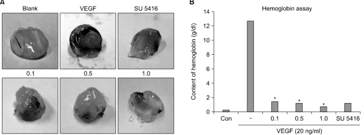
Fig. 1. The effect of crude extract of ark shell on in vivo angiogenesis. (A) Matrigel plugs were photographed (×40). Concentration of materials; VEGF (20 ng/ml), SU5416 (20 uM), extracts (0.1, 0.5, 1.0 mg/ml). (B) Quantification of hemoglobin content. Each value was generated from three different experiments. Blank; Matrigel alone, SU5416; VEGF receptor inhibitor. *p<0.05 versus hemoglobin content of VEGF-induced implants.
9. Tube formation assay
HUVECs (2×10⁴ cells) were seeded on a layer of previously polymerized Matrigel and then treated with crude extract of ark shell. After 24 h of incubation, the morphology of the cells was observed under a phase contrast microscope and photo- graphed at 40×magnification.
10. Gelatin-based zymography
The conditioned medium in which the HUVECs were cultured was analyzed by gelatin-based zymography, using the procedure described by Herron et al., with slight modification (1986). Conditioned medium were separated by SDS-PAGE using 10% acryl amide copolymerized with 0.33 mg/ml gelatin (Sigma, MO). After electrophoresis, the gel was rinsed twice with 2.5% Triton X-100 for 15 min and then incubated at 37oC for 24 h in incubation buffer (0.05 M Tris- HCl pH 7.5, 0.15 M NaCl, 0.01 M CaCl2, 1μM ZnCl2, and 0.02 % NaN3).
Gelatinase was identified following staining of the gel with coomassie-brilliant blue R250 and destaining with 7% acetic acid. The digested area appeared clear on a blue background, indicating the location of gelatinase.
11. Data analysis and statistics
Data were presented as the means±the SD or as a percentage of the control. Statistical comparisons between
groups were performed using the Student’s t test. *p<0.05 was considered to be statistically significant.
RESULTS
1. Crude extract of ark shell inhibits in vivo angiogenesis
To select a target sample that exhibited anti-angiogenic activity we initially used the Marine Bioactive Resource Database (MBRDB, http://portal.nfrdi.re.kr/mbrdb). First, we selected 6 samples (CM0520-110, CM0520-120, SN0540-260, SN0540-340, M60440-030 and 190430-230) that showed strong cytotoxic activity on the SNU-1 cell line and then narrowed these samples to CM0520-120, which showed a higher anti-angiogenic activity than the other 9 samples.
CM0520-120 (Scapharca subcrenata) was then collected from the East Sea of Korea on February 2005 and used for further experiments.
We used the mouse Matrigel plug assay, an established in vivo angiogenesis model, to evaluate the anti-angiogenic activity of the ark shell extract (Fig. 1),9) with SU5416, an angiogenesis inhibitor, as a positive control to compare the anti-angiogenic effects of ark shell extract. SU5416 (NSC 696819) is a small organic molecule that inhibits VEGF-mediated signaling through the Flk-1/KDR (VEGFR-2) tyrosine kinase receptor, which interferes with endothelial cell proliferation and the
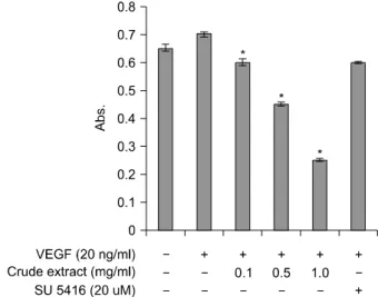
Fig. 2. Effect of crude extract of ark shell on the proliferation of HUVECs. Increasing the concentration of ark shell extract reduced the proliferation of HUVECs for 24 h. Data represent the mean±the SD of three independent experiments per- formed in triplicate. *p<0.05 versus control.
neovascularization required for the propagation of cancer.10) As shown in Fig. 1A, Matrigel plugs containing VEGF were abundantly filled with intact red blood cells, which indicate the formation of a functional vasculature inside the Matrigel, whereas vessels were not observed in the untreated Matrigel (blank). Furthermore, the Matrigel plug containing the ark shell extract produced fewer vessels than the plug containing VEGF, indicating that ark shell extract inhibits the formation of VEGF-induced neomicrovessels. We also measured the hemoglobin contents inside the Matrigel plugs to quantify the anti-angiogenic effect of the ark shell extract (Fig. 1B). The hemoglobin content of the VEGF-treated plug was found to be 13 g/dl, whereas that of ark shell treated plugs was only approximately 1 g/dl, which suggests that ark shell extract has strong in vivo anti-angiogenic activity.
2. Crude extract of ark shell inhibits proliferation of HUVECs
To determine the effects of ark shell extract on endothelial cell functions crucial to angiogenesis, its effect on VEGF-induced angiogenesis in vitro was investigated. We first examined the effect of ark shell extract on the proliferation of HUVECs by conducting an MTT assay using concentrations of the extract that ranged from 0 to 1.0 mg/ml (Fig. 2).
Treatment of the HUVECs with crude extract of ark shell for 24 h decreased their proliferation in a dose-dependent manner,
with cells treated with 1.0 mg/ml showing a 70% decrease in proliferation when compared with that of the control. These results suggest that ark shell extract inhibits the proliferation of HUVECs.
3. Crude extract of ark shell inhibits migration and invasion in HUVECs
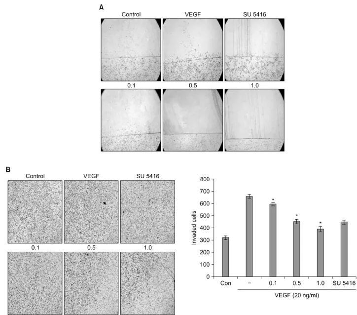
Migration and invasion of endothelial cells is one of the critical features in the formation of new blood vessels and the repair of injured vessels. Therefore, we investigated the effect of ark shell extract on the movement of HUVECs from a wounded edge to the open area using a wounding migration assay. Treatment with ark shell extract for 24 h profoundly decreased the migration of HUVECs, and this effect occurred in a dose-dependent manner (Fig. 3A).
To examine the effect of ark shell extract on the invasiveness of HUVECs, we conducted an invasion assay using a transwell system in which the lower and upper sides of the filter were coated with type IV collagen and Matrigel, respectively. Crude extract of ark shell suppressed the invasiveness of HUVECs in a dose-dependent manner after 24 h of incubation (Fig 3B), which indicates that ark shell extract inhibits migration and invasion in HUVECs.
4. Crude extract of ark shell reduces production and activation of MMP-2 in HUVECs
Regulation of extracellular proteolytic activity is important in the process of endothelial cell migration and invasion through the basement membrane, as well as in capillary morphogenesis. Therefore, we examined the effect of ark shell extract on the MMP-2 production of HUVECs using gelatin-based zymography. Treatment of HUVECs with ark shell extract for 24 h reduced the production of MMP-2 in a dose-dependent manner (Fig. 4), which indicates that ark shell extract reduces the production of MMP-2 in HUVECs.
5. Crude extract of ark shell inhibits the tube formation of HUVECs
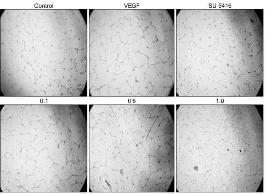
We conducted a tube formation assay to estimate the effect of ark shell extract on the differentiation of HUVECs.11) Briefly, HUVECs were placed on a growth factor-reduced Matrigel-coated plate and then incubated for 24 h. As shown in Fig. 5, HUVECs on Matrigel formed a blood vessel network
Fig. 3. Effect of crude extract of ark shell on the activity of migration and invasion. Ark shell extract inhibited the activity of migration (A) and invasion (B) of HUVECs in a dose-dependent manner. Data represent the mean±the SD of three independent experiments performed in triplicate. *p<0.05 versus control.
Fig. 4. Effect of crude extract of ark shell on the production of MMP-2. Ark shell extract reduced the production of MMP-2 in a dose-dependent manner.
in the absence of ark shell extract, whereas treatment with ark shell extract strongly inhibited the formation of tube-like structures in a dose-dependent manner.
DISCUSSION
Anticancer agents are often derived from the isolation of an active lead compound that occurs spontaneously in nature.
Until recently, natural product-derived anticancer drug development relied almost exclusively on the screening of terrestrial sources, such as plant extracts and fermentation products.12) However, the marine environment has proven to be a rich source of extremely potent compounds that have significant anti-tumor, anti-inflammatory, analgesic, immuno- modulating, anti-allergy and anti-viral effects.13)
Fig. 5. Effect of crude extract of ark shell on the tube formation of HUVECs. Ark shell extract inhi- bited the development of capillary- like structures (×40).
In cancer, tumor growth is dependent on the angiogenic growth of new blood vessels. Trials of anti-angiogenic therapy are based on strategies that interfere with angiogenic ligands, their receptors or downstream signaling. Angiogenesis is a complex process involving several cascades, a) enzymatic degradation of the vascular basement membrane and interstitial matrices by endothelial cells, b) directed migration and proliferation of endothelial cells to provide cells for the new vessels, c) invasion of endothelial cells and d) formation of capillary-like networks by endothelial cells.14,15)
To evaluate the utilization of ark shell extract as an anti-angiogenesis agent, we first determined the anti- angiogenic activity of ark shell extract by performing an in vivo mouse Matrigel-plug assay. The results of this assay showed that ark shell extract highly inhibited the formation of neomicrovessels in Matrigel (Fig. 1A), and we also noted that these newly synthesized vessels in the Matrigel plugs containing ark shell extract were not abundantly filled with intact red blood cells (Fig. 1B). In addition, we observed the effects of ark shell extract on angiogenesis in each step using in vitro angiogenesis assays. Furthermore, we observed the inhibition of proliferation by ark shell extract (Fig. 2), and found that this decrease in proliferation occurred in a dose-dependent manner.
The migration and invasive activity of HUVECs was also evaluated by wounding migration (Fig. 3A) and invasion (Fig.
3B) assays, since the mobility of endothelial cells, such as migration and invasion, is one of the initiation steps in angiogenesis. The results of these assays indicated that ark shell extract significantly reduced both of these activities in a dose-dependent manner.
Matrix Metalloproteinases (MMPs), which are secreted as proenzymes, are key regulators of extracellular matrix turnover that function through the degradation of a wide variety of extracellular matrix proteins.16) The regulation of extracellular proteolytic activity is important in the processes of endothelial cell migration and invasion, which occur during angiogenesis, and is also an essential step in tumor invasion and metastasis.17) Because the motility of the HUVECs was profoundly suppressed by the extract, we tested the effect of ark shell extract on the production of MMP-2 using gelatin-based zymography. As expected, ark shell extract reduced the production of MMP-2 (Fig. 4), which indicates that the inhibitory effects of ark shell extract on angiogenesis may be at least in part dependent on the reduction of MMP-2. In addition, the results of these in vitro angiogenesis assays indicated that ark shell extract has strong inhibitory effects on a series of angiogenic processes, including endothelial cell proliferation, migration, invasion, and tube formation. One aspect of endothelial cell chemotaxis in vitro that is important to the process of angiogenesis is the ability to promote
morphological differentiation into capillary-like structures. In the absence of ark shell extract, HUVECs that were placed on the Matrigel formed short and thick capillary-like networks that appeared to be indicative of angiogenic development, whereas HUVECs in the presence of ark shell extract showed reduced tube-like structures (Fig. 5).
In summary, that the results of this study indicated that ark shell extract inhibited the in vitro angiogenesis of HUVECs as well as in vivo VEGF-induced neovascularization in mouse Matrigel-plugs, and that these effects were associated with the decreased releases of -MMP-2. Therefore, we suggest that ark shell extract exhibits strong anti-angiogenic actions and also has the potential to be a useful inhibitor of a large number of serious diseases that are characterized by up-regulated angiogenesis. However, further study is required to identify and purify the responsible compound from the ark shell extract as well as to determine the molecular mechanisms by which a purified compound produced by the ark shell modulates endothelial cell function.
CONCLUSION
The ark shell extract exhibits strong anti-angiogenic actions and also has the potential to be a useful inhibitor of a large number of serious diseases that are characterized by up-regulated angiogenesis.
ACKNOWLEDGEMENTS
This study was supported in part by a grant from the 2003 Korean National Cancer Control Program (0220060-3), Ministry of Health & Welfare, R.O.K.
REFERENCES
1) Folkman J, Shing Y. Angiogenesis. J Biol Chem 267, 10931-10934, 1992.
2) Siddiqui FA, Desai H, Siddiqui TF, Francis JL. Hemoglobin induces the expression and secretion of vascular endothelial growth factor from human malignant cells. Hematol J 3,
264-270, 2002.
3) Savani RC, Cao G, Pooler PM, Zaman A, Zhou Z. Differential involvement of the Hyaluronan (HA) Receptors CD44 and Receptor for HA-mediated Motility in Endothelial Cell Function and Angiogenesis. J Biol Chem 276, 36770-36778, 2001.
4) Miao RQ, Agata J, Chao L, Chao J. Kallistatin is a new inhibitor of angiogenesis and tumor growth. Blood 100, 3245-3252, 2002.
5) Lee MS, Moon EJ, Lee SW, Kim MS, Kim KW, Kim YJ.
Angiogenic activity of pyruvic acid in in vivo and in vitro angiogenesis models. Cancer Res 61, 3290-3293, 2001.
6) Folkman J. What is the evidence that tumors are angiogenesis dependent? J Natl Cancer Inst 82, 4-6, 1990.
7) Da Rocha AB, Lopes RM, Schwartsmann G. Natural products in anticancer therapy. Curr Opin Pharm 1, 364-369, 2001.
8) Drabkin DS, Ausin JH. Spectrophotomeric constans for common hemoglobin derivaties in human, dog and rabbit blood. J Biol Chem 98, 719, 1932.
9) Passaniti A, Taylor RM, Pili R, et al. A simple, quantitative method for assessing antiogenesis and antiangiogenic agents using reconstituted basement membrane, heparin and fibroblast growth factor. Lab Investig 67, 519-528, 1992.
10) Fong TA, Shawver L, Sun L, et al. SU5416 is a potent and selective inhibitor of the vascular endothelial growth factor receptor (Flk-1/KDR) that inhibits tyrosine kinase catalysis, tumor vascularization and growth of multiple tumor types.
Cancer Res 59, 99-106, 1999.
11) Folkman J, Haudenschild C. Angiogenesis in vitro. Nature 288, 551-556, 1980.
12) Schwartsmann G, Da Rocha AB, Mattei J, Lopes R. Marine- derived anticancer agents in clinical trails. Expert Opin Investig Drugs 12, 1367-1383, 2003.
13) Newman DJ, Cragg GM. Marine natureal products and related compounds in clinical and advanced preclinical trails. J Nat Prod 67, 1216-1238, 2004.
14) Bussolino F, Mantovani A, Persico G. Molecular mechanisms of blood vessel formation. Trends Biochem Sci 22, 251-256, 1997.
15) Carmeliet P, Jain RK. Angiogenesis in cancer and other diseases. Nature 407, 249-257, 2000.
16) Giavazzi R, Taraboletti G. Preclinical development of metallo- proteasis inhibitors in cancer therapy. Ciritical Rev Oncol/
Hematol 37, 53-60, 2001.
17) Liekens S, Clercq ED, Neyts J. Angiogenesis: regulators and clinical applications. Biochem Pharm 61, 253-270, 2001.