• 검색 결과가 없습니다.

The nutlet morphology of the genus Glechoma L. (Lamiaceae) and its related taxa

N/A
N/A
Protected

Academic year: 2021

Share "The nutlet morphology of the genus Glechoma L. (Lamiaceae) and its related taxa"

Copied!
9
0
0

로드 중.... (전체 텍스트 보기)

전체 글

(1)

긴병꽃풀속(Glechoma L. 꿀풀과) 및 근연분류군의 소견과 형태에 관한 연구

장태수·홍석표*

경희대학교 이과대학 생물학과 & 기초과학연구소

The nutlet morphology of the genus Glechoma L. (Lamiaceae) and its related taxa

Tae-Soo Jang and Suk-Pyo Hong*

Laboratory of Plant Systematics, Department of Biology, Institute of Basic Sciences, Kyung Hee University, Seoul, 130-701, Korea (Received 3 March 2010 : Accepted 16 March 2010)

적 요: Glechoma 속 4종(G. grandis, G. hederacea, G. hirsuta, G. longituba)과 Nepetinae아족 중 Glechoma속과 가장 근연 분류군인 Agastache속, Dracocephalum속, Marmoritis속, Meehania속, Nepeta속 일부 분류군의 소견과 특징을 주사전자현미경과 광학현미경을 통하여 분류학적 유용성을 검토하였다. 연구된 분류군의 소견과 모양은 난형에서 타원형으로 나타났다. 소견과 크기는 길이 1.36−3.83 mm, 폭 0.80−2.65 mm로, Marmoritis rotundifolia가 가장 크고, Agastache nepetoides가 가장 작았다. 소견과의 표면은 세 가지 유형으로 나타났다. Type I은 Agastache 속으로 표면은 불규칙적으로 돌출되며, 장방형의 세포가 연결되는 유형이고, Type II는 Dracocephalum속으로 표면은 불규칙적이며, 원형의 세포가 움푹 들어간 유형이다. Type III은 Glechoma속, Marmoritis속, Meehania속,

Nepeta 속으로 표면은 매끄러우며, 세포가 능선으로 연결된 망상형이다. 모용(다세포성 비선모, 단세포성 비선모,

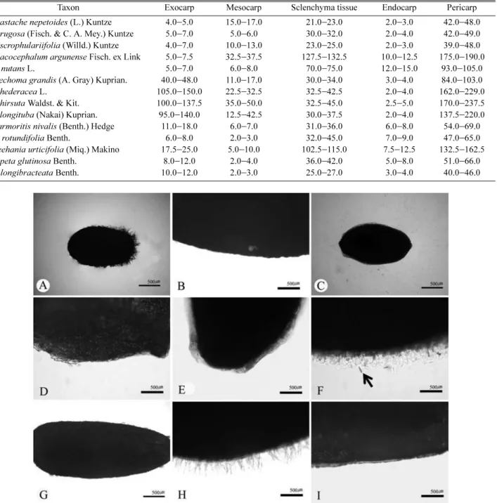
방패형 선모)은 일부 분류군에서 관찰되었다. 과피는 외과피와 중과피, 내과피로 구성되며, 과피의 두께(39.0−

237.5 µm)가 분류군간 큰 차이를 보였다. Glechoma속과 근연속 일부 근연분류군에 대한 소견과의 형태학적·

해부학적 형질(소견과의 모양, 표면 무늬, 선모와 모용 분포, 해부학적 구조)을 기재하였고, 그 분류학적 유용 성에 대해 논의하였다.

주요어: 긴병꽃풀속 및 근연분류군, 꿀풀과, 소견과 형태, 해부학적 형질

ABSTRACT: The nutlet morphology and pericarp structure of 14 taxa (4 taxa of the genus Glechoma and related taxa) were studied by light and scanning electron microscopy, and a detailed description of nutlet morphological features for all examined taxa is provided. The shape of the nutlet is ovate to elliptic. Its size is 1.36 −3.83 mm in length and 0.80−2.65 mm in width. The largest one was found in Marmoritis rotundifolia, while the smallest one was in Agastache nepetoides. Three different surface types were recognized in the studied taxa based on dif- ferences in cell outline and cell boundary relief (rectangular, pitted, and ridged negative reticulate-cell). Three types of trichome (uni-cellular non-glandular trichome, multi-cellular non-glandular trichome, and peltate glan- dular trichome) were distributed in the nutlets. The pericarp is formed in three layers (i.e., exocarp, mesocarp, and endocarp including sclenchyma tissue), and the pericarp in the studied species is 39.0 −237.5 µm thick. The morphological and anatomical features (the nutlet shape, surface type, and trichome distribution) in the genus Glechoma and related taxa are described and discussed.

Keywords: Glechoma and related taxa, Lamiaceae, nutlet morphology, anatomical characters

*Author for correspondence: [email protected]

(2)

꿀풀과(Lamiaceae), 박하족(Mentheae Dumort.)에 속하는 긴병꽃풀속(Glechoma L.)은 다년생 초본으로, 약 8종이 아 시아, 유럽에 분포한다(Li and Hedge, 1994). 본 속 식물의 줄기는 사각형으로, 기거나 비스듬히 서고, 잎은 심장형 으로 대생한다. 꽃은 액생하며 2−6개가 달리고, 악은 통형 에서 종형이며 15맥이다. 화관은 종형으로 윗부분이 팽대 하고 가장자리는 입술모양이다. 네 개의 수술 중 두 개는 화관 밖으로 돌출되어 있으며, 암술대는 두 갈래로 길이 가 거의 같고, 소견과는 암갈색으로 타원형에서 난형으로 털이 없다(Lee, 2003; Lee, 2006; Jang and Hong, 2007).

긴병꽃풀속은 포복경을 가지고, 2−6 개의 꽃이 액생하며, 약이 열리는 각도가 90

o

에서 두 갈래로 분지하는 특징들에 의해 근연속(Agastache Clayton ex Gronov., Dracocephalum L., Marmoritis Benth., Meehania Britton ex Small & Vail, Nepeta L.) 과 구별된다(Harley et al., 2004). 그러나 긴병꽃풀속을 포함한 일부 분류군의 분자생물학적 분석(rbcL, cpDNA, ITS 구간에 대한 염기서열 분석)에 의하면 속간의 한계가 불명확하게 존재하기도 하며, 선행 연구에서는 본 속내 일부 분류군(ITS - A. foeniculum, D. moldavica, G. hederacea;

cpDNA - A. nepetoides, G. hederacea, Meehania cordata) 만 이 다루어져 정확한 속간 유연관계가 파악되지는 못하였다 (Cantino et al., 1992; Wagstaff et al., 1995; Walker et al., 2004;

Walker and Sytsma, 2007).

긴병꽃풀속내 분류군에 대한 진단형질 대부분이 화관과 악의 비율 및 영양기관의 정량적인 형질에만 국한되어 종간 한계를 설정하는데 어려움이 있다(Li and Hedge, 1994). 본 속내 분류군 중 G. grandis와 G. longituba의 경우 연구자들에 따라서 동종으로 통합되기도 하고, 분리되기도 하는 등 종의 한계가 학자에 따라 많은 견해 차이를 보이고 있다 (Murata and Yamazaki, 1993; Li and Hedge, 1994).

피자식물에서 소견과(nutlet)의 외부 형태, 모양, 크기 등은 분류학적 진단형질로 매우 유용하다(Duletiæ-Lauševiæ and Marin, 1999). 특히 꿀풀과의 경우는 소견과의 표면무늬에 대한 많은 연구가 이루어졌고(Turner and Delprete, 1996;

Moon et al., 2009), 과피의 해부학적 구조 또한 여러 속 및 종간에 중요한 형질로 다루어졌다(Bilimovitch, 1935;

Wojciechowska, 1958; Makarova, 1967; Hedge, 1970; Ryding, 1982; Moon and Hong, 2006).

긴병꽃풀속과 근연 분류군이 속하는 Nepetinae아족에 대한 소견과의 해부학적, 형태학적인 선행연구가 이루어졌 으나(Makarova, 1967; Ryding, 1992a; Budantsev and Lobova, 1997; Moon et al., 2009), 긴병꽃풀속 일부 분류군(G. hederacea, G. longituba) 만 다루었을 뿐 속 전반에 걸친 분류학적 유연 관계에 대해서는 수행되지 않았다. 따라서 본 연구에서는 광학현미경과(LM)과 주사전자현미경(SEM)을 통해 긴병 꽃풀속과 근연속내 일부 분류군의 소견과에 대하여 형태 학적 및 해부학적 특징을 상세하게 기재하며 소견과 형태의 분류학적 유용성을 조사 연구하였다.

재료 및 방법

본 연구에 사용된 Glechoma속 4분류군 및 근연속내 일부 분류군(Agastache속 3분류군, Dracocephalum속 2분류군, Marmoritis속 2분류군, Meehania속 1분류군, Nepeta속 2분 류군)의 석엽표본 혹은 생체표본을 국내외 표본관인 F, G, K, KHUS, KRIB, KYO, NY, UPS, US, S, P (Holmgren and Holmgren, 1998)로부터 대여하여 연구하였고, 실험된 확증 표본은 appendix에 제시하였다.

소견과의 외부형태학적 연구를 위하여 생체표본 또는 석엽표본으로부터 성숙한 소견과를 각 분류군마다 10개씩 떼어낸 후 해부현미경(Nikon AZ100)하에서 크기를 측정 하고, 연화제(water : agepon solution = 20 : 1)에 넣어 24시간 동안 담가 조직을 부드럽게 한 후, 다시 FAA에 고정하였다.

그리고 알콜(70%, 80%, 90%, 100%에서 각 약30분−1시간 정도 보관)을 거처 탈수한 후 TBA (tert Butyl-alchohol: 100%) 를 넣고 동결 건조 처리하여 완전 건조시켰다. 건조된 시료는 알루미늄 스터브 위에 올린 다음 이온 증착기(HITACHI- E-1010) 를 이용하여 백금(Pt)으로 이온증착하고, 주사전 자현미경(HITACHI-S-3400N)으로 가속전력 20 kV에서 관 찰하고 사진 촬영하였다.

소견과의 해부학적 연구를 위해서는 파라핀 매몰법을 실시하였다(Ruzin, 1999). 자동조직처리기(Citadel 2000)를 이용하여 TBA로 탈수하고, 녹는점이 56−58

o

C인 파라핀과 치환시켜 조직포매기(Histocentre 3)로 embedding cassettes에 매몰한 후, 자동조직절편기(Finesse E)를 이용하여 5−8 µm 두께로 절단하였다. 절단된 절편을 40

o

C 의 물위에 부유 시킨 후 슬라이드위에 부착시켜 신전기(J-HSW)에 올려 신장시 키고 완전 건조시켰다. 준비된 슬라이드를 자동염색장치 (Varistain 24-4) 를 이용하여 Safranin O와 Fastgreen FCF로 이중염색하고, entellan으로 봉입하여 광학현미경하에서 관찰하고 사진 촬영하였다.

소견과의 외부형태학적 및 해부학적 기재에 사용된 용 어는 Ryding (1992a)과 Budantsev and Lobova (1997)를 따랐다.

Glechoma 속의 소견과는 점액성 과피층을 포함하고 있기 때문에 과피층에서 일정하게 존재하는 후벽조직 층과 소 견과 크기와의 관계를 나타내기 위해 피어슨 상관관계 (Pearson correlation) 를 통하여 분석하였다.

결 과

소견과의 모양 및 크기

연구된 분류군의 과실은 소견과로서 한 꽃에 4개를 생성 하며, 미성숙한 과실은 녹색을 띄나 성숙하면 갈색을 나타 낸다. 소견과의 모양은 난형(ovate; Figs. 1A−H, 2A−F), 광 타원형(widely elliptic; Figs. 2G−H, 3A−B), 타원형(elliptic;

Fig. 3C −J)을 보였다(Table 1). Agastache속, Glechoma속은

모두 난형이고, Dracocephalum속과 Meehania속은 광타원형

(3)

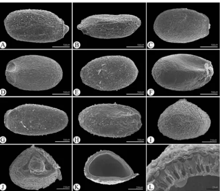
Fig. 1. SEM micrographs of nutlets in Glechoma taxa. A − B, I, K: G. grandis; C − D, J: G. longituba; E − F: G. hederacea; G − H, L: G. hirstua.

(A, C, E, G: dorsal side, B, D, F, H: ventral side, I: apical portion, J: hilum portion, K: cross section of middle portion, L: sectioned pericarp).

Fig. 2. SEM micrographs of nutlets in Agastache and Meehania taxa. A − B, I: A. rugosa; C − D, J: A. nepetoides; E − F, KL: A.

scrophulariefolia; G − H, M − O: M. urticifolia. (A, C, E, G: dorsal side, B, D, F, H: ventral side, I, M: apical portion, J, N: hilum portion, K:

cross section of middle portion, L − O: sectioned pericarp).

(4)

Fig. 3. SEM micrographs of nutlets in Dracocephalum, Marmoritis and Nepeta taxa. A: Dracocephalum nutans; B, K − N: D. arguense; C-D:

M. nivalis; E − F, O − P: M. rotundifolia; G − H, Q − R: N. glutinosa; I − J: N. longibracteata. (A, B, D, F, H, J: ventral side, C, E, G, I: dorsal side, K, O, Q: apical side, L, P, R: hilum, M: cross section of middle portion, N: sectioned pericarp).

Table 1. Morphological characters of nutlet of 16 taxa in Glechoma and its related taxa (n = 10 for all taxa).

Taxon Length (mm) Width (mm) L/W Shape TS NT GT ST

Agastache nepetoides (L.) Kuntze 1.36 (1.43) 1.48 0.82 (0.88) 0.96 1.52 (1.63) 1.78 ovate t d, v a re

A. rugosa (Fisch. & C. A. Mey.) Kuntze 1.59 (1.78) 1.94 0.87 (1.06) 1.27 1.48 (1.67) 1.94 ovate t d, v a re

A. scrophulariifolia (Willd.) Kuntze 1.50 (1.70) 1.81 0.80 (0.86) 0.92 1.67 (1.99) 2.23 elliptic t d, v a re

Dracocephalum argunense Fisch. ex Link 2.55 (2.71) 2.85 2.05 (2.18) 2.25 1.17 (1.24) 1.31 widely elliptic str a v p

D. nutans L. 2.21 (2.55) 2.66 1.62 (1.72) 1.88 1.32 (1.48) 1.58 widely elliptic str a a p

Glechoma grandis (A. Gray) Kuprian. 1.54 (1.84) 2.13 0.84 (0.98) 1.16 1.72 (1.87) 2.03 ovate str a d, v r

G. hederacea L. 1.76 (1.85) 1.97 1.16 (1.22) 1.29 1.43 (1.52) 1.58 ovate str a d, v r

G. hirsuta Waldst. & Kit. 1.88 (2.19) 2.56 0.95 (1.14) 1.34 1.69 (1.93) 2.35 ovate str a d, v r

G. longituba (Nakai) Kuprian. 1.79 (1.89) 2.00 1.15 (1.24) 1.31 1.40 (1.52) 1.68 ovate str a d, v r

Marmoritis nivalis (Benth.) Hedge 2.46 (2.79) 3.07 1.15 (1.27) 1.41 1.76 (2.19) 2.50 elliptic t a a r

M. rotundifolia Benth. 3.33 (3.67) 3.83 1.29 (1.54) 1.64 2.24 (2.38) 2.58 elliptic t a d r

Meehania urticifolia (Miq.) Makino 3.16 (3.32) 3.50 2.02 (2.31) 2.65 1.22 (1.44) 1.57 widely elliptic lens d, v d, v r

Nepeta glutinosa Benth. 2.21 (2.44) 2.70 1.16 (1.21) 1.27 1.80 (2.02) 2.29 elliptic t a a r

N. longibracteata Benth. 1.73 (1.85) 1.99 0.82 (0.93) 1.00 1.86 (2.00) 2.17 elliptic t a a r

List of abbreviations: TS, Shape of transversal section (t, trigonous, str, slightly trigonous); NT, non-glandular trichome (a, absent, d, dorsal

side, v, ventral side); GT, glandular trichome (a, absent, d, dorsal side, v, ventral side); ST, sexine ornamentation of nutlet surface (p, pitted-

cell, r, ridged negative reticulate-cell, re, rectangular-cell).

(5)

이며, Marmoritis속과 Nepeta속은 타원형이었다(Table. 1).

소견과의 횡단면은 Glechoma속, Dracocephalum속은 다소 둥근 형태의 삼각형(slightly trigonous; Figs. 1I−K, 3K−M)이 었으며, Meehania속은 렌즈형(lens; Fig. 2M−N), 그리고 Agastache속과 Marmoritis속, Nepeta속은 삼각형(trigonous;

Figs. 2I −K, 3O−R)을 보였다(Table 1).

조사된 분류군에서 소견과의 길이는 1.36−3.83 mm, 폭 0.80−2.65 mm로 다양하였으며, Marmoritis rotundifolia (3.67 × 1.54 mm)의 소견과 길이가 가장 길고, Agastache nepetoides (1.43 × 0.88 mm) 가 가장 짧았다(Table 1). 연구된 분류군 중 소견과의 크기는 일반적으로 Glechoma속, Agastache속, N. longibracteata ( 길이: 1.36−2.56 mm, 폭: 0.80−1.34 mm)가 나머지 분류군들 보다 작은 것으로 나타나 일부 분류군들 사이의 종식별에는 유용하였다(Figs. 1−3, Table 1).

소견과의 미세구조학적 형질(표면무늬 및 모용)

조사된 분류군에서 소견과의 배면(ventral side)은 볼록 하고, 기부에는 제(hilum)가 있다(Figs. 1−3). 소견과의 등면 (dorsal side) 은 뚜렷한 돌출선(Figs. 2A, C, E, 3C, E, G, I)이 있고, 그 위로 결절(tubercle)이 존재하는 속(Agastache, Marmoritis, Nepeta) 과 그렇지 않은 속(Glechoma, Meehania, Dracocephalum) 이 있었으며, 분류군마다 돌출선의 개수 와 모양에서 차이가 있었다(Figs. 1−3).

소견과의 표면은 세 유형으로 나타났다(Fig. 4). Type I은 Agastache속으로 표면은 불규칙적으로 돌출되었으며, 장방

형의 세포가 연결되어 나타나는 유형이고(Rectangular-cell;

Fig. 4A), Type II 는 Dracocephalum속으로 표면은 불규칙적 이며, 원형의 세포가 움푹 들어간 유형이다(Pitted-cell; Fig. 4B).

Type III은 Glechoma속, Marmoritis속, Meehania속, Nepeta 속으로 표면은 매끄러우며, 세포가 능선으로 연결된 망상형 이다(Ridged negative reticulate-cell; Fig. 4C−F).

모용(비선모: non-glandular trichome와 선모: glandular trichome)은 일부 분류군에서 각각 다르게 관찰되었다 (Fig. 4G−I, Table 1). Agastache속과 Meehania속은 비선모가 존재하였으며, Agastache속은 정단(apical)에 밀집되고 단 세포 모용(uni-cellular non-glandular trichome; Fig. 4I)을 보인 반면, Meehania속은 소견과 전체에 비슷한 밀도로 다세포 모용(multi-cellular non-glandular trichome; Fig. 4H)이 분포 하고 있었다(Table 1). 선모의 경우 Glechoma속, Meehania속, Dracocephalum arguense, Marmoritis rotundifolia 에서 존재 하였다. Glechoma속과 Meehania속은 소견과 전체에서 선모 가 분포하였으나(Fig. 4G, Table 1), Dracocephalum nutans, Marmoritis rotundifolia 는 배면과 정단 부분에만 존재하는 특 징을 보였으며 형태적인 차이를 보이지는 않았다(Table 1).

소견과의 해부학적 형질

소견과의 해부학적 형질을 관찰하였을 때, 과피의 표피층 은 점액성과피(myxocarpy)로 점액질을 형성하는 분류군과 그렇지 않은 분류군으로 구분되었다(Table 2). Glechoma속 모 든 분류군에서 점액성 과피층을 관찰할 수 있었으며(Fig. 6C−

Fig. 4. SEM micrographs of nutlet cell ornamentation in Glechoma and its related taxa. A: Agastache rugosa. B: Dracocephalum arguense.

C: G. hederacea, D: Marmoritis rotundifolia, E: Meehania urticifolia, F: Nepeta glutinosa, G: peltate glandular trichome (G. longituba). H:

multicellular non-glandular trichomes (M. urticifolia). I: unicellular non-glandular trichome (Agastache nepetoides).

(6)

H), 특히 G. hirsuta의 경우에 점액성 과피의 반응이 강했다 (Fig. 5F). 그러나 Glechoma속을 제외한 대부분의 분류군 에서는 점액성 과피층이 나타나지 않았다(Figs. 5−6, Table 2).

소견과의 과피는 외과피와 중과피, 내과피로 구성되어 있으며, 종피는 한 층으로 과피와 떨어져 자엽을 밀접하 게 싸고 있었다(Figs. 1F, 2L, O, 3M−N, 6). 외과피에는 공통 적으로 탄닌이 침적되어 있었다(Fig. 6). 과피의 두께는

Dracocephalum argunense 분류군에서 가장 두꺼웠으며, Agastache scrophulariifolia에서 가장 얇았다. 후벽조직의 두께는 D. argunense에서 가장 두꺼웠으며, A. nepetoides에서 가장 얇았다(Table 2). Glechoma속의 과피는 모두 점액성 과피(Figs. 5F, 6C−H)를 가지고 있기 때문에 전체 과피의 두께는 두꺼운 반면, 후벽조직의 두께는 상대적으로 얇았다 (Table 2). 그러나 Dracocephalum속, Marmoritis속, Meehania속 Table 2. Selected characteristic anatomical features of the nutlets in the genus Glechoma and its related taxa (unit : µ m).

Taxon Exocarp Mesocarp Sclenchyma tissue Endocarp Pericarp

Agastache nepetoides (L.) Kuntze 4.0 − 5.0 15.017.0 21.023.0 2.03.0 42.048.0 A. rugosa (Fisch. & C. A. Mey.) Kuntze 5.0 − 7.0 5.06.0 30.032.0 2.04.0 42.049.0 A. scrophulariifolia (Willd.) Kuntze 4.0 − 7.0 10.013.0 23.025.0 2.03.0 39.048.0 Dracocephalum argunense Fisch. ex Link 5.0 − 7.5 32.537.5 127.5132.5 10.012.5 175.0190.0

D. nutans L. 5.0 − 7.0 6.08.0 70.075.0 12.015.0 93.0105.0

Glechoma grandis (A. Gray) Kuprian. 40.0 − 48.0 11.017.0 30.034.0 3.04.0 84.0103.0

G. hederacea L. 105.0 − 150.0 22.532.5 32.542.5 2.04.0 162.0229.0

G. hirsuta Waldst. & Kit. 100.0 − 137.5 35.050.0 32.545.0 2.55.0 170.0237.5 G. longituba (Nakai) Kuprian. 95.0 − 140.0 12.542.5 30.037.5 2.04.0 137.5220.0 Marmoritis nivalis (Benth.) Hedge 11.0 − 18.0 6.07.0 31.036.0 6.08.0 54.069.0

M. rotundifolia Benth. 6.0 − 8.0 2.03.0 32.045.0 7.09.0 47.065.0

Meehania urticifolia (Miq.) Makino 17.5 − 25.0 5.010.0 102.5115.0 7.512.5 132.5162.5

Nepeta glutinosa Benth. 8.0 − 12.0 2.04.0 36.042.0 5.08.0 51.066.0

N. longibracteata Benth. 10.0 − 12.0 2.03.0 25.027.0 3.04.0 40.046.0

Fig. 5. LM micrographs showing the mucilaginous reaction of Nutlet in Glechoma amd its related taxa. A: Agastache rugosa; B:

Dracocephalum arguense; C: Glechoma hederacea; D: G. grandis; E: G. longituba; F: G. hirsuta; G: Marmoritis rotundifolia; H: Meehania

urticifolia; I: Nepeta glutinosa (arrow indicates the mucilage reaction).

(7)

의 후벽조직 두께는 과피의 두께에서 큰 비중을 차지하였다 (Fig. 6A−B, I−L).

고 찰

Nepetinae아족에 속하는 Glechoma속과 근연분류군의 소견과 연구 결과 크기와 모양, 미세형태학 및 해부학적 형질들이 다양하게 관찰되었다(Figs. 1−6, Tables 1−2).

연구된 분류군들은 작은 크기(1.36−3.83 × 0.80−2.65 mm) 의 소견과를 가졌고, 모양은 난형에서 타원형으로 나타났 으며(Figs. 1−3, Table 1), Nepetinae아족의 소견과 크기 및 모 양을 기재한 선행 연구와 유사한 범위를 보였다(Budantsev and Lobova, 1997; Moon et al., 2009).

소견과의 표면무늬는 크게 세 유형(Type I - Agastache속:

표면은 불규칙적으로 돌출되었으며, 장방형의 세포가 연 결되어 나타나는 유형, Type II - Dracocephalum속: 표면은 불규칙적이며, 원형의 세포가 움푹 들어간 유형, Type III - Glechoma 속, Marmoritis속, Meehania속, Nepeta속: 표면은 매끄러우며, 세포가 능선으로 연결된 망상형)으로 나타났다 (Fig. 4A−F). 본 연구에서 소견과의 표면무늬는 Glechoma속 과 Meehania속에서 세포가 능선으로 연결된 망상형을 보이 고, 근연 속내 일부분류군에서 관찰되어진 소견과 표면무 늬가 선행연구의 결과와 일치하였다(Budantsev and Lobova, 1997; Moon et al., 2009). 소견과의 표면무늬는 속간 한계에 중요한 형질로 Glechoma속과 Meehania속이 분자계통학 적으로 매우 근연 속이라는 결과를 지지해 주었다(Wagstaff et al., 1995).

Budantsev and Lobova(1997) 는 Nepetinae아족에 대한 소 Fig. 6. LM micrographs of anatomical structure of nutlet in Glechoma and its related taxa. A − B: Dracocephalum nutans; C − D: G. grandis;

E − F: G. hederacea; G: G. longituba; H: G. hirsuta; I − J: Marmoritis nivalis; K: Meehania urticifolia; L: Nepeta glutinosa. (MC: mucilaginous

cell, EX: excocarp, ME: mesocarp, EN: endocarp).

(8)

견과를 다루었으나 해부학적 구조를 언급했을 뿐, 모용에 대한 기재는 이루어지지 않았다. 본 결과에서 Glechoma속, Meehania 속, Marmoritis rotundifolia는 모두 선모가 분포하 였으나, 나머지 속에서는 선모가 분포하지 않았고, 비선모 는 Agastache속, Meehania속을 제외하고 존재하지 않았다 (Fig. 4G −I, Table 1).

Glechoma속내 분류군들의 공통적인 특이점으로 과피에 서 점액질이 형성되는 것을 관찰 할 수 있었다(Figs. 5F, 6C−H).

많은 꿀풀과의 소견과가 물에 젖었을 때 종자를 보호하기 위해서 ‘점액성과피(myxocarpy)’라는 특성을 갖는 것으로 알려져 있다(Zohary, 1962; Ryding, 1992b; Wagstaff et al., 1995; Ryding, 2001; Jeon and Hong, 2006; Moon et al., 2009).

건조한 지역에서 점액성과피가 더욱 빈번하게 출현하기 때문에 서식지 특성이 점액질 형성과 상호 관련이 있다고 판단된다(Ryding, 1992a). 또한 점액성과피는 수분 부족에 의한 종자의 건조를 막아주어 소견과의 발아를 부분적으로 조절하는 기능을 가지기도 한다(Fahn, 1990). Ryding (1992b) 은 G. hederacea에서 점액성 과피가 나타난다고 보고하였 으나, 본 실험에서는 G. hederacea보다는 G. hirsuta에서 오 히려 반응을 보였다(Fig. 5F). 또한 Nepeta속은 점액성과피가 약하게 형성된다고 기록되었으나(Duletiæ-Lauševiæ and Marin, 1999), 본 실험에서는 전혀 생성되지 않았다(Fig. 5I).

Glechoma 속과 근연 분류군의 과피는 외과피, 중과피, 그리고 내과피로 구성되어 있다(Fig. 6). 외과피의 경우 탄 닌이 침적되어 있는 것을 볼 수 있었는데, 탄닌은 방충효 과와 탈수 및 부패 방지에 효과가 있으며, 소견과를 보호 하기 위한 기작이다(Fahn 1990). 과피의 두께는 약 70 µm 보다 얇은 분류군과 약 80 µm 이상의 분류군으로 나누어 볼 수 있지만, 속 및 종간을 구별할 수 있는 형질로는 미약 하였다(Table 2). 소견과의 길이와 후벽조직층의 두께는 피 어슨 상관관계 분석에 의하면 큰 상관관계를 보이지는 못 하였으나(r = 0.47), Marmoritis속을 제외한 대부분의 속에 서 소견과의 크기가 클수록 후벽조직의 두께는 커짐을 확 인하였다(Tables 1-2). 꿀풀과내에서 소견과 후벽조직의 두 께는 과피의 두께 및 소견과의 크기와 일정한 상관관계를 보인다고 보고된 바 있으며(Duletiæ-Lauševiæ and Marin, 1999; Moon and Hong, 2006), 본 연구에서도 Duletiæ- Lauševiæ and Marin(1999) 의 결과와 부분적으로 일치하였다.

Nakai(1921) 는 Glechoma속의 기본종인 G. hederacea과 악편 및 화관의 미세한 차이를 들어 변종(G. hederacea var.

longituba) 을 제안했으나, 이 후 Kuprianova(1948)는 악편의 깊이와 모용의 밀도, 그리고 화관의 길이 차이를 근거로 종 (G. longituba) 으로 승격시켰다. 또한, G. grandis와 G. longituba 의 경우 연구자들에 따라서 동종으로 통합되기도 하고, 분 리되기도 한다(Murata and Yamazaki, 1993; Li and Hedge, 1994). G. hirsuta의 경우 식물체 전체에 털이 밀생하고, 악 편의 갈라짐 정도가 G. hederacea에 비해 길다는 차이가 있다 (Pojarkova, 1954). 그러나 소견과의 모양 및 크기, 표면 무늬,

모용 등의 미세구조학적 형질, 후벽조직, 중과피, 내과피 등의 해부학적 형질은 종간에 큰 차이를 나타내지 않았다 (Tables 1−2). 따라서 소견과의 형태학 및 해부학적 연구에 의하면, G. grandis, G. hederacea, G. hirsuta, G. longituba 모두 동일 분류군이거나 종 이하 분류군이라 판단 하지만, 정 확한 종간 한계를 설정하기 위해서는 외부 형태학적 자료를 통한 PCA분석 및 분자계통분류학적 연구가 동시에 수반 되어야 한다고 생각된다(Jang et al., unpubl. data).

결론적으로, Nepetinae아족에 속하는 Glechoma속과 근연 속 일부 분류군에 대한 소견과의 형질은 모양과, 표면 무늬, 선모와 비선모의 분포, 점액성 과피의 존재 유무, 그리고 해 부학적 구조에서 분류군간 구별에 다소 유용하게 나타났다.

사 사

본 연구를 위해 귀중한 표본을 대여해 주신 F, G, K, KHUS, KRIB, KYO, NY, UPS, US, S, P의 각 표본관의 관계자께 감 사를 드립니다. 또한 논문심사 과정 중 많은 유용한 조언과 보다 정확한 논문이 되도록 교정을 해주신 익명의 두 분 심 사자들께 감사드립니다. 본 연구는 2006년도 경희대학교 연구비지원 결과임(KHU-20060411).

인용문헌

Bilimovitsch, O. 1935. On the structure of the pericarp in the Labiatae from a systematic view point. Trudy Voronezhsk. Gosud. Univ.

7: 68-84 (in Russian with English summary).

Budantsev, A. L. and T. A. Lobova. 1997. Fruit morphology, anatomy and taxonomy of tribe Nepeteae (Labiatae). Edinburgh J. Bot.

54: 183-216.

Cantino, P. D., R. M. Harley and S. J. Wagstaff. 1992. Genera of Labiatae: Status and Classification. In Advances in Labiate Science. Harley, R. M. and T. Reynolds (eds.), Royal Botanic Gardens, Kew, Pp. 511-522.

Duletiæ-Lauševiæ, S. and P. D. Marin. 1999. Pericarp structure and myxocarpy in selected genera of Nepetoideae (Lamiaceae).

Nord. J. Bot. 19: 435-446.

Fahn, A. 1990. Plant anatomy, 4

th

ed. Pergamon Press, Oxford.

Harley, R. M., S. Atkins, A. L. Budantsev, P. D. Cantino, B. J. Conn, R. Grayer, M. M. Harley, R. De Kok, T. Krestovskaja, R.

Morales, A. J. Paton, O. Ryding and T. Upson. 2004. Labiatae.

In Flowering Plants, Dicotyledons: Lamiales (except Acan- thaceae including Avicenniaceae). Kadereit, J. W. (ed.), Springer- Verlag, Berlin-Heidelberg, Pp. 167-275.

Hedge, I. C. 1970. Observation on the mucilage of Salvia fruits.

Notes Roy. Bot. Gard. Edinburgh. 30: 79-95.

Holmgren, P. K. and N. H. Holmgren. 1998. (continuously updated):

Index herbariorum. - http://sciweb.nybg.org/science2/Index-

(9)

Herbariorum.asp

Jang, T.-S. and S.-P. Hong. 2007. The taxonomic consideration of leaf epidermal microsturcture in Glechoma L. (Nepetinae, Lamiaceae). Korean J. Pl. Taxon. 37: 239-254.

Jeon, Y.-C. and S.-P. Hong. 2006. A systematic study of Elsholtzia Willd. (Lamiaceae) in Korea. Korean J. Pl. Taxon. 36: 309-333 (in Korean).

Kupriyanova, L. 1948. On the kind Glechoma L. and its forms.

Bot. Zhurn. 33: 230-238 (in Russian).

Lee, T. B. 2003. Coloured flora of Korea. Hyangmunsa, Seoul. Pp.

120-123 (in Korean).

Lee, Y. N. 2006. New Flora of Korea. Kyohaksa, Seoul, Pp. 141- 144 (in Korean).

Li, X. and I. C. Hedge. 1994. Lamiaceae. In Flora of China, Vol. 17, Wu, Z. Y. and P. H. Raven. (eds.), Science Press and Missouri Botanical Garden, Beijing and St. Louis, Pp. 50-299.

Makarova, Z. I. 1967. On the significance of the anatomical struc- ture of the pericarp for the taxonomy of the tribe Nepeteae (Labiatae). Bot. J. USSR. 52: 33-41.

Moon, H.-K. and S.-P. Hong. 2006. Nutlet morphology and anat- omy of the genus Lycopus (Lamiaceae: Mentheae). J. Plant Res. 119: 633-644.

Moon, H.-K., S.-P. Hong, E. Smets and S. Huysmans. 2009. Micro- morphology and character evolution of nutlets in tribe Mentheae (Nepetoideae, Lamiaceae). Syst. Bot. 34: 760-776.

Murata, G. and T. Yamazaki. 1993. Lamiaceae (Labiatae). In Flora of Japan, Vol. IIIa, Iwatsuki, K., T. Yamazaki, D. E. Boufford and H. Ohba (eds.), Kodansha Ltd. Tokyo, Pp. 272-321.

Nakai, T. 1921. Labiatae Coreanae. Bot. Mag. 35: 173-174.

Pojarkova, A. I. 1954. Nepetinae. In Flora of the U.S.S.R., Vol. 20, Shihkin, B. K. (ed.), Academy of Science of the USSR, Moskova, Leningrad, Pp. 273-492.

Ryding, O. 1982. The Aeolanthus abyssinicus group (Labiatae). Nord.

J. Bot. 2: 219-229.

Ryding, O. 1992a. The distribution and evolution of myxocarpy in Lamiaceae. In Advances in Labiate Science. Harley, R. M. and T. Reynolds (eds.), Royal Botanic Gardens, Kew, Pp. 85-96.

Ryding, O. 1992b. Pericarp structure and phylogeny within Lamiaceae subfamily Nepetoideae tribe Ocimeae. Nord. J. Bot. 12: 273-298.

Ryding, O. 2001. Myxocarpy in the Nepetoideae (Lamiaceae) with notes on myxodiaspory in general. Syst. Geogr. Pl. 71: 503-514.

Ruzin, S. E. 1999. Plant microtechnique and microscopy. Oxford University Press, Oxford.

Turner, B. L. and P. G. Delprete. 1996. Nutlet sculpturing in Scutellaria sect. Resinosa (Lamiaceae) and its taxonomic utility. Pl. Syst.

Evol. 199: 109-120.

Wagstaff, S. J., R. G. Olmstead and P. D. Cantino. 1995. Parsimony analysis of cpDNA restriction site variation in subfamily

Nepetoideae (Labiatae). Amer. J. Bot. 82: 886-892.

Walker, J. B., K. J. Sytsma, J. Treuttlein and M. Wink. 2004. Salvia (Lamiaceae) is not monophyletic: Implications for the systematics, radiation, and ecological specializations of Salvia and tribe Mentheae. Amer. J. Bot. 91: 1115-1125.

Walker, J. B. and K. J. Sytsma. 2007. Staminal evolution in the genus Salvia (Lamiaceae): Molecular phylogenetic evidence for multiple origins of the staminal lever. Ann. Bot. 100: 375-391.

Wojciechowska, B. 1958. Taxonomy, morphology, and anatomy of seeds in the genus Salvia L. Monogr. Bot. 6: 1-56. (Polish with English summary).

Zohary, M. 1962. Plant Life of Palestine (Israel and Jordan). Ronald Press, New York.

Appendix: Specimens investigated.

Agastache nepetoides (L.) Kuntze: U.S.A., Ohio, 5 Aug.

1992, R.M. Lowden 5013 (UPS). - A. rugosa (Fisch. & C. A.

Mey.) Kuntze: KOREA, Mt. Kwangdeok, 26 Aug. 2005, S.P.

Hong & T.S. Jang 123 (KHUS). - A. scrophulariaefolia (Willd.) Kuntze: U.S.A., Georgia, without detailed date, 1982, Hoscelyn W. Hill 3425 (F). - Dracocephalum argunense Fisch.

ex Link: KOREA, Mt. Deokyu, 15 June 2004, G.R. Kim &

J.K. Kim s.n. (KRIB). - D. nutans L.: CHINA, without specific locality, Sep. 1921, S.W. Szechuan, 20655 (US). - Glechoma hederacea L.: SWITZERLAND, Geneva, without detailed date, 1973, Curchod & Hainard s.n. (S) - G. hirsuta Waldst.

& Kit.: AUSTRIA, Vindobonam, without detailed date, 1914, I’Herbier Delessert s.n. (G). - G. grandis (A. Gray) Kuprian.:

JAPAN, Honshu, 19 May 1982, Deguchi K, Ozaki T, Yamaguchi M. 3446 (KYO) - G. longituba (Nakai) Kuprian.:

KOREA, Moonsan, 14 May 2006, T.S. Jang 5071 (KHUS) - Marmoritis nivalis (Benth.) Hedge: Without specific locality, Feb. 1900, J.E. Winterbottom 1848 (K) - M. rotundifolia Benth.: NEPAL, western Himalaya, 23 Aug. 1931, Walter Koelz, 2692 (NY) - Meehania urticifolia (Miq.) Makino:

KOREA, Mt. Gong-Jak. 18 May 2006, S.P. Hong & T.S. Jang 5181 (KHUS) - Nepeta glutinosa Benth.: INDIA, without specific locality, 16 Jul. 1933, Walter Koelz, 5656 (NY). - N.

longibracteata Benth.: NEPAL, Himalaya, without detailed

date, Jan. 1990. DeloucheI. 16 (P).

수치

Fig. 1. SEM micrographs of nutlets in Glechoma taxa. A − B, I, K: G. grandis; C − D, J: G
Table 1. Morphological characters of nutlet of 16 taxa in Glechoma and its related taxa (n = 10 for all taxa).
Fig. 4A), Type II 는  Dracocephalum속으로 표면은 불규칙적 이며, 원형의 세포가 움푹 들어간 유형이다(Pitted-cell; Fig
Fig. 5. LM micrographs showing the mucilaginous reaction of Nutlet in Glechoma  amd its related taxa

참조

관련 문서

Biofilm architecture was also analyzed by scanning electron microscopy (SEM), and changes in gene expression related to biofilm formation were studied by quantitative RT-PCR.

이외 에도 오토클레이브 양생한 경량기포 콘크리트(Autoclaved aerated concrete) 의 제조에 CFBC 비산재를 활용한 연 Fig. Morphology of coal ashes observed

The phase structure and morphology of the phosphors were measured using X-ray diffraction (XRD) and field emission-scanning electron microscopy (FE-SEM), respectively..