ISSN 2288-1069 (Online)
http://dx.doi.org/10.12925/jkocs.2020.37.4.744
지구성 운동과 MitoQ 섭취가 MPTP로 유도된 파킨슨 질환 생쥐의 병리학적 특징에 미치는 영향
김동철1.*
․엄현섭
2․오은택
3․조준용
4․장용철
1,✝1한국체육대학교 운동생화학실, 석사
2건양대학교 스포츠의학과, 교수
3우송대학교 스포츠건강재활학과, 교수
4한국체육대학교 운동건강관리학과, 교수
1한국체육대학교 운동생화학실, 연구원
(2020년 7월 28일 접수: 2020년 8월 18일 수정: 2020년 8월 19일 채택)
The effect of endurance exercise and MitoQ intake on pathological characteristics in MPTP-induced animal model of Parkinson’s disease
Dong-Cheol Kim
1,*․Hyun Seob Um2․Eun-Tak Oh3․Joon-Yong Cho4․Yongchul Jang1,✝1
Exercise biochemistry Laboratory, Korea National Sport University
2
Department of Sports Medicine, Konyang University
3
Department of Sports Science & Rehabilitation, Woosong University
4
Department of Health and Exercise Science, Korea National Sport University (Received July 28, 2020; Revised August 18, 2020; Accepted August 19, 2020)
요 약 : 본 연구는 파킨슨 질환(Parkinson’s disease) 마우스 모델을 대상으로 지구성 운동과 MitoQ 섭
취가 뇌의 흑질의 미토콘드리아 기능에 미치는 영향을 확인하는데 목적이 있다. 파킨슨 질환을 유도하기 위해 C57BL/6 수컷 마우스를 대상으로 복강 내 1-methyl-4-phenyl-1,2,3,6 tetrahydropyridine (MPTP) 25mg/kg과 흡수를 돕기 위한 probenecid 250mg/kg을 이용하여 주 2회 5주간 총 10회 투여하였다. 실험 집단은 생리식염수를 투여하는 집단(Normal Conrol (NC), n=10), MPTP 투여집단(MPTP Control (MC), n=10), MPTP 투여 + MitoQ 투여집단(MPTP + MitoQ (MQ), n=10), MPTP 투여 + 운동집단 (MPTP + Exercise (ME), n=10), MPTP 투여 + MitoQ 투여 + 운동집단(MPTP + MitoQ + Exercise (MQE), n=10) 총 5 집단으로 구성하였으며, 운동집단은 지구성 운동을 실시하였고 MitoQ집단은 점진적 으로 250μmol로 늘리면서 5주간 섭취하였다. 연구결과 Rotarod-test에서 MC 집단에 비해 처치 집단은 운동 기능 저하의 개선을 보였다. 또한 MC 집단에 비해 처치 집단은 tyrosine hydroxylase의 수준의 증가 와 알파시누클린(α-synuclein) 단백질 축적을 감소시켰다. 그리고 미토콘드리아 생합성에 주요조절 인자 인 PGC-1α와 항산화 효소인 Catalase 발현이 MC 집단에 비해 처치 집단에서 증가해 미토콘드리아 기능
✝
Corresponding author
(E-mail: [email protected])
*이 연구는 김동철(한국체육대학교)의 석사학위논문을 수정·보완하여 작성됨
을 개선했으며, 세포사멸 조절인자인 Bcl-2의 증가와 Bax의 감소를 통해 세포사멸을 완화했다. 따라서 5주 간의 지구성 운동과 MitoQ 섭취는 파킨슨 질환에서 나타나는 병리학적 특징을 완화하고 운동기능을 향상 시키는데 효과적인 것으로 나타났다.
주제어 : 파킨슨 질환, 지구성 운동, 마이토큐, 알파시누클린, 미토콘드리아
Abstract : We investigated the whether endurance exercise and MitoQ intake mediated
neuroprotection are associated with mitochondrial function in 1-methyl-4-phenyl-1,2,3,6 tetrahydropyridine(MPTP) -induced mice model of Parkinson’s disease. C57BL/6 male mice were randomly assigned to five groups: Normal Conrol(NC, n=10), MPTP Control(MC, n=10), MPTP + MitoQ(MQ, n=10), MPTP + Exercise(ME, n=10) and MPTP + MitoQ + Exercise(MQE, n=10).Exercise intervention groups performed the treadmill exercise for 5days/week for 5 weeks with gradual increase of intensity. MitoQ intake groups consumed the MitoQ at a concentration of 250μmol by dissolving with water during experiment period. Our data demonstrated that ME and MQE group restored MPTP-induced motor dysfunction. In addition, treatment groups(MQ, ME and MQE) increased tyrosine hydroxylase levels, and suppressed the accumulation of α-synuclein levels.
Futhermore, treatment groups modulated the mitochondrial function such as upregulated mitochondrial biogenesis, increased antioxidant enzyme, enhanced a anti-apoptotic protein(e.g., BCL2), and reduced a pro-apoptotic protein(e.g., BAX). Taken together, these results suggested that endurance exercise and MitoQ intake-mediated increase in mitochondrial function contributes to improvement of aggravated dopaminergic neuronal, resulting in attenuation of motor function of Parkinson’s disease.
Keywords : Parkinson’s disease, endurance exercise, MitoQ, α-synuclein, mitochondria
1. 서 론
파킨슨 질환은 중뇌의 흑질(substantia nigra) 부위의 도파민성 신경세포가 사멸되어 나타나는 신경퇴행성 질환으로 강직(Rigidity), 떨림 (Tremor), 운동 완만(Bradykinesia), 자세 불안정 (Postural instability) 등의 운동장애가 나타난다 [1]. 현재까지 발병원인에 대해 정확하게 밝혀지 지 않았지만 흑질내 알파시누클린(α-synuclein) 단백질의 비정상적인 축적이 보고되고 있다[2].
정상적인 알파시누클린 단백질은 신경연접 부위 에서 도파민성 소포들의 유지 및 신경전달에 관 여하지만, 비정상적인 축적은 미토콘드리아의 기 능이상과 산화스트레스를 유발해 결과적으로 도 파민 신경세포의 소실을 유발한다[3].
미토콘드리아는 세포 내 에너지원인 ATP를 생 성할 뿐 아니라 활성산소 생성의 주요 기관으로
과도한 활성산소의 생성은 세포예정사
(programmed cell death)를 유발시킨다. 미토콘 드리아의 기능이상은 도파민 신경세포의 사멸을
증가시키며, 미토콘드리아의 감소는 파킨슨 질환 의 발병을 증가시키는 것으로 보고되고 있다[4].
Peroxisome proliferator-activated receptor gamma coactivator-1α(PGC-1α)은 전사조절 도움인자(transcriptional coactivator)로서 미토콘 드리아 생합성과 항산화 방어시스템을 조절하여 미토콘드리아의 기능을 증가시킨다. 선행연구에 따르면 파킨슨 질환자는 PGC-1α의 감소가 나 타났으며, PGC-1α의 증가는 알파시누클린의 수 준을 감소시켜 결과적으로 도파민 신경세포의 수 준을 증가시킨다고 보고하여, 파킨슨 질환 치료의 중요한 요인으로 제시하고 있다[5].
파킨슨 질환의 치료방법으로 약물과 수술적 방 법이 사용되고 있으나 그에 따른 다양한 부작용 역시 보고되고 있으며, 운동은 부작용이 없는 방 법으로 파킨슨 질환을 예방 또는 지연하는데 효 과적이라고 제시하고 있다[6]. 지구성 운동은 신 경세포를 보호하고 뇌 기능을 증가 시키는 것으 로 보고되고 있으며, 특히 파킨슨 환자의 운동기 능을 향상시켜 삶의 질을 높여 준다고 보고하고
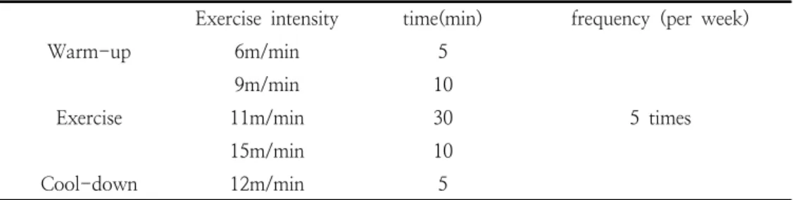
Table 1. Treadmill exercise protocol
Exercise intensity time(min) frequency (per week)
Warm-up 6m/min 5
5 times Exercise
9m/min 10
11m/min 30
15m/min 10
Cool-down 12m/min 5
있다[7]. 또한 파킨슨질환 동물모델을 대상으로
지구성 운동은 알파시누클린 단백질의 감소와 tyrosine hydroxylase(TH) 활성을 보고하였다[8].
특히 Jang 등[9]은 지구성 운동은 미토콘드리아 숫자의 증가와 산화스트레스를 감소시키는 효과 적인 방법으로 보고하고 있다.
운동뿐만 아니라 항산화제 섭취는 활성산소의 감소 및 미토콘드리아 기능을 개선시킨다[10]. 특 히 마이토큐(MitoQ)는 미토콘드리아 표적 항산 화제로써 혈액뇌장벽(Blood-brain barrier)과 신 경 세포막을 지나 미토콘드리아 막을 통과한다.
미토콘드리아 안으로 들어간 MitoQ는 불안정 상 태의 활성산소를 안정화시켜 다른 항산화물질보 다 효율적으로 미토콘드리아를 보호한다[11].
MitoQ의 섭취는 활성산소로부터 미토콘드리아를 보호하고 기능을 향상시키는 것으로 보고하고 있 으며[12], 이러한 관점에서 운동을 수행할 수 없 는 환자들에게 MitoQ의 섭취는 또 하나의 파킨 슨질환의 진전을 감소시키는 방법으로 제시할 수 있다고 생각된다. 하지만 아직까지 MitoQ에 의 한 파킨슨질환의 운동기능 향상 기전에 대한 연 구는 부족한 실정이며, MitoQ 섭취가 파킨슨질 환의 진전에 영향을 미치지 않는다는 상반된 연 구도 제시되고 있다[13]. 또한 운동과 MitoQ의 복합 처지가 파킨슨 질환에 미치는 분자생물학적 연구는 전무한 실정이다.
따라서 본 연구의 목적은 파킨슨질환 마우스 모델을 대상으로 5주간 지구성운동과 MitoQ의 섭취가 뇌의 흑질에서 알파시누클린 단백질 및 도파민성 신경세포의 수준과 PGC-1α 단백질 발현 및 신경세포사멸과 관련된 메커니즘이 운동 기능(motor function)에 미치는 영향을 규명하고 자 한다.
2. 실 험
2.1. 실험동물
본 연구에서 사용된 실험동물은 생후 8주령 된 C57BL/6 마우스(n=50)를 대상으로 온도(23±
1℃), 습도(40~60%) 및 명암주기 12시간의 환경 에서 사육하였으며, 실험기간 동안 동물용으로 제 조된 사료(Purina 쥐사료–5057)와 물은 자유 공 급하였다. 실험 집단은 생리식염수를 투여하는 집 단(Normal Conrol (NC), n=10), MPTP 투여집 단(MPTP Control (MC), n=10), MPTP 투여 + MitoQ 투여집단(MPTP + MitoQ (MQ), n=10), MPTP 투여 + 운동집단(MPTP + Exercise (ME), n=10), MPTP 투여 + MitoQ 투여 + 운동집단(MPTP + MitoQ + Exercise (MQE), n=10) 총 5 집단으로 구성하였으며, 모 든 실험방법 및 절차는 K 대학교 동물실험윤리위
원회의 승인을 거쳐서 진행하였다
(KNSU-IACUC-2018-01).
2.2. 파킨슨 질환 마우스 모델 및 운동 프로토콜 파킨슨 질환을 유도하기 위해 25mg/kg의 MPTP(Sigma aldrich)와 항보강제인 Probenecid (Sigma aldrich)를 250mg/kg을 주 2회 5주간 총 10회 복강 투여하였다. 5주간의 MPTP 투여 후 운동집단은 실험동물용 트레드밀(8Lanes, Daemyung Scientific Co, Korea)을 이용하여 1 주간의 사전 적응훈련(10min/day, 6m/min, 7days/week)을 마친 후 파킨슨 모델 쥐의 운동 수행 능력을 고려하여 Lau 등[14]이 제시한 운동 프로그램을 근거로 5주간 수행하였으며, 운동 프 로토콜은 (표 1)과 같다.
2.3. MitoQ 섭취 방법
MitoQ는 Dr. Mike Murphy(University of Cambridge, UK)를 통해 제공받아 사용을 하였으 며, MitoQ 섭취방법 및 농도 설정은 Rodriguez-Cuenca 등[15]의 선행연구를 참고하 여 MitoQ가 용해된 음수를 공급하는 방식으로 진행하였다. MitoQ가 용수된 음수의 맛으로 인 해 발생할 수 있는 실험동물의 거부반응을 중재 하기 위해 MitoQ의 농도는 100μmol에서 시작 하여 매주 점진적으로 증가시키고, 최종적으로 250μmol로 설정하였다. 각 케이지 3~4마리를 동시 사육하며 섭취량은 250mL씩 3일에 한 번 씩 MitoQ 음수를 교체하였다.
2.4. 운동 기능 검사
Rota-rod(fiveonin acclerating retarod;
JD-A-07, Jeung do, Korea) 트레드밀을 이용하 여 운동협응 기능과 균형감각을 검사하였다. 본 검사 전 2일간 Rota-rod 트레드밀 위에 1분간 적응시키고 120초 동안 10rpm 속도로 사전 적응 훈련을 하였다. 본 검사에서 300초 동안 10rpm 속도로 5번 떨어진 초를 확인하였다. 각 검사가 끝난 후 이전 동물의 냄새가 검사 결과에 영향을 줄 수 있으므로 에탄올(70%)을 이용하여 장비를 닦았다.
2.5. 뇌 분리 및 조직 고정
5주간 운동과 MitoQ 섭취 후 Rompun(바이엘 코리아 주식회사, Korea)과 Zoletil 50(Virbac, France)을 혼합해 50mg/kg을 복강 내 주입시켜 마취 시켰다. Western blot analysis를 위해 뇌 흑질(substantia nigra)을 적출하여 액화 질소에 동결시키고 -80℃의 초저온 냉동기(Bio-Freezer, Forma Science)에 분석시까지 냉동 보관하였다 (n=6, per group). Immunohistochemistry을 하기 위해 실험동물의 흉강을 열고 좌심실로 10mM phosphate buffered saline(PBS)를 4분 동안 주입 한 후, 0.1M PBS 녹인 4% paraformaldehyde 고정액을 4분 동안 관류시켰다. 이후 뇌 조직을 30% sucrose 용액에서 5일 동안 침전시키고 freezing microtome(Leica, Nussloch, Germany) 를 이용해 35㎛ 두께의 연속관상 절편을 하였다 (n=4, per group).
2.6. Western blotting
집단별로 분리된 흑질을 Protein Extraction
Solution-RIPA (ELPIS biothch, Korea)을 이용 하여 단백질을 추출하였고, Bradford[16]법에 따 라 단백질을 정량하였다. 단백질(30㎍)을 10-12% SDS-Polyacrylamide gel에 분주하여 전 기영동 한 후 polyvinylidene fluoride(PVDF) membrane으로 전사 시켰으며, 3% BSA를 통해 blocking 하였다. 본 실험에서 사용된 1차 항체는 Tyrosine hydroxylase(millipore, 1:1000); α -synuclein(Santa cruz, 1:1000); PGC-α(Santa cruz, 1:1000); Catalase(Santa cruz, 1:1000);
Bax(Santa cruz, 1:1000); Bcl-2(Santa cruz, 1:1000)을 4℃에서 12시간 반응시켰다. 다음날 TBS-T 용액으로 10분간 5회 세척 후 상온에서 2차 항체와(horseradish peroxidase-conjugated goat anti-rabbit, Invitrogen; horseradish peroxidase-conjugated goat anti-mouse, Santa Cruz, 1:5000)를 이용하여 90분 동안 반응시켰 다. 마지막으로 membrane을 WBLR solution (WBLUF0100; Millipore, USA)에 1분 동안 발 색하고 이미지 분석 시스템(Molecular Imager ChemiDoc XRS System, Bio-Rad, USA)을 이용 해 스캔하고, Quantity One 1-D Analysis Software(Bio-Rad, USA)을 이용하여 단백질 발 현 수준을 산출하였다.
2.7. Immunohistochemistry
면역조직화학법은 자유부유법(free floating method)을 사용하였으며, 집단별로 10장의 조직 을 선택하여 10mM PBS로 세척한 후 과산화수 소(H2O2)를 이용하여 peroxidase 제거하였다.
이후 10% Nomal Gogt Serum 및 0.3%Triton X-100을 이용하여 실온에서 60분 동안 반응 후 1차 항체인 Tyrosine hydroxylase(millipore, 1:200)을 4℃에서 12시간 동안 반응시켰다. 다음 날 뇌 조직을 PBS로 세척한 후 Biotinylated goat secondary antibody(Vector Laboratories, Burlingame, CA, USA)를 실온에서 2시간 반응 시켰다. 이후 avidin-biotin-peroxidase complex (Vectastain-Elite ABC kit, Vector Laboratories) 를 이용하여 30분간 반응시키고 diaminobenzidine (DAB,Vector, USA)를 이용하여 발색시켰다. 이 후 gelatin으로 코팅된 슬라이드에 뇌 조직을 올 려 건조 후 에탄올 70%, 80%, 90%, 100%를 이 용하여 탈수시키고, xylene을 이용하여 투명화시 켜 Premount를 사용하여 봉입한 후 광학 현미경 으로 관찰하였다.
2.8. Immunofluorescence
각 집단의 뇌 조직을 PBS로 세척한 후 antigen retrieval 과정을 위해 0.01M Sodium Citrate Buffer에 조직을 넣어 90℃에서 1시간 동안 반응 시켰다. 조직을 PBS로 세척한 후 과산화수소 (H2O2)를 통해 peroxidase 제거하였다. 10%
Nomal Donky Serum 및 0.3% Triton X-100을 이용하여 blocking한 후 1차 항체인 α -synuclein(Santacruz, 1:1000)을 4℃에서 12시 간 반응시켰다. 다음날 PBS로 세척한 후 2차 안 티 Alexa Fluor 488 이용하여 2시간 반응시켰다.
다음으로 조직을 슬라이드에 올린 후
VECTASHIELD Antifade Mounting Medium을 이용하여 봉입하였다. 마지막으로 Fluorescence microscope(Leica Microsystems, TCS SP8, Germany)을 통해 관찰하였다.
2.9. 자료처리방법
수집된 자료는 SPSS Statistics 24.0 통계 프로 그램을 이용하였으며 각 변인들에 대한 기술 통 계치는 평균±표준편차(Mean±SD)로 산출하였 다. 각 집단 간 변인의 차이를 확인하기 위해 일 원변량분석(One-wayANOVA)을 실시하였으며, 집단 간 유의한 차이가 있을 경우 Bonferroni를 이용하여 사후 검증을 실시하였다. 모든 검증의 통계적인 유의수준은 α=0.05로 설정하였다.
3. 결과 및 고찰
3.1. 지구성 운동과 MitoQ 섭취가 운동기능에 미치는 영향
파킨슨 질환은 떨림, 운동완서, 근육강직 등 이 상운동장애의 특징이 나타난다. MPTP를 이용하 여 파킨슨질환을 유도한 실험동물대상으로 5주간 지구성 운동과 MitoQ 섭취 후 운동기능 능력을 평가하기 위해 Rotarod-test 검사를 실시하였다 (Fig 1). 그 결과 집단 간 유의한 차이가 나타났 으며(F[4,20]=8.377, p=.001), 사후검증 결과 MC 집단은 NC 집단과 비교하여 유의하게 감소 하였다(p=.001). 이러한 결과는 MPTP의 투여로 인해 운동기능이 감소가 나타난 선행연구와 일치 한 결과를 나타냈다[17]. 반면 ME와 MQE 집단 에서 회전하는 원통에서 더 긴 시간 유지하여 운 동기능의 증가를 나타냈다(MC vs ME (p=.008);
MC vs MQE (p=.002)). 이는 MPTP로 파킨슨
질환을 유도한 마우스 모델에서 지구성 운동을 통해 운동 기능의 개선을 보고한 연구와 일치한 결과를 나타냈다[18]. 본 연구에서 운동과 MitoQ를 복합처치한 집단에서 운동기능의 향상 을 나타냈지만, MitoQ 섭취 집단은 원통에 있는 시간이 MC집단에 비해 증가하는 경향을 보였지 만 통계적으로 유의한 차이는 나타나지 않았다.
결과적으로 지구성 운동과 MitoQ의 복합 섭취는 파킨슨 질환의 운동기능을 개선하는 효과적인 방 법으로 사료된다.
Fig. 1. The effects of endurance exercise and MitoQ intake on motor balance and coordination in the Parkinson’s disease mice model. Latency time after endurance exercise and MitoQ intake (n=10 per group). Values are means±
SD. *Significant difference between groups(*
p
<.05, **p
<.01, ***p
<.001). NC:normal control, MC: MPTP control, MQ: MPTP injection + MitoQ administration, ME: MPTP injection + endurance exercise, MQE: MPTP injection + MitoQ administration + endurance exercise.
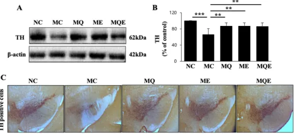
3.2. 지구성 운동과 MitoQ 섭취가 도파민성 신경세포에 미치는 영향
파킨슨 질환은 뇌의 흑질 부위의 도파민성 신 경세포가 소실되는 병리적인 특징이 나타나며, 그 로 인해 도파민수준의 감소로 운동기능장애가 나 타난다. TH는 도파민성 신경세포에서 아미노산 인 L-trosine을 L-dopa로 전환하여 도파민 합성 에 관여하는 속도제한 효소로서 많은 선행연구에 서 TH의 수준은 도파민성 신경세포의 수를 나타
Fig. 2. The effects of endurance exercise and MitoQ intake on tyrosine hydroxylase (TH) expression in the substantia nigra. A) Representative blot of western images for TH (n=6 per group). B) Quantification of TH levels. C) Representative immunohistochemistry staining of TH (n=4 per group). Values are means±SD.
*Significant difference between groups(*
p
<.05, **p
<.01, ***p
<.001). NC: normal control, MC: MPTP control, MQ: MPTP injection + MitoQ administration, ME: MPTP injection + endurance exercise, MQE: MPTP injection + MitoQ administration + endurance exercise.내는 중요 지표(marker)로 사용되고 있다[19]. 앞 서 확인된 운동과 MitoQ 섭취에 의한 운동기능 향상이 TH 효소와 관련이 있는지 확인하였다.
본 연구에서 Western blot 과 Immunohisto- chemistry 실험을 통해 TH 효소 단백질의 수준 을 분석한 결과(Fig 2), 집단 간 통계적으로 유의 한 차이가 나타났으며(F[4,25]=11.394, p=.001), 사후검증 결과 MC 집단은 NC 집단에 비해 유 의한 감소가 나타났다(p=.001). 이는 앞서 MC 집단에서 운동기능의 감소가 나타난 결과를 뒷받 침한다. 반면 MQ 집단(p=.004), ME 집단 (p=.003), MQE 집단(p=.005)은 MC 집단에 비 교하여 유의하게 증가하였다(Fig 2B). 이는 운동 이 MPTP로 파킨슨 질환을 유도한 마우스 모델 에서 흑질 부분 TH 효소 발현이 증가를 보고한 연구와 MitoQ 섭취로 흑질 부위의 TH 효소 발 현의 증가를 보고한 연구와 일치하였다[20,21].
결과적으로 운동과 MitoQ 섭취는 MPTP로 인한 TH 단백질의 감소를 회복해 운동기능을 향상시 키는 것으로 생각된다.
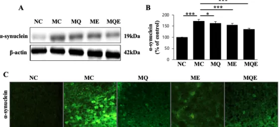
3.3. 지구성 운동과 MitoQ 섭취가 알파시누클린 단백질 수준에 미치는 영향
비정상적인 알파시누클린 단백질의 축적은 도 파민 신경세포의 사멸을 촉진한다. 따라서 최근 알파시누클린 단백질의 감소 또는 제거는 파킨슨 질환의 중요 치료 방법으로 제시되고 있다[22].
앞서 운동과 MitoQ 섭취는 도파민성 신경세포의 수준을 증가시켰고 이러한 결과가 알파시누클린 단백질 축적과 관련이 있는지 확인하였다(Fig 3A). 그 결과 집단 간 통계적으로 유의한 차이가 나타났으며 (F[4,25]=149.142, p=.001), 여러 선 행연구와 유사하게 MC 집단이 NC 집단과 비교
하여 통계적으로 유의한 증가를 나타냈다
(p=.001, Fig 3B). 반면 MC 집단에 비해 MQ 집단(p=.016), ME 집단(p=.001), MQE 집단 (p=.001)은 유의하게 감소하였다(Fig 3B). 이러한 결과는 파킨슨질환 동물모델을 대상으로 지구성 운동을 통해 알파시누클린을 감소를 보고한 연구 와 일치하였다[23]. 아직까지 MitoQ 섭취로 인 해 알파시누클린의 감소 원인을 알 수 없지만 coenzyme Q10과 같은 항산화제의 섭취가 알파
Fig. 3. The effects of endurance exercise and MitoQ intake on α-synuclein expression in the substantia nigra. A) Representative blot of western images for α-synuclein (n=6 per group). B) Quantification of α-synuclein levels. C) Representative Immunofluorescence staining of α-synuclein (n=4 per group). Values are means±SD. *Significant difference between groups(*
p
<.05, **p
<.01, ***p
<.001). NC: normal control, MC:MPTP control, MQ: MPTP injection + MitoQ administration, ME: MPTP injection + endurance exercise, MQE: MPTP injection + MitoQ administration + endurance exercise.
시누클린 수준을 감소시킨다는 결과[24]를 바탕 으로 coenzyme Q10과 triphenylphosphonium의 결합 물질인 MitoQ 섭취 역시 알파시누클린의 축적을 억제하는 것으로 생각된다. 이를 종합해보 면, 지구성 운동과 MitoQ의 섭취에 의한 알파 시누클린의 감소는 도파민 신경세포를 증가시켰 을 것으로 생각된다. 하지만 아직까지 운동과 MitoQ의 섭취에 의한 알파시누클린의 감소 기전 에 대한 연구는 부족한 실정이며 추후 연구가 필 요할 것으로 사료된다.
3.4. 지구성 운동과 MitoQ 섭취가 PGC-1α 단백질 수준과 신경세포사멸에 미치는 영향 미토콘드리아의 기능이상은 파킨슨질환의 주된 원인으로 알려져 있다. 사후 파킨슨 질환자의 뇌 에서 미토콘드리아 복합체 I의 감소가 나타났으 며, PINK, DJ1 및 Parkin등과 같은 미토콘드리 아의 중요한 기능을 담당하는 유전자는 파킨슨 질환의 발병을 유발하는 것으로 알려져 있다[25].
지구성 운동과 MitoQ의 섭취가 어떠한 기전으로 알파시누클린을 감소시키는지 정확히 밝혀지지 않았지만 최근 알파시누클린은 미토콘드리아 외
막과 결합하여 미토콘드리아의 분절을 증가시켜 미토콘드리아의 사멸을 촉진시킨다고 보고되고 있다[26]. PGC-1α는 미토콘드리아의 생합성에 핵심적인 역할을 하며 PGC-1α의 감소는 알파 시누클린 축적을 증가시키며, 그 수준에 따라 알 파시누클린을 조절하는 것으로 보고되고 있다[5].
따라서 운동과 MitoQ 섭취에 따른 알파시누클린 단백질 감소가 PGC-1α 수준과 관련이 있는지 확인하였다(Fig 4A). 그 결과 집단 간 통계적으로 유의한 차이가 나타났으며(F[4,25]=44.88, p=.001), MC 집단이 NC 집단과 비교하여 통계적으로 유 의한 증가를 나타냈다(p=.001, Fig 4B). 반면 MC 집단에 비해 MQ 집단(p=.001), ME 집단 (p=.001), MQE 집단(p=.001)은 유의하게 감소 하였다(Fig 4B). 이는 파킨슨질환 동물모델을 대 상으로 지구성 운동을 통해 PGC-1α 발현의 증 가를 보고한 연구와 MitoQ 섭취로 PGC-1α의 증가를 나타낸 선행연구와 일치한 결과를 나타냈 다[27,28]. 또한, 비정상적인 알파시누클린의 축 적은 산화스트레스를 유발시키는 것으로 알려져 있으며, 특히 PGC-1α는 미토콘드리아 내 항산 화 효소를 조절하여 활성산소를 감소시키기 때문
Fig. 4. The effects of endurance exercise and MitoQ intake on α-synuclein expression and apoptosis-related factors in the substantia nigra. A) Representative blot of western images for PGC-1α and Catalase (n=6 per group). B-C) Quantification of PGC-1α and Catalase levels. D) Representative blot of western images for BCL2 and BAX.
B-C) Quantification of BCL2 and BAX levels. Values are means±SD. *Significant difference between groups(*
p
<.05, **p
<.01, ***p
<.001). NC: normal control, MC:MPTP control, MQ: MPTP injection + MitoQ administration, ME: MPTP injection + endurance exercise, MQE: MPTP injection + MitoQ administration + endurance exercise.
에 앞선 운동과 MitoQ 섭취에 따른 PGC-1α의 증가가 항산화 효소에 미치는 영향을 확인하기 위해 미토콘드리아에 존재하는 Catalase 수준을 확인하였다. 그 결과 집단 간 통계적으로 유의한 차이가 나타났으며(F[4,25]=10.540, p=.001), MC 집단은 NC 집단에 비교하여 유의하게 감소 하였다(p=.001, Fig 4C). 또한 MC 집단에 비해 MQ 집단(p=.02), ME 집단(p=.018), MQE 집단 (p=.004)는 유의하게 증가하였다(Fig 4C). 이러한 결과는 운동, MitoQ 그리고 복합섭취에 따른 알 파시누클린 단백질의 감소는 PGC-1α의 증가에 따른 것을 생각되며, Catalase의 증가를 통해 활 성산소의 발생을 억제하여 결과적으로 미토콘드 리아 기능을 개선시켰을 것으로 생각된다. 또한, 알파시누클린 단백질 축적은 신경세포의 사멸을 유도하기 때문에 본 연구에서 신경세포사멸 관련 단백질 수준을 확인한 결과(Fig 4D), 알파시누클 린의 증가를 보인 MC 집단에서 NC 집단에 비 해 세포사멸억제 단백질인 B-cell lymphoma 2(BCL2)의 감소가 나타났고(p=.001, Fig 4E), 세 포사멸을 촉진하는 인자인 BCL2-associated
X(BAX)의 유의한 증가를 나타냈다(p=.001, Fig 4F). 이러한 결과는 MC 집단에서 축적된 알파시 누클린 단백질이 신경세포사멸을 증가시키고 결 과적으로 운동기능의 장애를 유발시킨 것으로 생 각된다. 하지만 BCL2의 경우 MC 집단에 비해 MQ 집단(p=.001), ME 집단(p=.001), MQE 집 단(p=.001)은 유의하게 증가하였으며(Fig 4E), BAX의 경우 MC 집단에 비해 MQ 집단 (p=.002), ME 집단(p=.028), MQE 집단 (p=.002)은 유의하게 감소하였다(Fig 4F). 이는 MPTP로 인한 증가된 신경세포사멸 기전을 운동 과 MitoQ 섭취로 인해 완화한 것으로 생각되며, 파킨슨 동물모델에서 지구성 운동을 통해 BCL2 의 증가와 BAX의 감소[29]를 보고한 연구와 일 치하였다. 또한 MitoQ 섭취를 통해 세포사멸을 억제한다는 연구[21]와 일치하는 결과를 얻었다.
이를 종합해보면, 지구성운동, MitoQ 그리고 복 합처치는 PGC-1α와 Catalase 수준을 증가시켜 미토콘드리아의 기능을 향상시키고, 신경세포사멸 을 억제하는 것으로 나타났다.
4. 결 론
본 연구는 MPTP로 유도된 파킨슨질환 동물모 델을 대상으로 5주간의 지구성운동, MitoQ 그리 고 복합처치는 PGC-1α와 Catalase 수준을 증가 시키고 세포사멸관련 인자를 감소시켜 결과적으 로 알파시누클린 단백질 수준을 감소시킨 것으로 생각된다. 또한 지구성운동, MitoQ섭취에 의한 알파시누클린 단백질 감소는 도파민성 신경세포 를 증가시켜 운동기능을 개선시킨 것으로 나타났 다. 따라서, 지구성 운동과 미토콘드리아를 표적 으로 하는 MitoQ의 섭취는 파킨슨 질환의 진전 을 감소시키는 효과적인 방법일 것으로 생각된다.
References
1. J.A. Obeso, M.C. Rodriguez-Oroz, C.G.
Goetz, C. Marin, J.H. Kordower, M.Rodriguez, E.C. Hirsch, M. Farrer, A.H. Schapira, G. Halliday, “Missing piecesin the Parkinson's disease puzzle”,
Nature Medicine
, Vol.16, No.6 pp.653-661, (2010).
2. L.V. Kalia,A.E. Lang, “Parkinson's disease”,
The Lancet
, Vol.386, No.9996 pp.896-912, (2015).
3. A.B. Singleton,M. Farrer, J. Johnson, A.
Singleton, S. Hague, J. Kachergus, M.
Hulihan, T.Peuralinna, A. Dutra, R.
Nussbaum, S. Lincoln, A. Crawley, M.
Hanson, D.Maraganore, C. Adler, M.R.
Cookson, M. Muenter, M. Baptista, D.
Miller, J.Blancato, J. Hardy, K.
Gwinn-Hardy, “alpha-Synuclein locus triplication causesParkinson's disease”,
Science
, Vol.302, No5646 pp. 841, (2003).4. S.W. Tait, D.R.Green, “Mitochondrial regulation of cell death”,
Cold Spring Harbor Perspectives in Biology
, Vol.15, No.9 a008706, (2013).5. C. Ciron, L.Zheng, W. Bobela, G.W.
Knott, T.C. Leone, D.P. Kelly, B.L.
Schneider, “PGC-1αactivity in nigral dopamine neurons determines vulnerability to α-synuclein”,
Acta Neuropathologica
Communications
, Vol.3, No.16 s40478- 015-0200-8, (2015).6. G.M. Petzinger,B.E. Fisher, S. McEwen, J.A. Beeler, J.P. Walsh, M.W. Jakowec,
“Exercise-enhanced neuroplasticity targeting motor and cognitive circuitry in Parkinson's disease”,
The Lancet Neurology
, Vol.12, No7 pp. 716-726, (2013).7. J.H. Koo, Y.C.Jang, D.J. Hwang, H.S. Um, N.H. Lee, J.H. Jung, J.Y. Cho, “Treadmill exercise produces neuroprotective effects in a murine model of Parkinson's disease by regulating the TLR2/MyD88/NF-κB signaling pathway”,
Neuroscience
, Vol.356, pp. 102-113, (2017).8. Y. Jang, J.H. Koo, I. Kwon, E.B. Kang, H.S. Um, H. Soya, Y. Lee, J.Y. Cho,
“Neuroprotective effects of endurance exercise against neuroinflammation in MPTP-induced Parkinson's disease mice”,
Brain Research
, Vol.1655, pp. 186-193, (2017).9. Y. Jang, I.Kwon, W. Song, L.M. Cosio- Lima, S. Taylor, Y. Lee, “Modulation of mitochondrial phenotypes by endurance exercise contributes to neuroprotection against a MPTP-induced animal model of PD”,
Life Science
, Vol.209, pp. 455-465, (2018).10. A.O. Oyewole, M.A. Birch-Machin,
“Mitochondria-targeted antioxidants”,
The FASEB Journal
, Vol.29, No.12 pp.4766-4771, (2015).
11. M.J. McManus, M.P. Murphy, J.L.
Franklin, “The mitochondria-targeted antioxidant MitoQprevents loss of spatial memory retention and early neuropathology in atransgenic mouse model of Alzheimer's disease”,
Journal of Neuroscience
, Vol.31, No.44 pp.15703-15715, (2011).
12. H. Jin, A. Kanthasamy, A. Ghosh, V.
Anantharam, B. Kalyanaraman, A.G.
Kanthasamy, “Mitochondria-targeted antioxidants for treatment of Parkinson's disease:preclinical and clinical outcomes”,
Biochimica et Biophysica Acta
, Vol.1842, No.8 pp. 1282-1294, (2014).13. B.J. Snow,F.L. Rolfe, M.M. Lockhart, C.M. Frampton, J.D. O'Sullivan, V. Fung, R.A. Smith,M.P. Murphy, K.M. Taylor, “A double-blind, placebo-controlled study to assessthe mitochondria-targeted antioxidant MitoQ as a disease-modifying therapy in Parkinson's disease”,
Movement Disorders
, Vol.25, No.11 pp. 1670-1674, (2014).14. Y.S. Lau, G. Patki, K. Das-Panja, W.D.
Le, S.O. Ahmad, “Neuroprotective effects and mechanisms of exercise in a chronic mouse model of Parkinson's disease with moderate neurodegeneration”,
European Journal of Neuroscience
, Vol.33, No.7 pp.1264-1274, (2011).
15. S.Rodriguez-Cuenca, H.M. Cochemé, A.
Logan, I. Abakumova, T.A. Prime, C.
Rose, A.Vidal-Puig, A.C. Smith, D.C.
Rubinsztein, I.M. Fearnley, B.A. Jones, S.
Pope,S.J. Heales, B.Y. Lam, S.G. Neogi, I.
McFarlane, A.M. James, R.A. Smith, M.P.Murphy, “Consequences of long-term oral administration of the mitochondria- targeted antioxidant MitoQ to wild-type mice”,
Free Radical Biology & Medicine
, Vol.48, No.1 pp. 161-72, (2010).16. M.M Bradford, “A rapid and sensitive method for the quantitation of microgram quantities of protein utilizing the principle of protein-dye binding”,
Analytical Biochemistry,
Vol.72, No.1-2 pp.248-254, (1976).
17. G.E. Meredith,D.J. Rademacher, “MPTP mouse models of Parkinson's disease: an update”,
Journal of Parkinson's Disease
, Vol.1, No.1 pp. 19-33, (2011).18. G.M.Petzinger, J.P. Walsh, G. Akopian, E.
Hogg, A. Abernathy, P. Arevalo, P.Turnquist, M. Vucković, B.E. Fisher, D.M. Togasaki, M.W. Jakowec, “Effects of treadmill exercise on dopaminergic transmission in the1-methyl-4-phenyl- 1,2,3,6-tetrahydropyridine-lesioned mouse model of basal ganglia injury”,
Journal of
Neuroscience
, Vol.27, No.20 pp. 5291- 5300, (2007).19. H.F. Shu, T.Yang, S.X. Yu, H.D. Huang, L.L. Jiang, J.W. Gu, Y.Q. Kuang,
“Aerobic exercisefor Parkinson's disease: a systematic review and meta-analysis of randomized controlled trials”,
PLoS One
, Vol.9, No.7 pp. e100503, (2014).20. H.S. Cho, M.S.Shin, W. Song, T.W. Jun, B.V. Lim, Y.P. Kim, C.J. Kim, “Treadmill exercise alleviates short-term memory impairment in 6-hydroxydopamine- induced Parkinson's rats”,
Journal of Exercise Rehabilitation
, Vol.9, No.3 pp.354-364, (2013).
21. A. Ghosh, K.Chandran, S.V. Kalivendi, J.
Joseph, W.E. Antholine, C.J. Hillard, A.Kanthasamy, A. Kanthasamy, B.
Kalyanaraman, Neuroprotection by amitochondria-targeted drug in a Parkinson's disease model,
Free Radical Biology & Medicine
, Vol.49, No.11 pp.1674-1684, (2010).
22. L. Xu, J. Pu, “Alpha-Synuclein in Parkinson's Disease: From Pathogenetic Dysfunction to Potential Clinical Application”,
Parkinson’s Disease
, Vol.2016, 1720621, (2016).23. W. Zhou, J.C.Barkow, C.R. Freed,
“Running wheel exercise reduces α -synuclein aggregation and improves motor and cognitive function in a transgenic mouse model of Parkinson's disease”,
PLoS One
, Vol.12, No.12 e0190160, (2017).24. L. Yang, N.Y.Calingasan, E.J. Wille, K.
Cormier, K. Smith, R.J. Ferrante, M.F.
Beal, “Combination therapy with coenzyme Q10 and creatine produces additiveneuro- protective effects in models of Parkinson's and Huntington's diseases”,
Journal of Neurochemistry
, Vol.105, No.5 pp.1427-1439, (2009).
25. M.R. Cookson, “Parkinsonism due to mutations in PINK1, parkin, and DJ-1 and oxidative stress and mitochondrial
pathways”,
Cold Spring Harbor Perspectives in Medicine
, Vol.2, No.9 a009415, (2012).26. J.H. Park,J.D. Burgess, A.H. Faroqi, N.N.
DeMeo, F.C. Fiesel, W. Springer, M.
Delenclos, P.J. McLean, “Alpha-synuclein- induced mitochondrial dysfunction is mediated via a sirtuin 3-dependent pathway”,
Molecular Neurodegeneration
, Vol.15, No.1 s13024-019-0349, (2020).27. J.H. Koo, J.Y.Cho, “Treadmill Exercise Attenuates α-Synuclein Levels by Promoting Mitochondrial Function and Autophagy Possibly via SIRT1 in the Chronic MPTP/P-Induced Mouse Model of Parkinson's Disease”,
Neurotoxicity Research
, Vol.32, No.3 pp. 473-486, (2017).28. Y. Xi, D.Feng, K. Tao, R. Wang, Y. Shi, H. Qin, M.P. Murphy, Q. Yang, G. Zhao,
“MitoQ protects dopaminergic neurons in a 6-OHDA induced PD model by enhancing Mfn2-dependent mitochondrial fusion via activation of PGC-1α”,
Biochimica et Biophysica Acta-Molecular Basis of Disease
, Vol.2864, No.9 pp.2859-2870, (2018).
29. Y.C. Jang ,D.J. Hwang, J.H. Koo, H.S.
Um, N.H. Lee, D.C. Yeom, Y. Lee, J.Y.
Cho, “Association of exercise-induced autophagy upregulation and apoptosiss uppression with neuroprotection against pharmacologically induced Parkinson's disease”,