The Anti-obesity Effect of Aureobasidium pullulans SM-2001 Extract (Polycan ® ) on 3T3-L1 Preadipocytes and Adipocytes
Young-Suk Kim
1, Jong-Min Lim
1, Bon-Hwa Ku
1, Seung-Bae Moon
1, Hyung-Rae Cho
1, Seon-Min Lee
2and Jung-Hee Kwon
3*
lGlucan Co. Ltd., 25-15, Worasan-ro 950 beon-gil, Munsan-eup Jinju-si, Gyeongsangnam-do 52840, Korea
2Biological Resources Research Group, Gyeongnam Department of Environment Toxicology and Chemistry, Korea Institute of Toxicology (KIT), 17 Jeigok-gil, Jinju 52834, Korea
3NPChemBio Co. Ltd., Venture Support Facilities, Jinju Bioindustry Promotiom Foundation, 991 Worasan-ro, Munsan-eup, Jinju-si, Gyeongsangnam-do 52839, Korea
Received January 21, 2020 /Revised August 24, 2020 /Accepted September 15, 2020
Obesity, the world’s leading metabolic disease, is a serious health problem in both industrialized and developing countries. Natural substances are of great interest in preventative medicine, especially in the field of metabolic syndromes—from insulin resistance to obesity and diabetes. In the present study, we investigated the effect of A. pullulans SM-2001 Extract (Polycan
®) on the adipocyte differ- entiation of 3T3-L1 preadipocytes and the anti-obesity effect of 3T3-L1 adipocytes. Although β-glucan has been found to have health benefits in the regulation of the immune system and blood cholesterol levels, its role in obesity has not been fully investigated. Polycan
®suppressed lipid accumulation and glycerol-3-phosphate dehydrogenase (GPDH) activity without affecting cell viability in 3T3-L1 pre- adipocytes and adipocytes. Polycan
®also inhibited cellular lipid accumulation through down-regu- lation of transcription factors, such as PPARγ and C/EBPα, and induced dose-dependent phosphor- ylation of AMP-activated protein kinase (AMPK)—a cellular energy sensor—while the total AMPK protein content remained unchanged. Taken together, this shows that the activation of AMPK by Polycan
®in adipocytes plays a critical role in Polycan
®-induced inhibition of adipocyte differentiation.
Our results show that Polycan
®has an anti-obesity action in vitro, suggesting a potential novel pre- ventative agent for obesity and other metabolic diseases.
Key words : Adipocyte differentiation, AMPK, beta-glucan, obesity, PPARγ
*Corresponding author
*Tel : +82-55-291-9383, Fax : +82-55-754-1102
*E-mail : [email protected]
This is an Open-Access article distributed under the terms of the Creative Commons Attribution Non-Commercial License (http://creativecommons.org/licenses/by-nc/3.0) which permits unrestricted non-commercial use, distribution, and reproduction in any medium, provided the original work is properly cited.
Journal of Life Science 2020 Vol. 30. No. 10. 835~843 DOI : https://doi.org/10.5352/JLS.2020.30.10.835
Introduction
Obesity has become a public health crisis worldwide, and the prevalence has steadily increased over the past few decades. Recent studies estimated that obesity increases the risk of numerous of chronic diseases, such as dyslipidemia, hypertension, cardiovascular diseases, and type II diabetes [4, 34]. Adipose tissue plays a critical role in lipid metabo- lism and energy balance. Adipocyte differentiation, known as adipogenesis, is the anabolic process of fat cell develop- ment [30]. There is evidence that a group of closely related nuclear receptors, called peroxisome proliferator-activated receptors (PPARs), are involved in obesity. Among three
PPAR isotypes identified as α, β, and γ, PPARγ is mainly expressed in adipose tissue and have been revealed to be re- quired for the adipocyte differentiation, with the CCAAT-en- hancer-binding proteins (C/EBPs) transcription factors [39].
Besides, most of the PPARγ target genes in adipose tissue are directly involved in lipogenic pathways, including lip- oprotein lipase (LPL), adipocyte fatty acid binding protein (aP2), uncoupling protein-2 (UCP-2), and glucose-transporter 4 (Glut4) [24].
Preadipocyte cell lines are useful models for investigating the adipogenesis process. 3T3-L1 preadipocyte which can be induced to differentiate into adipocyte cells, is one of the most studied preadipocyte cell lines [16, 32, 33]. During dif- ferentiation into adipocyte, PPARγ and C/EBPs are involved in the sequential expression of adipocyte differentiation [1, 11, 26], whereas expression of PPARγ and C/EBP, which trigger the expression of adipocyte-specific proteins, are in- duced during terminal differentiation of the adipocyte line- age [14, 29].
Many natural extracts have drawn attention because of
addition, some reports revealed the hypoglycaemic effect of β-glucan extracts from plants or mushroom in animal ex- periments [9, 27] and clinical trials [37], whereas β-glucan derived from other origins showed no hypoglycaemic effects on the STZ-induced diabetes [5, 40, 41]. However, Polycan
®extracted from a UV-induced mutant of A. pullulans (SM- 2001), mainly containing β-1,3/1,6-glucan [18] showed rela- tively favourable effects against diabetic complications, par- ticularly diabetic nephropathy and hepatopathy [41], and ab- normalities in lipid metabolism [5]. Versatile roles of β-glu- cans have to be further investigated in obesity and other metabolic syndrome.
In the present study, we explored the effect of Polycan
®on adipocyte differentiation in 3T3-L1 preadipocyte model to gain further insight into the role of β-glucan in adipo- genesis and obesity.
Materials and Methods
Chemicals and materials
Polycan
®were supplied by Glucan Corp. (Korea) and were stored in a refrigerator at 4℃ to protect from light and degradation. 3T3-L1 cells were purchased from the Amer- ican Type Culture Collection (Manassas, VA, U.S.A.). Dul- becco's Modified Eagle Medium/Ham's F-12 nutrient mix- ture (DMEM/F12), fetal bovine serum (FBS), calf bovine se- rum (CBS), penicillin-streptomycin, phosphate buffered sal- ine (PBS) and transferrin were obtained from Gibco BRL (Rockville, MD., U.S.A.). Dexamethasone (DEX), insulin, 1- methyl3-isobutylxanthine (IBMX), GPDH assay kit and Monoclonal Anti-β-actin were purchased from Sigma Chem- ical Co. (St. Louis, MO., U.S.A.). C/EBPα (D56F10) XP
®Rabbit mAb and PPARγ (81B8) Rabbit mAb were purchased from Cell Signaling Technology (Danvers, MA., U.S.A.). The lip-
Glucan Assay Kit (Megazyme, Chicago, USA). In Polycan , around 90% of the total glucan consists of β-1,3/1,6-glucans and the rest is α-glucan (pullulan).
3T3-L1 cell culture and cell viability assay Mouse embryo preadipocyte (3T3-L1) cell lines was ob- tained from ATCC (American Type Culture Collection, Manassas, VA, U.S.A.) and cultured in Dulbecco’s modified Eagle’s medium (DMEM) containing 10% calf serum, 100 U/ml penicillin and 100 μg/ml streptomycin in a humidified atmosphere of 5% CO
2at 37℃. Cell viability was measured using the CCK-8 kit (Dojindo, Kumamoto, Japan), according to the manufacturer’s instructions. Briefly, the cells were plated in 96-well plates at a density of 1×10
4cells/well. After overnight incubation, the cells were treated with the 0, 100, 200, 400 ug/ml Polycan
®to be tested (at the concentration as indicated) and cultured for 24 hr. After incubation, the CCK-8 solution was added to each well and incubated for 3 hr at 37℃. The absorbance at 450 nm was measured using a microplate reader (Bio-Tek Company, Winooski, VT, U.S.A.). The experiments were performed in triplicate.
3T3-L1 adipocyte differentiation and Oil Red O staining
Adipocyte differentiation was induced in 100% confluent
3T3-L1 cells with adipogenic medium (DMEM containing
10% CBS with 0.5 mM IBMX, 1 μM dexamethasone, and 1
μg/ml insulin) treated with different concentration of Poly-
can
®(0, 100, 200, 400 μg/ml). Two days after induction, the
medium was changed to DMEM including 10% FBS and 1
μg/ml insulin with the samples for additional two days. The
cells were then maintained in DMEM with 10% FBS for an-
other four days. Lipid accumulation in adipocytes was esti-
mated by staining with Oil Red O. Six days after the ini-
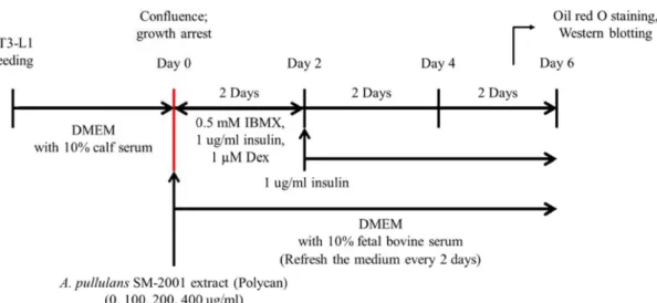
Fig. 1. Experimental scheme. For the induction of adipocyte differentiation, 3T3-L1 preadipocytes were seeded. At confluence (day 0), the cultured preadipocytes were induced to differentiate by the addition of differentiating medium containing 0.5mM methylisobutylxanthine (IBMX), 1 μg/ml insulin, and 1 μM dexamethasone (Dex) from day 0 to day 2. At day 2, medium was changed with medium containing 1 μg/ml insulin for an additional 2 days from day 2 to day 6. The medium was refreshed every 2 days. At day 6, differentiated 3T3-L1 cells were subjected to Oil red O solution or used for Western blot analysis. Polycan® was added to the cell culture medium at concentrations of 0, 100, 200 and 400 ug/ml from day 0 to day 6. DMEM, Dulbecco’s modified Eagle’s medium.
tiation of differentiation, 3T3-L1 cells were washed with PBS and fixed in 60% ethanol as described previously [23](Fig.
1). The cells were stained with 0.3% Oil red O dye for 1 hr to show accumulated cytoplasmic lipid. The visualization of lipids was performed by Olympus IX50 microscope (Olympus, Tokyo, Japan).
Lipolysis assay
Amounts of glycerol released from cells into the medium were measured to analyze the lipolytic effect of Polycan
®on the triacylglycerol accumulated in adipocytes. Medium was collected from the culture plate and heated at 65℃ for 8 min to inactivate enzymes released from the cells. The glycerol was measured with a commercial lipolysis assay kit (AMS biotechnology, Abingdon, OX., U.K.). Cellular protein content was analyzed with a BCA protein assay kit (Pierce, Rockford, IL., U.S.A.) using BSA as a standard.
GPDH activity
The 3T3-L1 adipocytes were harvested 48 hr after ini- tiation of differentiation or 6 days after differentiation with 0, 100, 200, 400 ug/ml Polycan
®. Cells were carefully washed twice with ice-cold PBS and collected with a scraper into 300 ul of 100 mM tri-ethanolamine/HCl buffer, pH 7.5, 2.5 mM EDTA. The harvested cells were sonicated in ice at 25 ultrasonic bursta of 10 sec each in a DU-250 Bioruptor with
a maximum output power of 250 W (Tosho Denki, Co. Ltd., Japan). After centrifugation at 13,000× g for 5 min at 4℃, the supernatants were assayed for GPDH activity according to the method of Wise and Green [17]. GPDH activity was measured under zero-order kinetics and optimal substrate and cofactor conditions at 25℃ for 180 sec in a spectropho- tometer (Beckman Coulter, DU 530, Indianapolis, IN, U.S.A.).
The standard reaction mixture contained 100 mM triethanol- amine/HCl buffer (pH 7.5), 2.5 mM EDTA, 0.1 mM/2-mer- captoethanol, and 0.12 mM NADH. The reaction was ini- tiated by the addition of 0.2 mM dihydroxyacetone phos- phate, and the rate of NADH oxidation was measured by a change in absorbance at 340 nm for 60 sec. Enzyme activity (%) was expressed as percent against control (100%) [18].
Western blot analysis
For Western blot analysis, cells (3×10
5) were cultured in
3 ml of DMEM and differentiated to the adipocytes by in-
cubating in DMEM containing 10% CBS, 0.5 mM IBMX, 1
μM dexamethasone, and 1 μg/ml insulin. The cells were har-
vested 48 hours after initiation of differentiation or 6 days
after differentiation with 0, 100, 200, 400 ug/ml Polycan
®,
washed twice in PBS and then dissolved for 30 min with
lysis buffer [150 mM NaCl, 50 mM Tris (pH 7.2), 1 mM
EDTA, 0.5% sodium deoxycholate, 1% Nonidet P-40, 1 mM
sodium vanadate, 1 mM NaF, 20 ug/ml aprotinin, 50 ug/ml
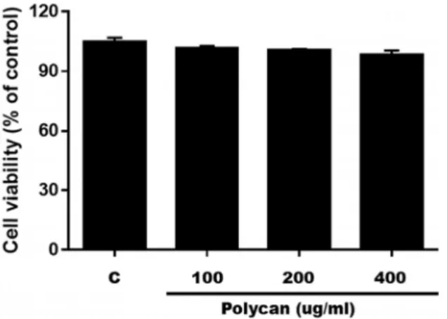
Fig. 2. Cytotoxic effect of Polycan® on 3T3-L1 cells. Cell viability was measured using CCK-8 staining. The data represent the mean ± SD of three separate experiments
secondary antibodies for 1 hr at room temperature. Bands were visualized using ECL solution (Thermo Scientific) and quantified using the Chemidoc Imaging System (Bio-Rad;
Hercules, CA, U.S.A.).
Statistical analysis
The results were analyzed using Prism version 5.00 soft- ware (GraphPad Software, San Diego, CA, U.S.A.). One-way ANOVA was applied to calculate the significance between the groups. Statistical significance was indicated by a p value of <0.05. Data were expressed as the mean ± SEM of three independent experiments.
Results and Discussion
Proliferation of preadipocytes
To identify whether Polycan
® (A. pullulans SM-2001 Ex-tract) inhibited the proliferation of 3T3-L1 cells, preadipocyte were treated with Polycan
®at the concentration of 0, 100, 200, or 400 μg/ml. As shown in Fig. 2, cell viability was
in vitro [12].
Effect of Polycan
®on lipid accumulation
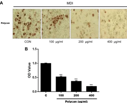
The effect of Polycan
®on intracellular lipid accumulation was examined by oil red O staining (Fig. 3A). To test the inhibitory effects of Polycan
®on lipid accumulation in 3T3- L1 cells, insulin, DEX and IBMX were used to induce 3T3-L1 pre-adipocytes to differentiation. At the concentration of 0, 100, 200 and 400 μg/ml Polycan
®inhibited the adipocyte differentiation (Fig. 3B). Quantitative data was obtained through extraction of Oil-Red O-stained cells with isopro- panol and spectrophotometric analysis. The O.D. absorbance significantly decreased (p<0.05) in cells treated with Polycan
®in dose-dependent manner (Fig. 3B), and the decrease rates were 51.2%, 36.03%, and 18.3% at concentrations of 100, 200 and 400 μg/ml of Polycan
®, respectively. These results in- dicated that Polycan
®could inhibit adipocyte differentiation, thus inhibit intracellular lipid accumulation in 3T3-L1 cells.
Effects of Polycan
®on glycerol release
To clarify the direct effect of Polycan
®on lipolysis, the amount of glycerol released into the medium was measured (Fig. 4). The amount of glycerol in the medium was in- creased by 78% in the presence of 400 ug/ml Polycan
®. In the present study, it was clear that Polycan
®treatment de- creased the inracellular lipid content in 3T3-L1 adipocytes (Fig. 3) as well as increased the amount of glycerol released into the medium, indicating activation of lipolysis.
Effect of Polycan
®on GPDH activity
To ascertain the inhibition of the accumulation of intra-
cellular lipid in 3T3-L1 preadipocytes, we examined the ef-
fect of Polycan
®on GPDH activity. GPDH is an index of
differentiation as one of the lipid-synthesizing enzymes ex-
pressed in adipocytes differentiated from preadipocytes.
A
MDICON 100 μg/ml 200 μg/ml 400 μg/ml
B
Fig. 3. Effects of Polycan® on intracellular lipid accumulation in 3T3-L1 cells. 3T3-L1 cells were treated with polycan (0, 100, 200, 400 μg/ml) for day 6. (A) The ma- ture adipocytes were stained with Oil- red O, and the (B) OD value were meas- ured to quantify intracellular lipid con- tent. Three independent experiments were performed and the data were shown as mean ± SD. Values do not share the same letter are significantly different (p<0.05). ***p<0.001.
Fig. 4. Effects of Polycan® on glycerol release in 3T3-L1 adipo- cytes. The differentiated 3T3-L1 adipocytes were treated in serum-free medium with Polycan® (0, 100, 200, 400 ug/ml). The medium was collected and assayed for glyc- erol content. Three independent experiments were per- formed and the data were shown as mean ± SD. Values do not share the same letter are significantly different (p<0.05). ***p< 0.001, **p<0.01, *p<0.05.
Fig. 5. Effect of Polycan® on GPDH activity in cultured 3T3-L1 adipocytes. The 3T3-L1 adipocytes were harvested 6 days after the initiation of differentiation with 0, 100, 200, 400 μg/ml Polycan®. Three independent experiments were performed and the data were shown as mean ± SD. Values do not share the same letter are significantly different (p<0.05). *** p<0.001.
Cultured 3T3-L1 preadipocytes and adipocytes were ex- posed to Polycan
®at various concentrations, and then the cells were differentiated with a differentiation medium or DMEM after differentiation. As shown in Fig. 5, the treat- ment of 3T3-L1 preadipocytes and adipocytes with Polycan
®significantly inhibited GPDH activity dose dependently.
This result demonstrates for the first time, to the best of our knowledge, Polycan
®causes a significant decrease in the ac-
tivity of GPDH in 3T3-L1 preadipocytes without eliciting cell cytotoxicity, suggesting that Polycan
®may block adipo- genesis, at least in part, by down-regulating key adipogenic transcription factors in 3T3-L1 preadipocytes and may have antiatherogenic, anti-inflammatory, and antidiabetic effects through down-regulation of GPDH in 3T3-L1 adipocytes.
Effect of Polycan
®on expression and activity of adipogenic proteins
Above results demonstrated that Polycan
®inhibits intra-
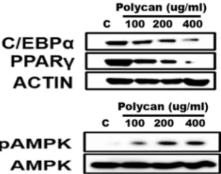
Fig. 6. Effect of Polycan® on the protein levels of the PPARγ, C/EBPα, AMPK, and pAMPK in 3T3-L1 adipocytes by Western blotting. 3T3-L1 adipocytes were harvested 6 days after the differentiation with 0, 100, 200, 400 ug/ml Polycan®. β-actin was used as a housekeeping protein control.
treatment with Polycan also dose-dependently decreased the protein levels of PPARγ, C/EBPα whereas increased the phosphorylation of AMPK (Fig. 6). PPARγ expression was significantly decreased concomitantly with reduction of C/
EBPα protein expression in 400 ug/ml polycan group com- pared to the control group (Fig. 6). Polycan
®effectively in- hibited the differentiation medium-induced increase of PPAR γ expression in 3T3-L1 adipocytes at concentrations greater than 100 ug/ml. Adipogenesis is the process by which pre- cursor stem cell differentiate into lipid laden adipocytes adi- pocytes [15]. This process is regulated by transcriptional acti- vators such as PPARγ and C/EBPα [6, 33]. These tran- scription factors were known to regulate the middle and late stages of adipocyte differentiation [20]. Also, FABP4 is a dif- ferentiated adipocyte marker gene that is transcriptionally regulated by PPARγ [43, 44]. Although C/EBPα is an im- portant factor in terminal differentiation of adipocytes, knockout of C/EBPα in adipocytes did not show insulin sen- sitivity [10, 38]. It means that C/EBPα is an essential factor in which adipocyte acquires insulin sensitivity. As obesity
and regulates glucose and lipid homeostasis in adipocytes [28]. Once activated, AMPK phosphorylates a number of proteins and modulates the transcription of genes implicated in the regulation of energy metabolism to switch on catabolic pathways that produce ATP and switch off anabolic path- ways that consume ATP [8].
Several studies already have reported that β-glucan in- hibits adipocyte differentiation and improves serum lipid levels in high fat diet-induced obese rat models [22, 25].
However, they all used β-1,3-glucan, not β-1,3/1,6-glucan evaluated in the present study. Although both β-1,3-glucan and β-1,3/1,6-glucan have suppressive function in obesity, the latter is more structurally stable compared to β-1,3-glu- can because of its innate glycosidic bond. Therefore, β-1,3/
1,6-glucan has merits in higher yields in the production process as anti-obesity materials. To the best of our knowl- edge, the results of the present study demonstrate, for the first time, that Polycan
®consisting mainly of β-1,3/1,6-glu- can have anti-obesity effects in 3T3-L1 adipocyte.
The nuclear receptor PPARγ and members of the C/EBPα complex synergistically activate downstream promoters of adipocyte-specific genes, such as acetyl-CoA carboxylase, ac- yl CoA synthase and GPDH. Polycan
®inhibits cellular lipid accumulation through down-regulation of transcription fac- tors such as PPARγ and C/EBPα and up-regulation of phos- phorylated AMPK. Consistent with our data showing that the expression of cytosolic GPDH was enhanced by high lev- els of PPARγ in adipocyte [36], there were reduced activity of GPDH and decreased level of PPARγ in Polycan
®treated adipocyte.
In conclusion, we report that Polycan
®exerts an anti-obe-
sity effect through inhibition of the expression of key tran-
scription factors and genes responsible for adipocyte differ-
entiation. In addition, we have shown that activation of
AMPK by Polycan
®in adipocytes plays a critical role in Polycan
®-induced inhibition of adipocyte differentiation.
Taken together, Polycan
®may have novel preventative po- tential for obesity and other metabolic diseases, warranting further investigation for the precise mechanism.
The Conflict of Interest Statement
The authors declare that they have no conflicts of interest with the contents of this article.
References
1. Amri, E. Z., Bonino, F., Ailhaud, G., Abumrad, N. A. and Grimaldi, P. A. 1995. Cloning of a protein that mediates tran- scriptional effects of fatty acids in preadipocytes. Homology to peroxisome proliferator-activated receptors. J. Biol. Chem.
270, 2367-2371.
2. Bell, S., Goldman, V. M., Bistrian, B. R., Arnold, A. H., Ostroff, G. and Forse, R. A. 1999. Effect of beta-glucan from oats and yeast on serum lipids. Crit. Rev. Food Sci. Nutr.
39, 189-202.
3. Bhathena, S. J. and Velasquez, M. T. 2002. Beneficial role of dietary phytoestrogens in obesity and diabetes. Am. J.
Clin. Nutr. 76, 1191-1201.
4. Cani, P. D., Bibiloni, R., Knauf, C., Waget, A., Neyrinck, A.
M., Delzenne, N. M. and Burcelin, R. 2008. Changes in gut microbiota control metabolic endotoxemia-induced inflam- mation in high-fat diet-induced obesity and diabetes in mice.
Diabetes 57, 1470-1481.
5. Choi, J., Kim, J., Jung, G., Moon, S., Cho, H., Ku, S. and Sohn, J. 2018. Hypoglycemic and hypolipemic effect of β-glucan originated from Aureobasidium in STZ-induced diabetic rats. J. Anim. Plant Sci. 28, 9.
6. Cowherd, R. M., Lyle, R. E. and McGehee, R. E. Jr. 1999.
Molecular regulation of adipocyte differentiation. Semin. Cell Dev. Biol. 10, 3-10.
7. Czop, J. K. 1986. The role of beta-glucan receptors on blood and tissue leukocytes in phagocytosis and metabolic activa- tion. Pathol. Immunopathol. Res. 5, 286-296.
8. Daval, M., Foufelle, F. and Ferre, P. 2006. Functions of AMP- activated protein kinase in adipose tissue. J. Physiol. 574, 55-62.
9. De Paula, A. C., Sousa, R. V., Figueiredo-Ribeiro, R. C. and Buckeridge, M. S. 2005. Hypoglycemic activity of poly- saccharide fractions containing beta-glucans from extracts of Rhynchelytrum repens (Willd.) C.E. Hubb., Poaceae. Braz.
J. Med. Biol. Res. 38, 885-893.
10. El-Jack, A. K., Hamm, J. K., Pilch, P. F. and Farmer, S. R.
1999. Reconstitution of insulin-sensitive glucose transport in fibroblasts requires expression of both PPARgamma and C/EBPalpha. J. Biol. Chem. 274, 7946-7951.
11. Elberg, G., Gimble, J. M. and Tsai, S. Y. 2000. Modulation of the murine peroxisome proliferator-activated receptor
gamma 2 promoter activity by CCAAT/enhancer-binding proteins. J. Biol. Chem. 275, 27815-27822.
12. Estrada, A., Yun, C. H., Van Kessel, A., Li, B., Hauta, S. and Laarveld, B. 1997. Immunomodulatory activities of oat beta- glucan in vitro and in vivo. Microbiol. Immunol. 41, 991-998.
13. Finck, B. N. 2018. Targeting metabolism, insulin resistance, and diabetes to treat nonalcoholic steatohepatitis. Diabetes 67, 2485-2493.
14. Freytag, S. O., Paielli, D. L. and Gilbert, J. D. 1994. Ectopic expression of the CCAAT/enhancer-binding protein alpha promotes the adipogenic program in a variety of mouse fi- broblastic cells. Genes Dev. 8, 1654-1663.
15. Green, H. and Kehinde, O. 1975. An established preadipose cell line and its differentiation in culture. II. Factors affecting the adipose conversion. Cell 5, 19-27.
16. Gregoire, F. M., Smas, C. M. and Sul, H. S. 1998. Under- standing adipocyte differentiation. Physiol. Rev. 78, 783-809.
17. Haslam, D. W. and James, W. P. 2005. Obesity. Lancet 366, 1197-1209.
18. Jeon, J. R. and Kim, J. Y. 2006. Effects of pine needle extract on differentiation of 3T3-L1 preadipocytes and obesity in high-fat diet fed rats. Biol. Pharm. Bull. 29, 2111-2115.
19. Jeon, T., Hwang, S. G., Hirai, S., Matsui, T., Yano, H., Kawa- da, T., Lim, B. O. and Park, D. K. 2004. Red yeast rice ex- tracts suppress adipogenesis by down-regulating adipo- genic transcription factors and gene expression in 3T3-L1 cells. Life Sci. 75, 3195-3203.
20. Johmura, Y. 2007. Characterization of novel genes regulating adipocyte differentiation. Yakugaku Zasshi 127, 135-142.
21. Kanagasabapathy, G., Chua, K. H., Malek, S. N., Vikines- wary, S. and Kuppusamy, U. R. 2014. AMP-activated pro- tein kinase mediates insulin-like and lipo-mobilising effects of beta-glucan-rich polysaccharides isolated from Pleurotus sajor-caju (Fr.), Singer mushroom, in 3T3-L1 cells. Food Chem.
145, 198-204.
22. Kang, S. A., Jang, K., Hong, K., Choi, W., Jung, K. and Lee, I. 2002. Effects of dietary β-Glucan on adiposity and serum lipids levels in obese rats induced by high fat diet. J. Kor.
Soc. Food Sci. Nutr. 31, 1052-1057.
23. Kasturi, R. and Joshi, V. C. 1982. Hormonal regulation of stearoyl coenzyme A desaturase activity and lipogenesis during adipose conversion of 3T3-L1 cells. J. Biol. Chem. 257, 12224-12230.
24. Kersten, S., Desvergne, B. and Wahli, W. 2000. Roles of PPARs in health and disease. Nature 405, 421-424.
25. Kim, M., Kim, O., Chung, C., Jang, K., Kim, C. and Kang, S. A. 2015. β-Glucan from Aureobasidum species inhibits fat accumulation in 3T3-L1 adipocyte differentiation. Food Sci. Biotechnol. 24, 1147-1150.
26. Kim, W. K., Lee, C. Y., Kang, M. S., Kim, M. H., Ryu, Y.
H., Bae, K. H., Shin, S. J., Lee, S. C. and Ko, Y. 2008. Effects of leptin on lipid metabolism and gene expression of differ- entiation-associated growth factors and transcription factors during differentiation and maturation of 3T3-L1 preadipo- cytes. Endocr. J. 55, 827-837.
27. Kim, Y. W., Kim, K. H., Choi, H. J. and Lee, D. S. 2005. Anti-
Na, M., Hattori, M. and Bae, K. 2010. Lanostane triterpenes from Ganoderma lucidum suppress the adipogenesis in 3T3-L1 cells through down-regulation of SREBP-1c. Bioorg.
Med. Chem. Lett. 20, 5577-5581.
31. Lia, A., Hallmans, G., Sandberg, A. S., Sundberg, B., Aman, P. and Andersson, H. 1995. Oat beta-glucan increases bile acid excretion and a fiber-rich barley fraction increases cho- lesterol excretion in ileostomy subjects. Am. J. Clin. Nutr.
62, 1245-1251.
32. Liu, L. H., Wang, X. K., Hu, Y. D., Kang, J. L., Wang, L.
L. and Li, S. 2004. Effects of a fatty acid synthase inhibitor on adipocyte differentiation of mouse 3T3-L1 cells. Acta Pharmacol. Sin. 25, 1052-1057.
33. MacDougald, O. A. and Lane, M. D. 1995. Transcriptional regulation of gene expression during adipocyte differ- entiation. Annu. Rev. Biochem. 64, 345-373.
34. Mark, D. H. 2005. Deaths attributable to obesity. JAMA 293, 1918-1919.
35. Masihi, K. N., Madaj, K., Hintelmann, H., Gast, G. and Kaneko, Y. 1997. Down-regulation of tumor necrosis fac- tor-alpha, moderate reduction of interleukin-1beta, but not interleukin-6 or interleukin-10, by glucan immunomodula- tors curdlan sulfate and lentinan. Int. J. Immunopharmacol.
1999. PPAR gamma is required for the differentiation of adi- pose tissue in vivo and in vitro. Mol. Cell 4, 611-617.
40. Seo, H., Kim, J., Shin, H., Kim, T., Chang, H., Park, B. and Lee, J. 2002. Production of β-1,3/1,6-glucan by Aureobasi- dium pullulans SM-2001. Kor. J. Biotechnol. Bioeng. 17, 5.
41. Sohn, J., Kim, J., Lim, J., Cho, H., Ku, S. and Choi, J. 2018.
Effects of β-glucan from Aureobasidium pullulans in a streptozotocin-induced rat diabetes model. Curr. Nutr. Food Sci. 15, 12.
42. Soltys, J. and Quinn, M. T. 1999. Modulation of endotoxin- and enterotoxin-induced cytokine release by in vivo treat- ment with beta-(1,6)-branched beta-(1,3)-glucan. Infect. Immun.
67, 244-252.
43. Takahashi, N., Kawada, T., Goto, T., Yamamoto, T., Taimatsu, A., Matsui, N., Kimura, K., Saito, M., Hosokawa, M., Miyashita, K. and Fushiki, T. 2002. Dual action of isoprenols from herbal medicines on both PPARgamma and PPARalpha in 3T3-L1 adipocytes and HepG2 hepatocytes. FEBS Lett.
514, 315-322.
44. Thompson, G. M., Trainor, D., Biswas, C., LaCerte, C., Berger, J. P. and Kelly, L. J. 2004. A high-capacity assay for PPARgamma ligand regulation of endogenous aP2 expre- ssion in 3T3-L1 cells. Anal. Biochem. 330, 21-28.
초록:3T3-L1세포에서 흑효모 SM-2001 추출물(Polycan
®)의 항비만 효과
김영숙
1․임종민
1․구본화
1․문승배
1․조형래
1․이선민
2․권정희
3*
(1(주)글루칸, 2안전성평가연구소 생명자원연구그룹, 3(주)엔피켐바이오)