Copyright ⓒ 2014, The Microbiological Society of Korea
Fluorescence Characteristics of a Tryptophan Mutant of Leucine-responsive Regulatory Protein (Lrp)
Robert Pokoo, Eui Ho Lee, and Chan Yong Lee*
Department of Biochemistry, Chungnam National University, Daejon 305-764, Republic of Korea
트립토판 돌연변이 루신-반응 조절 단백질의 형광 특성
로버트 포쿠․이의호․이찬용*
충남대학교 생화학과
(Received November 10, 2014 / Accepted December 3, 2014)
Leucine-responsive Regulatory Protein (Lrp) from Escherichia coli is an 18.8 kDa protein composed of 164 amino acids. Wild type Lrp (Lrp Wt) does not possess any tryptophan amino acid which has strong intrinsic fluorescence, whereas the mutant Lrp R145W contains a single tryptophan at the position 145 in the leucine-responsive domain.
To investigate the fluorescence character, the Lrp R145W and Lrp Wt proteins were purified. The fluorescence intensity of Lrp R145W is much higher than that of wild type protein, and the intensity of Lrp R145W was decreased by binding to its specific DNA designed from ilvIH operon and to
L-leucine. In addition, the tryptophan fluorescence intensity of Lrp R145W was strongly quenched by addition of acrylamide even in the least amount of concentration as well as by urea. The data obtained from this study may give valuable information on the three dimensional structure of Lrp R145W.
Keywords: fluorescence, global regulatory protein (Lrp), leucine
*For correspondence. E-mail: [email protected]; Tel.: +82-42-821-5482;
Fax: +82-42-822-7548
Leucine-responsive regulatory protein (Lrp) is a global regulatory protein and is widely known to regulate a lot of metabolic and functional activities of operons in enteric bacteria such as Escherichia coli (Calvo and Matthews, 1994;
Newman et al., 1996). It stimulates the expression of operons involved actively in amino acid biosynthetic pathways and also represses operons in amino acid catabolism (Ersting et al., 1992; Brinkman et al., 2003). Previous researches on Lrp protein have shown that it has an isoelectric point (pI) of 9.3 and a monomeric molecular mass of 18.8 kDa. It has also been postulated that at a concentration of 10 µM, Lrp protein exists as a dimer in solution and it binds to double stranded DNA in a homodimeric conformation state (Wang and Calvo, 1993).
This protein is classified under a family of bacterial regulatory protein that plays vital roles in regulating mechanisms by which enteric-bacteria control their metabolic activities in order to adapt to the constantly changing physical and chemical environment (Newman et al., 1992; Calvo and Matthews,
1994). Despite that, Lrp from Escherichia coli shows little similarities with other bacterial regulatory proteins such as the LysR family or the two-component family of bacterial regulatory proteins (Brinkman et al., 2003). Similarly, they also show only about 10% relationship with well-known regulatory proteins such as Crp or Fnr and to other DNA-binding proteins such as Integration Host Factor (IHF) or the Histone-like regulatory proteins (H-NS) (Brinkman et al., 2003).
Lrp regulon consists of about 75 transcriptional units, which are either activated or repressed in response to the presence or absence of the effector, leucine (Calvo and Matthew, 1994;
Newman and Lin, 1995). This amino acid influences the oligomeric state of Lrp, with the dimer-dimer interaction being favored in its presence (Chen et al., 2001, 2005; Ettema et al., 2002; Perterson et al., 2007). The Lrp protein is also known to mediate transitions between "feast and famine" in enteric bacteria because of its reciprocal regulatory functions of amino acid metabolism (Ernsting et al., 1992; Newman and Lin, 1995;
Landgraf et al., 1996).
Tryptophan is an important intrinsic fluorescent probe, which can be used to estimate the nature of micro-environment
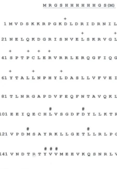
Fig. 1. Amino acid sequence of wild type Lrp protein. The positions of the Lrp DNA binding mutants and leucine responsive mutants indicated by the symbols + and #, respectively (She-pin et al., 2002;
Brinkman et al., 2003). The number of amino acids from original Lrp wild type is marked in left column and the underlined amino acids of MRGSHHHHHGS were originated from pQE30 vector.
The gene coding for the first amino acid Met (M) in Lrp Wt (pCV 294) and Lrp R145W (pCV305) was replaced with Ser (S) as well as Arg 145 (R) with Trp (W) in pCV 305.
of the tryptophan within the protein. The wavelength of tryptophan fluorescence is widely used as a tool to monitor changes in proteins and also to make inferences regarding their local three dimensional structures (Lakowicz, 1983). Hence, the fluorescence of a protein molecule can be used as a diagnostic tool to identify the conformational state of protein under investigation. The objective of this study with the changed form to tryptophan of Leucine-responsive regulatory protein (Lrp R145W) is to elucidate more information on its binding and spectroscopic characteristics through fluorescence spectroscopy, and by using the method of tryptophan fluorescence quenching analysis at the addition of acrylamide and urea, respectively.
Materials and Methods
Strains, enzymes, and chemicals
E. coli XL-1 Blue competent cell and BL21 expressing strains were purchased from RBC Bioscience Co (Korea). The oligonucleotides were synthesized from Genotech (Korea). г -32P-ATP was obtained from Du Pont-NEN (USA) and RNase A and DNase 1 from Sima (USA). T4 polynucleotide kinase, L-leucine, Bradford protein concentration determination reagents as well as others were purchased from Bio-Rad (USA).
Protein purification by 6X His-tag Lrp Wt and Lrp R145W The construction of the wild type (pCV 294) and mutant (pCV 305) genes coding for the 6X His-tagged wild type Leucine-responsive regulatory protein (Lrp Wt) and Lrp 145W, respectively, were described in author’s previous paper (Lee et al., 2010). Frozen cells of mass 5 g, obtained from the culture were thawed 30 min on ice and then resuspended in 25 ml of lysis buffer (50 mM NaH2PO4, 300 mM NaCl, 10 mM imidazole at pH 8.0). Protease inhibitor PMSF was added at a concentration of 1 mg/ml and the cell suspension was sonicated on ice 3 times within a period of 5 min. Due to viscous nature of the solution after sonication, RNase A (10 µg/ml) and DNase 1 (5 µg/ml) were added and incubated on ice for 15 min. The detailed conditions for the expression and purifications of proteins were the same as the previous paper (Lee et al., 2010).
Fluorescence spectroscopy
Fluorescence Spectrometer FS2000 (Hitachi, Japan) was used in measuring (scanning) the emission spectra of the mutant protein containing just one tryptophan amino acid residue at 277 nm of fixed excitation. The fluorescence intensity of the Lrp R145W alone and the Lrp R145W in the presence of the oligomeric DNA (21-mer) were measured. Lrp R145W were bound together with the complementary double
stranded DNA formed by thermal annealing of the two synthetic oligomers 5′-AAGGAGAATATTATGCTATGG-3′
and 5′-CCATAGCATAATATTCTCCTT-3′. Effect of leucine on fluorescence emission spectra of Lrp R145W was investigated by measuring the relative fluorescence intensity according to increasing concentrations of leucine (thus, 0–3 mM) in the presence or absence of 21-mer DNA. Quenching analysis with acrylamide and urea were performed for analyzing the extent to which the tryptophan fluorescence of Lrp R145W.
Gel retardation assay
DNA samples were labeled with г-32P-ATP (3,000 Ci/mmole) and T4 polynucleotide kinase, incubated with Lrp, and fractionated by electrophoresis as previously described (Rica et al., 1989). The specific activity of the DNA fragment was determined as described previously (Cui et al., 1996).
Results and Discussion
The recombinant wild type plasmids containing the gene coding for the Leucine-responsive regulatory protein (pCV 294) and mutant lrp R145W (pCV 305) were generated according to the procedures described in author’s previous
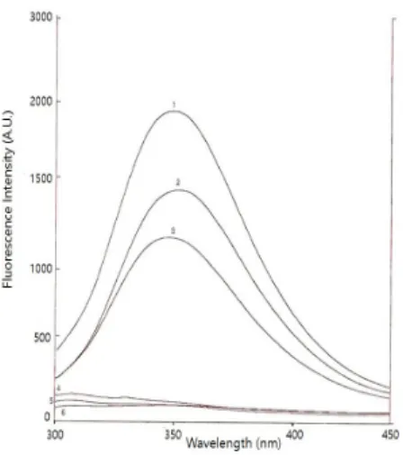
Fig. 2. Fluorescence emission spectra of Lrp R145W. 1, Lrp R145W itself; 2, Lrp R145W with DNA; 3, Lrp R145W with DNA and L-leucine; 4, double stranded 21 mer DNA; 5, Lrp Wt; 6. TE buffer.
Excitation at 277 nm. The complementary double stranded DNA was formed by thermal annealing of two synthetic oligomers in TE buffer. The protein concentrations of Lrp and DNA were 2 μM, respectively, L-leucine was 2 mM.
paper (Lee et al., 2010). The plasmids containing the lrp genes were ligated with pQE30 vector because the cloning vector introduces 6X His-Tag to the N-terminal region of the protein.
The derivatives of Lrp with 6X His tags allow proteins to be purified by affinity chromatography on Ni-NTA resin. The yield of soluble 6X His-Lrp per g wet weight cells after purification was about 10 mg and nearly 100% of the product was capable of binding to DNA, as measured by titrating a quantity of the protein with DNA (data not shown). The nucleotide DNA sequencing was confirmed by automated sequencing and its translated amino acid sequences are shown in Fig. 1. The 11 amino acid extension at the N-terminus does not appear to have major negative effects on the function of Lrp as evidenced by the binding studies (Cui et al., 1996).
In the previous paper (Lee et al., 2010), it was confirmed that increasing concentration of Lrp R145W correspondingly led to an increase in fluorescence intensity. Hence, there is a directly proportional relationship between the concentration of Lrp R145W and its fluorescence intensity. Lrp Wt protein do not posses any tryptophan amino acid, whereas the mutant Lrp R145W containing the only one tryptophan amino acid residue which has strong intrinsic fluorescence. Hence, tryptophan fluorescence is widely used in fluorescence spectroscopic studies on the three dimensional structure of protein. And this will give more light on the region that tryptophan occupies on the Lrp protein, and observe whether it will lead to a change in the conformation of tryptophan-mutated form of the protein.
Therefore, the Lrp R145W and Lrp Wt proteins were purified
to investigate the fluorescence character.
In Fig. 2, the fluorescence emission spectra of Lrp R145W were tested under different conditions to check the effect of binding with the DNA and/or leucine on its fluorescence activity at an excitation wavelength of 277 nm. Fluorescence intensity of Lrp R145W shows much higher intensity than that of wild type protein. Three amino acids in protein including tryptophan, tyrosine, and phenylalanine exhibit fluorescence properties attributed to the presence of the benzene ring in their primary structures. The tryptophan emission spectrum is dominant over the weaker fluorescence of tyrosine and phenylalanine (Lakowicz, 1983). Although phenylalanine and tyrosine are present in the wild type protein, the fluorescence spectra of wild type Lrp was very low at this condition (scan number 5 in Fig. 2).
In the following experimental analysis, the fluorescence spectra of Lrp R145W unbound or bound to the complementary double stranded DNA were plotted. The two complementary DNA (5′-AAGGAGAATATTATGCTATGG-3′ and 5′-CCAT AGCATAATATTCTCCTT-3′) were designed from the specific Lrp binding DNA sequences from ilvIH operon (Cui et al., 1996). The fluorescence spectrum of Lrp R145W was also measured in the presence DNA and leucine that are bound together. As a control, wild type Lrp, DNA, and buffer themselves were separately measured to test fluorescence intensity. For the analysis above, this experiment was repeated several times to obtain results of best fit to plot a graph.
It was also observed that the Lrp Wt protein recorded a very low fluorescence spectrum, even whether or not it was bound to 21-mer DNA and leucine (data not shown). The reason for this characteristic feature is attributed to fact that the wild type Lrp does not contain tryptophan. As shown in Fig. 2, Lrp R145W itself recorded the highest maximum peak of fluorescence intensity compared to the fluorescence spectra produced by the complex of Lrp R145W with DNA. In the same aspect, fluorescence spectra emitted by Lrp R145W with DNA in the presence of leucine was low compared to the above two fluorescence spectra recorded. Additionally, it was observed that the maximum peak of emission by Lrp R415W changed from 349 nm to 353 nm by binding to the DNA (spectra 1 and 2 in Fig. 2). From these results, it was inferred that the binding of DNA to Lrp R145W resulted in a change of conformation of the mutant protein especially at its binding region and the surface groups.
In order to get more characteristics of Lrp R145W, the effect of increasing of leucine on its relative fluorescence intensity of the protein was tested. For the investigation, leucine was introduced to Lrp R145W in the presence or absence of the 21-mer DNA at an excitation 277 nm. The experiment was repeated several times to obtain results that were used to plot
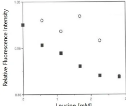
Fig. 3. Effect of leucine on fluorescence intensity of Lrp R145W.
The relative fluorescence intensities were calculated with the value of the each fluorescence intensity of Lrp R145W with adding L-leucine divided by the intensity of Lrp R145W itself emitted at 344 nm, respectively. ○, Relative fluorescence intensity of Lrp R145W without DNA; ■, Relative fluorescence intensity of Lrp R145W-DNA complex. The protein concentrations of Lrp and DNA were 2 μM, respectively.
Fig. 4. Effect of the increasing of leucine concentration on the binding Lrp R145W to the specific DNA from ilvIH operon. To determine stoichiometry, a 20 µl sample containing 20% glycerol, 20 mM Tris-HCl (pH 8.8), 2.0 µg bovine serum albumin, 0.1 mM EDTA, 0.2 mM DTT, 50 mM NaCl, 1.0 mM MgCl2,0.5 pmol DNA, and 1.0 pmol Lrp R145W was incubated for 20 min at room temperature. The region containing the Lrp-DNA complex was excised and counted for
32P as described previously (Cui et al., 1996).
graph of best fit. It was inferred that the complex of Lrp R145W with DNA in the presence of leucine showed a further significant reduction in fluorescence emission spectra with changing of maximal peak of emission from 353 nm to 348 nm (spectra 2 and 3 in Fig. 2). These results might be due to the fact that the binding of leucine also leads to a change in structural conformation of the mutant protein. It was stated that the N-terminal domain, also referred to helix-turn-helix domain, is the DNA binding unit of the Lrp protein consisting of three α-helices with a conserved tight turn between the second and third helices (Ernsting et al., 1992).
In Fig. 3, it was also identified that the relative fluorescence intensities from Lrp R145W itself were generally higher than that of Lrp R145W bound to 21-mer DNA, and shown that the intensities from Lrp R145W both in the presence or absence of DNA decreased steadily as the concentration of leucine increased correspondingly. These results are quite correlated well with the gel retardation assay to check the formation of Lrp and DNA complex. As shown in Fig. 4, the bound Lrp R145W-DNA complex was decreased by the addition of leucine. The Lrp monomer is shown to consist of three regions namely; the N-terminal HTH domain, flexible linker peptide, and the C-terminal domain (Brinkman et al., 2003). It is also known that the transcriptional regulation activities of Lrp protein are sometimes influenced by leucine which binds to it at the C-terminal domain (Roesch and Bloomfield, 1998).
The C-terminal domain of Lrp consists of a fold similar to that of RAM (regulation of amino acid metabolism) domain;
which is involved in the allosteric regulation of amino acid metabolism in prokaryotes (Ettema et al., 2002; Thaw et al., 2003). The RAM domain of Lrp contains the binding site for the effector leucine and is also referred to as the leucine
response domain (Willins et al., 1991). From this experiment and the features of structure in the literature, tryptophan 145 in Lrp may be located in the leucine binding domain at the C-terminal, inferring that leucine has a significant effect on the structure of Lrp R145W whether it is bound to 21-mer DNA or not. And this led to a significant reduction in relative fluorescence intensity.
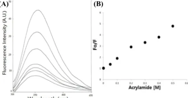
Tryptophan quenching analysis was performed to identify the specific location on the protein and also to identify the general conformation of Lrp R145W. In order to achieve this aim, the quenching agents such as acrylamide and urea were used. Acrylamide is a polar, uncharged compound that has been shown to quench the fluorescent of indole derivatives by collision process. Acrylamide were increasingly added to dialyzed Lrp R145W and the mixtures were excited into ultra-violet light at 277 nm and their fluorescence intensities were measured. The fluorescence intensity decreases correspondingly from 44 to 9 arbitrary units (Fig. 5A). A graph showing the effect of the acrylamide concentration on the fluorescence intensity from Lrp R145W was plotted (Fig. 5B).
The fluorescence intensity of the emission at 344 nm from the mutant protein in absence of acrylamide (Fo) was recorded and used to compare those values that were obtained in the increasing of acrylamide concentration (F).
According to the results obtained, it was inferred that acrylamide was a strong quencher of tryptophan fluorescence of Lrp R145W. It was shown that the extent of quenching of tryptophan fluorescence intensity also increases steadily as concentration of acrylamide increased. The extent of fluorescence
(B) (A)
Fig. 6. (A) Fluorescence spectra of Lrp R145W with increasing concentration of urea. As shown the plotted lines from top to down, urea concentration was increased to 0‒10 M. The protein concentration was 2 μM. Excitation at 277 nm. (B) Effects of urea quenching on the tryptophan fluorescence of Lrp R145W. Fo, the fluorescence intensity without urea; F, the fluorescence intensity at the concentration of urea emitted at 344 nm.
(A) (B)
Fig. 5. (A) Effects of acrylamide on tryptophan fluorescence of Lrp R145W. As shown in plotted lines from top to down, acrylamide concentration was increased from 0 to 0.5 M. The protein concentration was 2 μM. Excitation at 277 nm. (B) Acrylamide quenching of tryptophan fluorescence of Lrp R145W. Fo, the fluorescence intensity of the mutant protein in absence of acrylamide; F, the fluorescence intensity at the concentration of acrylamide emitted at 344 nm.
quenching by acrylamide in this experiment gives preliminary information that tryptophan at position 145 in the amino acid sequence is located on the surface of the Lrp R145W. Acrylamide diminishes the fluorescence of tryptophan through the collision with tryptophan or complex formation, therefore, the extent of conformational change affecting the accessibility of individual tryptophan residues could be tested with acrylamide through the quenching experiment. Further structural studies by X-ray diffraction techniques will lead to identification of the specific region occupied by tryptophan and the overall structure of Lrp R145W.
In order to a draw a conclusive characteristic on quenching analysis of Lrp R145W, urea was also used to analyze the quenching effect on tryptophan fluorescence shown in Fig 6A.
Urea is known to be a natural denaturant of proteins and therefore it is expected that the protein can be denatured signifying a complete change in its native conformation at the end of the experiment. During this analysis, desalted Lrp R145W was mixed with different increasing concentrations of urea and their respective fluorescence intensity was measured.
Similarly, as the acrylamide quenching analysis on Lrp R145W, the fluorescence intensity of the mutant protein in the absence of urea (Fo) was recorded and used to compare those values that were obtained in the presence of urea (F). Results obtained in this analysis were used in plotting a graph of fluorescence intensity of Lrp R145W against increasing urea concentration.
From the graph in Fig. 6A, as the concentration of urea added to the protein increased, the fluorescence intensity decreased slowly. This means that an increase in the rate of quenching of tryptophan fluorescence also increased accordingly. However, the rate at which urea quenched fluorescence intensity in Lrp R145W was slow when the results were compared to that of acrylamide quenching analysis of the
mutant protein (Figs. 5B and 6B). The difference identified was owing to the different mode of activity of quenching by the two quenching agents. In Fig. 6B, it was observed that quenching analysis by adding urea, the initial fluorescence intensity value (Fo) and that of the final value (F) were far apart and diagrammatically showed a different pattern of consistent reduction in fluorescence intensity. This pattern was different from that of the acrylamide fluorescence quenching analysis.
From these results, it was expected that high concentrations might have denatured the Lrp R145W as it did to all proteins at such concentrations.
L-Leucine amino acid has been identified as the most abundant and readily available building-block of proteins, so it serves as a good reporter to study the activity and presence of proteins in enteric bacteria (She-pin et al., 2002; Brinkman et al., 2003). The fluorescence properties of endogenous tryptophan residue of Lrp can serve as local intrinsic probes for investigation of the dynamic nature of the protein by binding of leucine. In conclusions, wild type protein exhibits very low fluorescence intensity due to the absence of tryptophan. Lrp R145W, however, exhibits high fluorescence intensity due to presence of tryptophan at position 145. Fluorescence properties of Lrp R145W respectively were analyzed and the following are conclusive results obtained. 1) The binding of DNA to Lrp R145W, significantly decreased the protein’s fluorescence intensity due to changes in its conformation. In addition, the intensity in the presence or absence of DNA was also decreased by increasing of leucine. 2) Acrylamide quenching analysis had huge effects on the fluorescence property on Lrp R145W.
These results give information on the three dimensional structure such as orientation of the region W 145 on the Lrp occupying a location either on the surface of the protein or close to the DNA binding site.
적 요
루신-반응 조절 단백질(Lrp)은 18.8 kDa의 분자량을 갖는 164개의 아미노산으로 이루어진 글로벌 조절 단백질으로서, 야 생형의 단백질(Lrp Wt)에는 아미노산 중 가장 강한 자체 형광을 띠는 트립토판이 존재하지 않는다. Lrp 단백질의 구조변이에 대 한 정보를 줄 수 있는 형광분석을 위하여 Lrp Wt과 트립토판이 루신-반응 영역에 단지 하나 존재하는 돌연변이 단백질(Lrp R145W)을 분리·정제하였다. Lrp R145W 단백질은 이들 ilvIH 오페론에서 고안된 Lrp 결합 특정 DNA와 아미노산 루신과의 결합 후에 형광이 감소하였으며 acrylamide, urea 등에 의해서도 급격히 쇄광하는 양상을 보였다. 이들 형광 실험 결과는 Lrp의 3 차원적 구조 및 배향을 연구에 중요한 정보를 제공하여 줄 수 있 을 것이다.
Acknowledgements
This work was supported by the research fund from Chungnam National University. We thank Prof. Eunice Lee of Eulji University for her careful reading and correction in preparing the manuscript.
References
Brinkman, A.B., Ettema, G.J.T., de Vos, W.M., and van der Oost, J. 2003.
The Lrp family of transcriptional regulators. Mol. Microbiol. 48, 287 –294.
Calvo, J.M. and Matthews, R.G. 1994. The leucine-responsive regulatory protein, a global regulator of metabolism in Escherichia coli. Microbiol. Rev. 58, 466–490.
Chen, S., Rosner, M.H., and Calvo, J.M. 2001. Leucine-regulated self-association of leucine-responsive regulatory protein (Lrp) from Escherichia coli. J. Mol. Biol. 312, 625–635.
Chen, S., Iannolo, M., and Calvo, J.M. 2005. Cooperative binding of the leucine-responsive regulatory protein (Lrp) to DNA. J. Mol. Biol.
345, 251–264.
Cui, Y., Midkiff, M.A., Wang, Q., and Calvo, J.M. 1996. The leucine responsive regulatory protein (Lrp) from Escherichia coli.
Stoichiometry and minimal requirements for binding to DNA. J.
Biol. Chem. 271, 6611–6617.
Ernsting, B.R., Atkinson, M.R., Matthews, R.G., and Ninfa, A.J. 1992.
Characterization of the regulon controlled by the leucine responsive
regulatory protein in Escherichia coli. J. Bacteriol. 174, 1109–1118.
Ettema, T.J.G., Brinkman, A.B., Tani, T.H., Rafferty, J.B., and van der Oost, J. 2002. A novel ligand-binding domain involved in regulation of amino acid metabolism in prokaryotes. J. Biol. Chem. 277, 37464–
37468.
Lackowicz, J.R. 1983. Principles of fluorescence spectroscopy. pp. 111–
153. Plenum press, New York, USA.
Landgraf, J.R., Wu, J., and Calvo, J.M. 1996. Effects of nutrition and growth rate on Lrp levels in Escherichia coli. J. Bacteriol. 178, 6930–
6936.
Lee, C.Y., Kim, S.C., and Seo, C.H. 2010. Purification and fluorometric analysis of leucine-responsive regulatory protein from Escherichia coli. Kor. J. Microbiol. 46, 104–108.
Newman, E.B., D’Ari, R., and Lin, R.T. 1992. The leucine-Lrp regulon in Escherichia coli: A global response in search of a raison d'etre.
Cell 68, 618–620.
Newman, E.B. and Lin, R. 1995. Leucine-responsive regulatory protein:
a global regulator of gene expression in Escherichia coli. Annu. Rev.
Microbiol. 49, 747–775.
Newman, E.B., Lin, R.T., and D’Ari, R. 1996. The leucine/Lrp regulon.
Annu. Rev. Microbiol. 49, 1513–1525.
Peterson, S.N., Dahlquist, F.W., and Reich, N.O. 2007. The role of high affinity non-specific DNA binding by Lrp in transcriptional regulation and DNA organization. J. Mol. Biol. 369, 1307–1317.
Rica, E., Aker, D.A., and Calvo, J.M. 1989. A protein that binds to the regulatory region of the ilvIH operon of Escherichia coli. J.
Bacteriol. 171, 1658–1664.
Roesch, P.L. and Bloomfield, I.C. 1998. Leucine alters the interaction of the leucine-responsive regulatory protein (Lrp) with the firm switch to stimulate site-specific recombination in Escherichia coli.
Mol. Microbiol. 27, 751–761.
She-pin, H., Pierre, B., and Wesley, H.G. 2002. Global gene expression profiling in Escherichia coli K-12. The effects of Leucine-responsive regulatory protein. J. Biol. Chem. 277, 40309–40323.
Thaw, P., Sedelnikova, S.E., Muranova, T., Wiese, S., Ayora, S., Alonso, J.C., Brinkman, A.B, Akerboom, J., van der Oost, J., and Rafferty, J.B. 2003. Structural insight into gene transcriptional regulation and effector binding by the Lrp/AsnC family. Nucleic Acids Res. 34, 1439 –1449.
Wang, Q. and Calvo, J.M. 1993. Lrp, a global regulatory protein of Escherichia coli, binds cooperatively to multiple sites and activates transcription of ilvIH. J. Mol. Biol. 229, 306–318.
Willins, D.A., Ryan, C.W., Platko, J.V., and Calvo, J.M. 1991.
Characterization of Lrp, an Escherichia coli regulatory protein that mediates a global response to leucine. J. Biol. Chem. 26, 10768–
10774.