사염화탄소로 섬유화가 유도된 흰쥐 간에서 털부처꽃 뿌리 추출물의 항산화 및 섬유화저해 활성
이승은*†·안태진*·김금숙*·김영옥*·한희선**·서진숙*·정해영***
박충범*·차선우*·박호기*·성낙술****
*충북 음성군 농촌진흥청 국립원예특작과학원 인삼특작부, **경기 수원시 국립식량과학원
***부산시 금정구 부산대학교 약학대학, ****충남 금산군 (재)금산국제인삼약초연구센터
Antioxidant and Anti-fibrotic Properties of Root Extract of
Lythrumsalicaria L. in CCL4-Induced Liver Fibrosis Rat Model
Seung Eun Lee
*†, Tae Jin Ahn
*, Geum Sook Kim
*, Young Ok Kim
*, Hee Sun Han
**, Jin Sook Seo
*, Hae Young Chung
***, Chung Berm Park
*, Sun Woo Cha
*, Ho Ki Park
*, and Nak Sul Seong
*****Department of Herbal Crop Research, National Institute of Horticultural and Herbal Science, Rural Development Administration, Eumsung, Chungbuk 369-873, Korea.
**National Institute of Crop Science, RDA, Suwon, Gyeonggi 441-857, Korea.
***Department of Pharmacy, Pusan National University, Pusan 609-735, Korea.
****International Ginseng and Herb Research Institute, Geumsan, Chungnam 312-701, Korea
ABSTRACT : Fifty percent ethanol extract of Lythrum salicaria Linne root (LSR) was tested in vitro on antioxidant activity, and furthermore was investigated on antioxidative and fibrosis protecting activities in CCL
4-induced liver fibrosis rat model.
Ratio of hepatic GSH/GSSG (reduced glutathione/oxidized glutathione) as bio-parameter of antioxidant level in CCL
4plus LSR-treated rats for 6 weeks significantly increased from 2.8- to 5.7-fold than that of CCL
4-treated rats at p < 0.05. Thiobarbi- turic acid reactive substances (TBARS) contents in CCL
4plus LSR-treated rats ranged from 1.57- to 2.19-fold of normal rats and were lower than those in CCL
4plus silymarin-treated rats (1.78~2.46-fold of normal rats) (p < 0.05). Amounts of hydrox- yproline of liver tissue showing the content of total collagen, a parameter of fibrosis, in CCL
4plus LSR-administrated rat livers were 4.9~8.8
㎍/
㎎(
−47 ~
−71%, compared with that in CCL
4-treated rat livers (16.6
㎍/
㎎tissue), which were significantly lower than those in CCL
4plus silymarin-administrated rats being 8.4~11.7
㎍/
㎎(
−30 ~
−50%). This collagen reducing effect of liver tissue in CCL
4plus LSR-treated rats was supported by histological observation using microscopy assay. From the results, we conclude that the root of L. salicaria have efficient antioxidant potential and effective antifibrotic activities.
Key Words :
Antioxidant, Antifibrotic, Collagen, Liver Fibrosis, ROSINTRODUCTION
Liver fibrosis known as a pathological state that progresses to liver cirrhosis and chronic liver disease (Oh et al., 2003) has occurred from the activation of hepatic stellate cells (HSCs) in damaged liver. HSCs play a role to produce collagen in the early liver fibrogenesis (Du et al., 1999) and the activation of HSCs producing a lot of extracellular matrix (ECM) materials such as collagen and fibronectin is triggered by reactive oxygen species (ROS)
(Matsui et al., 2004). Hence inhibiting of the activation of HSCs is a key point to reduce fibrotic progression. Articles have reported that plants or herbs having antioxidant activity had ameliorated several diseases or pathological conditions (Pospelova and Barnaulov, 2000; Rao et al., 2006).
Lythrum salicaria Linne, purple loose-strife or spiked loose-strife, of Lythracea family is a weedy plant that is distributed all over the world including Korea (Lee, 1996).
The activities of L. salicaria have been reported on antioxidant activity of leaves in vitro (Tunalier et al., 2007),
†Corresponding author: (Phone) +82-43-871-5586 (E-mail) [email protected] Received 2009 June 5 / Revised 2009 July 18 / Accepted 2009 July 29
on effect against hyperglycemic mice (Lamela et al., 1986), on antimicrobial effect (Becker et al., 2005), on antilisterial activity (Altanlar et al., 2006) and on anti-inflammatory and anti-nociceptive effects (Tunalier et al., 2007). From the above reports, none of the article has not reported about the antioxidant and anti-fibrotic activity of L. salicaria root in fibrosis. We selected root part of the plant from the result in preliminary experiments which have been reported as a paper (Lee et al., 2009). The study was performed to verify the activities of L. salicaria root extract against antioxidant and liver fibrosis-protection in rat fibrosis model.
MATERIALS AND METHOD
1. Chemicals
Carbon tetrachloride, corn oil, DAB (3,3'-diaminobenzidine tetrahydrochloride), fast green, MTT (3-(dimethylthiazol-2-yl)- 2,5-diphenyl tetrasodium bromide), silymarin, sirius red, and sodium nitrite were purchased from Sigma Chemical Co (USA). And H2DCFDA (2,7-di-chlorodihydrofluorescein diacetate, L-hydroxyproline and trolox were did from Aldrich (USA), and Molecular Probes (Eugene, OR, USA), and Wako (Japan), respectively.
2. Cell lines
SH-SY5Y cell line originated from human neuroblastoma, and YPEN1 cell line, prostate cell line of bulls, have been obtained from ATCC (American Type Culture Collection, Rockville, MD, USA).
3. Plant part and preparation of extract
L. salicaria root was collected at the medicinal plant experiment farm of National Institute of Crop Science (NICS), Rural Development Administration (RDA), August 2004. Powdered root (four kilograms) was extracted with 50% ethanol in refluxing apparatus at 85℃. Extract was evaporated in vacuum evaporator at 50℃ (EYELA N-1000, Japan). Finally, dark-yellow extract (603 g) was obtained and stored at −27℃ until using.
4. Assay of
in vitroantioxidant activity against reactive oxygen species (ROS) and peroxynitrite (ONOO
−) in chemical environments and cells
Total ROS scavenging activity of LSR was investigated in 2',7'-dichlorodihydrofluorescein diacetate (H2DCFDA) reaction
system using LPS-treated rat liver (Reddy et al., 2006).
Peroxynitrite scavenging activity of LSR was conducted by the method of Kooy et al. (1994). Effect of LSR on BHP- induced ROS production in YPEN1 cells was evaluated in DMEM media by the modified method of Kweon et al. (2006) and analyzed on the fluorescence at Ext485 nm/
Emm535 for 15 min. Effect of LSR on SH-SY5Y cells viability was analyzed by the modified method of Schmid et al. (2007) using MTT reagent.
5. Design of animal experiment
SD male rats with body weight from 100 g to 110 g were distributed for eight groups (n = 6) which were composed of normal group, CCL4 single treated group (negative control), CCL4 plus LSR-treated groups (three groups), CCL4 plus silymarin-treated groups (positive controls, three groups). All groups were accommodated at 23~24℃, 55% humidity, and were maintained on 12 hours dark/12 hours light rule, were administrated with commercial stock diets (Samtaco, Korea) and were allowed of tap water for six weeks.
6. Assay of antioxidant activity in liver fibrosis rat model
Liver collected from fibrosis rat model was homogenated in 0.1M potassium phosphate buffer (pH 7.4) with teflon homogenizer (Wheaton overhead stirrer, USA). Lipid peroxidation of liver homogenate was evaluated as TBARS using TEP (1,1,3,3-tetra ethoxypropane) as standard for calculation according to the method of Botsoglou et al. (1994) as below. Hepatic GSH/GSSG ratio was measured by the combined methods of Ellaman (1959) and S'wiergosz-Kowalewska et al. (2006). GST activity in liver cytosol fraction was analyzed according to the principle of Habig et al. (1974) using the molecular extinction coefficient (EnM/340 nm = 9.6 nM−1㎝−1) of GSH-2,4-dinitrochlorobenzene (DNCB) conjugate produced from the reaction of DNCB and reduced glutathione (GSH) at 25℃ for 20 min.
Protein quantification for showing GST activity was conducted by the method of Bradford [1976] using bovine serum albumin as standard.
7. Assay of inhibition activity on collagen production in liver fibrosis rat model
Total collagen content of fibrosis rat liver was quantified
by the method of Woessner (1961). Histological assay on collected rat liver lobes was performed by the method of Carmiel-Haggai et al. (2004) using staining with 0.1% sirius red and counterstaining with 0.1% fast green. Stained sections were dehydrated, were mounted with mounting medium and were below the microscopic observation using light microscope (Leica DE/DM 5000B, Germany).
8. Statistical analysis
The results were shown as mean±standard deviation (SD). The significance of data was analyzed with Duncan's multiple range test of one way ANOVA test using SAS program (Enterprise Guide 4) at p< 0.05.
RESULTS
1. Antioxidant activity of LSR on ROS and peroxynitrite in chemical environments
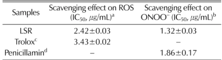
From the result of the experiment on in vitro antioxidant activity of LSR in chemical reaction system conducted on total ROS using with DCFDA, a fluorescent precursor compound, LSR showed more effective scavenging activity (IC50, 2.42㎍/mL) than trolox, a positive control compound, did (IC50, 3.43㎍/mL)(Table 1). LSR also showed effective scavenging activity on peroxynitrite (ONOO−, 1.32㎍/mL) in dihydrorhodamine (DHR) reaction system comparing to penicillamin (1.86㎍/mL), a control compound. These two data indicate that the extract of LSR exhibited scavenging activity on free radicals in chemical reaction systems.
2. Effect of LSR on antioxidant activity against cells
Inhibition activity of LSR on t-BHT-induced ROS production in YPEN1 cells was exhibited in Fig. 1. LSR (−40, −200, and −1,000㎍/mL) and t-BHT combination treatments showed fluorescence at Ext485 nm/Emm535 as 54.5, 54.5 and 62.8, respectively, which were the same degree of t-BHT-not treated control (52.4). However, t-BHT single treatment increased the value to 111.4, which was corresponding to 2.13-fold of control. Therefore, it is suggested that LSR exhibited very strong antioxidant activity on t-BHT induced oxidative stress. The cell viabilities of SH-SY5Y cells treated with LSR (1, 10, 50, 100, and 1,000㎍/mL) were tested by MTT assay, and were 91.6, 96.7, 90.6, 88.8, and 104.4%, compared with the value in control which was not treated with LSR, respectively (Fig.2). This explains that LSR has no cytotoxicity on SH- SY5Y cells and furthermore there are a possibility of increasing the cell viability under the concentration of 1,000㎍/mL.
Table 1. Scavenging effect of LSR against ROS and peroxynitrite (ONOO−) in chemical environments.
Samples Scavenging effect on ROS (IC50, ㎍/mL)a Scavenging effect on ONOO− (IC50, ㎍/mL)b
LSR 2.42±0.03 1.32±0.03
Troloxc 3.43±0.02 −
Penicillamind − 1.86±0.17
aScavenging effect on ROS was tested on DCFDA reaction system
bScavenging effect on ONOO− was tested in DHR reaction system
c,dPositive control compounds
Fig. 1.
Inhibition effect of LSR on BHP-induced ROS production in YPEN1 cells. Cont, control; t-BHP-10, tert-butyl hydroperoxide (10
µM); LSR-40, LSR-200 and LSR-1000 means that final concentrations of LSR in the reaction system were 40, 200 and 1000
㎍/mL.
Fig. 2.
Effect of added LSR on the viability of SH-SY5Y cells. LSR-
1, -10, -50, -100 and -1000 means that final
concentrations of LSR in the reaction system were 1, 10,
50 , 100 and 1000
㎍/mL, respectively. Values in each bar
are not significantly different at P < 0.05.
3. Antioxidant activity of LSR in liver fibrosis rat model
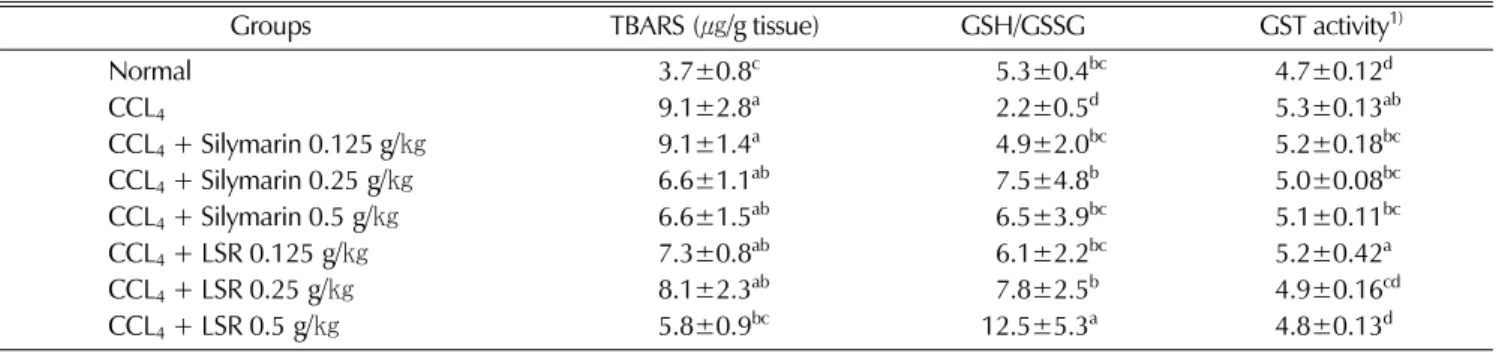
Antioxidant activity of LSR was verified in carbon tetrachloride-induced liver fibrosis rat model. The activity was compared with normal rats, negative control group (CCL4 single treated, no LSR administration or no silymarin administration) and CCL4 plus silymarin administrated groups in the aspect of lipid peroxidation production, glutathione maintenance, and glutathione-S-transferase (an antioxidant and hepatic phase II enzyme) activity.In fibrosis induced rat model, hepatic TBARS content as a marker of lipid peroxidation in CCL4 treated rats was 9.1㎍/g tissue, which was an increased value of 2.46-fold compared with that (3.7㎍/g tissue) of normal rats. LSR treatment (0.125, 0.25 and 0.5 g/㎏) showed liver lipid peroxidation of 7.3, 8.1 and 5.8㎍/g tissue respectively, which were 1.97-, 2.19- and 1.56-fold of normal rats.
Silymarin treatment (0.125, 0.25 and 0.5 g/㎏) showed 9.1, 6.6 and 6.6㎍/g tissue respectively in hepatic TBARS content, which were 2.46-, 1.78- and 1.78-fold compared with that of normal rats. Data exhibited that LSR treatment could more effectively reduce the hepatic lipid peroxidation than silymarin treatment. Effect of LSR on the ratio of hepatic GSH and GSSG tested in fibrosis rat was exhibited in Table 2. Carbon tetrachloride treated but not administrated with LSR or silymarin rats (NC) showed 2.2 in the ratio of GSH/GSSG, which was a reduced value into the degree of 58.5% compared with the value of normal rats (5.3).
The ratios of GSH/GSSG in fibrosis rats treated with 0.125, 0.25 and 0.5 g/㎏ of LSR were 6.1, 7.8, and 12.5, respectively. This data showed that LSR increased 1.15-, 1.47- and 2.36-fold in the ratio of GSH/GSSG compared with the value of normal rat. The values in the rats treated
with 0.125, 0.25 and 0.5 g/㎏ of silymarin, were 4.9, 7.5, and 6.5, and which were ranged from 0.92- to 1.42-fold compared with the value of normal rat. Our data showed that LSR treatment on fibrosis induced rats were more effective on keeping the level of glutathione than silymarin, a commercial liver protecting drug. Activity of hepatic glutathione S-transferase in the fibrosis rat model administrated with LSR was analyzed. Hepatic GST activity of the rat in negative control group was 5.3µM conjugated DNCB/㎎ protein/min, which was an increased value as 12.7% compared with the activities in normal group (4.7µM conjugated DNCB/㎎ protein/min). Treatments with 0.125, 0.25 and 0.5 g/㎏ of LSR showed 5.2, 4.9 and 4.8µM conjugated DNCB/mg protein/min respectively in hepatic GST activity, and these activities were increased values as 10.6, 4.3 and 2.1% compared with normal. The activities of hepatic GST in the silymarin (0.125, 0.25 and 0.5 g/㎏) treated fibrosis rats showed 5.2, 5.0 and 5.1µM conjugated DNCB/mg protein/min, which were increased values as 10.6, 6.4 and 8.5% compared with the normal group. Data in Table 2 showed that the degrees increased of the hepatic GST activity of LSR-administrated fibrosis rats were comparatively lower than those of negative control rats and silymarin-administrated rats.
4. Inhibition activity of LSR on collagen production of liver fibrosis rat model
Collagen production of fibrosis liver of rat was measured in two aspects of total collagen content as L-hydroxyproline and histological microscopic assay.
L-Hydroxyproline content in normal or fibrosis-induced rats which were administrated with LSR or silymarin or
Table 2. Effect of LSR on hepatic lipid peroxidation (as TBARS), GSH/GSSH and glutathione-S-transferase activity in fibrosis-induced liver of rat.Normal, vehicle-injected group; CCL4 (1 mL of 40% CCL4/㎏ body weight with 1.5 mL of corn oil plus 1 mL of distilled water, 2 times/week for 6 weeks)-injected group. Values with alphabet on each bar are significantly different at P < 0.05.
Groups TBARS(㎍/g tissue) GSH/GSSG GST activity1)
Normal 3.7±0.8c 95.3±0.4bc 4.7±0.12d
CCL4 9.1±2.8a 92.2±0.5d 5.3±0.13ab
CCL4+ Silymarin 0.125 g/㎏ 9.1±1.4a 94.9±2.0bc 5.2±0.18bc
CCL4+ Silymarin 0.25 g/㎏ 6.6±1.1ab 97.5±4.8b 5.0±0.08bc
CCL4+ Silymarin 0.5 g/㎏ 6.6±1.5ab 96.5±3.9bc 5.1±0.11bc
CCL4+ LSR 0.125 g/㎏ 7.3±0.8ab 96.1±2.2bc 5.2±0.42a
CCL4+ LSR 0.25 g/㎏ 8.1±2.3ab 97.8±2.5b 4.9±0.16cd
CCL4+ LSR 0.5 g/㎏ 5.8±0.9bc 12.5±5.3a 4.8±0.13d
1)Unit : µM conjugated DNCB/mg protein/min
none were shown in Fig. 3. The content of liver collagen in CCL4 single treated rats (NC) was 16.6㎍/g tissue, which was an elevated value of 3.6-fold compared with the content in normal rats (4.6㎍/g tissue). Feeding of 0.125, 0.25 and 0.5 g/㎏ LSR on liver fibrosis rats made collagen content of 7.1, 4.9 and 8.8㎍/g tissue, which were respectively 1.5-, 1.0- and 1.9-fold compared with the normal non-fibrotic rats. The content of liver collagen in 0.125, 0.25 and 0.5 g/㎏ silymarin fed fibrosis rats were 8.4, 11.7 and 11.3㎍/g tissue, which corresponded to 1.8-, 2.5- and 2.4-fold of normal rats, respectively. Data showed that both of LSR and silymarin have decreasing effect on the liver collagen content in fibrosis rat model when compared with the values in CCL4 single treated rats (NC), and furthermore treatment of LSR was more effective to decrease collagen synthesis than treatment of silymarin.
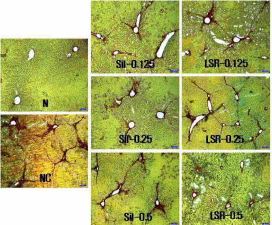
Histological assays for visualizing and for comparing collagen production in fibrosis rats which were treated or
Fig. 4.
Histological observation for explaining the effect of LSR on collagen production in fibrosis-induced liver of rat (×100).
N, vehicle-injected group; NC, negative control, CCL
4(1 mL of 40% CCL
4/
㎏body weight with 1.5 mL of corn oil plus 1 mL of distilled water, 2 times/week for 6 weeks) –injected group; Sil-0.125, -0.25 and -0.5, 0.125 g, 0.25 g and 0.5 g of silymarin/
㎏body weight/rat/day-supplemented & CCL
4-injected group; LSR-0.125, -0.25 and -0.5, 0.125 g, 0.25 g and 0.5 g of LSR/
㎏body weight of rat/day-supplemented & CCL
4-injected group.
Fig. 3.
Effect of LSR on total collagen content of fibrosis-induced
liver of rat. Total collagen content was indicated as L-
hydroxyproline. Normal, vehicle-injected group; NC,
negative control, CCL
4(1 mL of 40% CCL
4/
㎏body
weight with 1.5 mL of corn oil plus 1 mL of distilled
water, 2 times/week for 6 weeks) –injected group; Sil-
0.125, -0.25 and -0.5, 0.125 g, 0.25 g and 0.5 g of
silymarin/
㎏body weight/rat/day-supplemented & CCL
4-
injected group; LSR-0.125, -0.25 and -0.5, 0.125 g,
0.25 g and 0.5 g of LSR/
㎏body weight of rat/day-
supplemented & CCL
4-injected group. Values with alphabet
on each bar are significantly different at P < 0.05.
not treated with LSR or silymarin, were shown in Fig. 4.
Red-dark brown stain, made by sirius red, indicating amounts of collagen protein in fibrosis rat liver was increased in the adjacent area of central veins in CCL4
treated group (NC), which was the strongest among the groups. Stains in LSR and silymarin groups were decreased dose-dependently. Brown color of LSR groups was more little than those of silymarin groups, but was stronger than that in normal rat liver. From this simple observation via microscopy, LSR showed more effective in decreasing the collagen production than silymarin did, but quantification on the stain in all of the groups needed further study.
DISCUSSION
We have selected LSR in preliminary in vitro and in vivo screening experiments (data not shown). Fifty percent ethanol extract of LSR was retested on its capacity as antioxidant in several in vitro system. LSR indicated very effective scavenging activity on free radicals compared with trolox or penicillamin in H2DCFDA or in DHR or in YPEN1 cells. This study reveals that the cell viabilities of SH-SY5Y cells treated with LSR ranged from 89 to 104%
compared with the value of control not treated with LSR.
The cell viability test in SH-SY5Y treated with LSR explains that antioxidant activity of LSR mentioned above has not been arisen from cell toxicity or cell death. Next experiments were conducted to verify the antioxidant and hepatoprotective activity of LSR in liver fibrosis induced rat model.
Carbon tetrachloride (CCL4), a species of compounds evoking chemical hepatic injury, could be transformed to radical, trichloromethyl radical (CCL3 and/ or CCL3OO−) by the metabolism of hepatic microsomal P450, which is scavenged with antioxidants like glutathione (Jeong et al., 2002). Liver injury by carbon tetrachloride or trichloromethyl radical evokes changes in expression of antioxidant enzyme (Fountoulakis et al., 2002), decreases bio-antioxidants level such as glutathione (Lee et al., 2007), and increases lipid peroxidation materials (Campo et al., 2004).
In this study, the content of hepatic lipid peroxidation was elevated more on carbon tetrachloride group (TBARS in Fig. 3) than normal group. Compared with CCL4 single treated rats (NC, 9.1㎍/g) or silymarin-administrated fibrosis rats (6.6~9.1㎍/g), the degrees of lipid peroxidation in LSR
administrated fibrosis rats (5.8~8.1㎍/g) were significantly low. Status of glutathione (GSH/GSSG) in fibrosis rat model indicates that CCL4-injection decreases the ratio of reduced form glutathione/oxidized form glutathione (into 2.2), but LSR supplement increases significantly the ratio (6.1~12.5) into the degree of normal, and the effect is higher than silymarin supplement (4.9~7.5). Although opposite result has been reported by author (Amalia et al., 2007), activity of hepatic glutathione S-transferase, an antioxidant and hepatic phase II enzyme, was elevated in CCl4 or galatosamine (another chemical inducing liver injury)-treated animals compared with CCL4 non-treated normal animals, but was decreased by the additive administration of free radical scavenging compound or herb (Wang et al., 2008). This feature has been indicated in our study, that is, hepatic glutathione-S transferase activity in CCL4 treated rats (NC) was elevated than that of normal rats, but the enzyme activity in LSR (0.25 and 0.5 g/㎏) treated rats was significantly reduced. The result shows that LSR has effective antioxidant activity in carbon tetrachloride-intoxicated rat in order to the dose.
Administration of carbon tetrachloride not only induces oxidative stress but also affects the accumulation of collagen and fibrosis (Leung et al., 2008). Furthermore, hepatocyte lipid peroxidation plays a major role in the regulation of collagen alpha 1 gene expression and may be a link between hepatocyte injury and hepatic fibrosis (Bedossa et al., 1994). Between collagen deposition in liver and hepatic production of MDA (malondialdehyde) and HNE (4- hydroxynonenal) in a cholestatic liver injury, there is a positive linear correlation (Parola et al., 1996).
Collagen synthesis was significantly increased after CCL4- treatment (Luckey and Petersen, 2001). The amounts of 4- hydroxyproline to total protein, as index of hepatic collagen in CCL4 administrated rats, exceeded significantly those of normal rats, the progression of which is proportionate to the duration of CCL4 administration (Paakko et al., 1996). In our study, total hepatic collagen accumulation (16.6㎍/㎎
tissue) in CCL4-injected negative control rats increased 3.6- fold of normal rats (4.6㎍/㎎ tissue), but LSR treatment on fibrosis rat model decreased the collagen content (as 4.9~8.8㎍/㎎ tissue). Degrees of collagen reduction in LSR treated rats (1.0~1.9-fold) were higher than those (8.4 ~ 11.7㎍/㎎ tissue, 1.8~2.5-fold) in silymarin treated rats. This result was supported by histological test using sirius red
staining-fast green counterstaining. Red-brown stain around vein, which indicated collagen protein, in carbon tetrachloride treated rats (NC) was stronger than the stain in normal rats, but those of LSR or silymarin administrated fibrosis rats were weaker in order to doses. LSR showed more effective in decreasing the stain than silymarin.
Although microscopic observations were visualized, further study is needed to quantify collagen content on microscopic image.
In conclusion, it is suggested that antioxidant activity of LSR verified from in vitro and/or in vivo test on free radicals or lipid peroxidation have ameliorated collagen production in fibrosis rats and therefore, have affected antifibrotic and/or hepatoprotectic activity on fibrotic liver of rats. Further studies are required to illustrate the antifibrotic mechanism of LSR or others.
ACKNOWLEDGMENTS
This study was carried out with the support of National Joint Agricultural Research Project (No.20060101030022) of RDA, Republic of Korea. We thank Bio-Green21 project (No. 20050301034393 & 20070301034045) of RDA for providing the plant material used for this study.
LITERATURE CITED
Altanlar N, Citoglu GC and Yilmaz BS. (2006). Antilisterial activity of some plants used in folk medicine. Pharmaceutical Biology. 44:91-94.
Amalia PM, Possa MN, Augusto MC and Francisca LS. (2007).
Quercetin prevents oxidative stress in cirrhotic rats. Digestive Diseases and Science. 52:2616-2621.
Becker H, Scher JM, Speakman JB and Zapp J. (2005).
Bioactivity guided isolation of antimicrobial compounds from Lythrum salicaria. Fitoterapia. 76:580-584.
Bedossa P, Houglum K, Trautwein C, Holstege A and Chojkier M. (1994). Stimulation of collagen alpha 1 (1) gene-expression is associated with lipid peroxidation in hepatocellular injury : a link to tissue fibrosis. Hepatology. 19:1262-1271.
Botsoglou NA, Fletouris DJ, Papageorgiou GE, Vassilopoulos VN, Mantis AJ and Trakatellis AG. (1994). Rapid, sensitive and specific thiobarbituric acid method for measuring lipid peroxidation in animal tissue, food, and feedstuff samples.
Journal of Agricultural and Food Chemistry. 42:1931-1937.
Bradford MM. (1976). A rapid and sensitive method for the quantification of microgram quantities of protein utilizing the principle of protein-dye binding. Analytical Biochemistry.
72:248-254.
Campo GM, Avenoso A, Campo S, Ascola AD, Ferlazzo AM
and Calatroni A. (2004). The antioxidant and antifibrogenic effects of the glycosaminoglycans hyauronic acid and chodroitin-4-sulphate in a subchronic rat model of carbon tetrachloride-induced liver fibrogenesis. Chemico-Biological Interactions. 148:125-138.
Carmiel-Haggai M, Cederbaum AI and Nieto N. (2005). A high-fat diet leads to the progression of non-alcoholic fatty liver disease in obese rats. The Journal of the Federation of Ameri- can Societies for Experimental Biology 19:136-138
Du WD, Zhang WD, Zhai WR and Zhou XM. (1999). Dynamic changes of type I, II, and IV collagen synthesis and distribution of collagen-producing cells in carbon tetrachloride-induced rat liver fibrosis. World Journal of Gastroenterology. 5:397-403.
Ellman GL. (1959). Tissue sulfhydryl group. Archives of Biochemistry and Biophysics. 82:70-72.
Fountoulakis M, Vera MC, Crameri F, Boess F, Gasser R, Albertini S and Suter L. (2002). Modulation of gene and protein expression by carbon tetrachloride in the rat liver.
Toxicology and Applied Pharmacology. 183:71-80.
Habig WH, Pabst MJ and Jakoby WB. (1974). Glutahione-S- transferase : The first enzymatic step in mercapturic acid formation. Journal of Biological Chemistry. 249:7130-7139.
Jeong HG, You HJ, Park SJ, Moon AR, Chung YC, Kang SK and Chun HK. (2002). Hepatoprotective effects of 18β- glycyrrhetinic acid on carbon tetrachloride-induced liver injury:
inhibition of cytochrome P4502E1 expression. Pharmacological Research. 46:221-227.
Kooy NW, Royall JA, Ischiropoulos H and Beckman JS.
(1994). Peroxynitrite-mediated oxidation of dihydrorhodamine 123. Free Radical Biology and Medicine. 16:149-156.
Kweon MH, Park YI, Sung HC and Mukhtar H. (2006). The novel antioxidant 3-caffeoyl-1-methylquinic acid induces Nrf2- dependent phase II detoxifying genes and alters intracellular glutathione redox. Free Radical Biology and Medicine. 40:
1349-1361.
Lamela M, Cadavid I and Calleja JM. (1986). Effects of Lythrum salicaria extracts on hyperglycemic rats and mice.
Journal of Ethnopharmacology. 15:153-160.
Lee KJ, Choi JH and Jeong HG. (2007). Hepatoprotective and antioxidant effects of the coffee diterpenes kahweol and cafestol on carbon tetrachloride-induced liver damage in mice. Food and Chemical Toxicology. 45:2118-2125.
Lee SE, Park CG, Ahn YS, Son YD, Cha SW and Seong NS.
(2009). Antioxidative and hepatoprotective effects of Lythrum salicaria. Korean Journal of Medicinal Crop Science 17:1-7.
Lee WC. (1996). Lineamenta florae Korea. PP748, Academy Publishing Company, Seoul.
Leung TM, Tipoe GL, Liong EC, Lau TYH, Fung ML and Nanji AA. (2008). Endothelial nitric oxide synthase is a critical factor in experimental liver fibrosis. International Journal of Experimental Pathology. 89:241-250.
Luckey SW and Petersen DR. (2001). Activation of Kupffer cells during the course of carbon tetrachloride-induced liver injury and fibrosis in rats. Experimental and Molecular Pathology. 71:
226-240.
Matsui H, Ikeda K, Nakajima Y, Horikawa S, Imanishi Y and Kawada N. (2004). Sulfur-containing amino acids attenuate the
development of liver fibrosis in rats through down-regulation of stellate cell activation. Journal of Hepatology. 40:917-925.
Oh WY, Pyo S, Lee KR, Lee BK, Shin DH, Cho SI and Lee SM. (2003). Effect of Holotrichia diomphalia larvae on liver fibrosis and hepatotoxicity in rats. Journal of Ethnopharmacology.
87:175-180.
Paakko P, Anttila S, Sormunen R, AlaKokko L, Peura R, Ferrans VJ and Ryhanen L. (1996). Biochemical and morphological characterization of carbon tetrachloride induced lung fibrosis in rats. Archives of Toxicology. 70:540-552.
Parola M, Leonarduzzi G, Robino G, Albano E, Poli G and Dianzani MU. (1996). On the role of lipid peroxidation in the pathogenesis of liver damage induced by long-standing cholestasis. Free Radical Biology and Medicine. 20:351-359.
Pospelova MI and Barnaulov OD. (2000). The antioxidant and antihypoxant and antioxidant effects of medicinal plants as the basis for their use in destructive disease of the brain. Human Physiology. 26:100-106.
Rao BSS, Shanbhoge R, Upadhya D, Jagetia GC, Adiga SK, Kumar P, Guruprasad K and Gayathri P. (2006).
Antioxidant, anticlastogenic and radioprotective effect of Coleus aromaticus on Chinese hamster fibroblaset cells(V79) exposed to gamma radiation. Mutagenesis. 21:237-242.
Reddy MM, Mahipal SVK, Subhashini J, Reddy AC, Roy KR, Reddy GV, Reddy PRK and Reddanna P. (2006). Bacterial
lipopolysaccharide-induced oxidative stress in the impairment of steroidogenesis and spermatogenesis in rats. Reproductive Toxicology. 22:493-500.
Schmid M, Zimmermann S, Krung HFA and Sures B. (2007).
Influence of platinum, palladium and rhodium as compared with cadmium, nickel and chromium on cell viability and oxidative stress in human bronchial epithelial cells. Environment International. 33:385-390.
S'wiergosz-Kowalewska R, Bednarska A and Kafel A. (2006).
Glutathione levels and enzyme activity in the tissues of bank vole Clethrionomys glareolus chronically exposed to a mixture of metal contaminants. Chemosphere. 65:963-974.
Tunalier Z, Kosar M, Kupeli E, Calis I and Baser KHC.
(2007). Antioxidant, anti-inflammatory, anti-nociceptive activities and composition of Lythrum salicaria L. extracts. Journal of Ethnopharmacology. 110:539-547.
Wang T, Sun NL, Zhang WD, Li HL, Lu GC, Yuan BJ, Jiang H, She JH and Zhang C. (2008). Protective effects of dehydrocarvidine on carbon tetrachloride-induced acute hepatotoxicity in rats. Journal of Ethnopharmacology. 117:300- Woessner JF.308. (1961). The determination of hydroxyproline in tissue and protein samples containing small proportions of this imino acid. Archives of Biochemistry and Biophysics. 93:440- 447.