• 검색 결과가 없습니다.

First Report of Fusarium Wilt Caused by Fusarium proliferatum on Safflower

N/A
N/A
Protected

Academic year: 2021

Share "First Report of Fusarium Wilt Caused by Fusarium proliferatum on Safflower"

Copied!
5
0
0

로드 중.... (전체 텍스트 보기)

전체 글

(1)

Safflower (Carthamus tinctorius) is cultivated herbaceous annual plant of the family Asteraceae. Safflower has been widely cultivated in Korea, India, China, Egypt, Southern Europe, North America, and Australia (Lee, 1980). Safflower seeds contain α-linoleic acid, which used as cooking oil and clinically for the treatment of cataclasis, osteoporosis and rheumatoid arthritis in Korea (Kang et al., 1999; Kim, 1992; Lee et al., 2002). In addition, its flower has been utilized for folk medicine as an analgesic, antithrombotic and antihyperten- sive crude drug, as well as natural dye and food colorants (Han, 1988).

It has been reported that main pathogens on safflower are Alternaria alternata (leaf spot), Botrytis cinerea (gray mold), Colletotrichum acutatum (Anthracnose), Fusarium oxysporum (Fusarium wilt), Phytophthora cactorum (Phytophthora root

rot), Puccinia carthami (rust), Sclerotium rolfsii (collar rot), and Sphaerotheca fuliginea (powdery mildew) worldwide (Farr and Rossman, 2016). Recently, we observed wilt symptoms on saf- flower grown in green house in Jeonju, Korea, thus leading to identify the causal agent of the wilt disease in this study.

Isolation of the pathogen. Several wilted safflower were observed in May, 2015 in Jeonju, Korea. The symptoms ap- peared as brown discoloration of leaves on the lower part of stem (Fig. 1A). The infected plants were wilted with sudden dropping of leaves (Fig. 1B). Severely infected plants were covered by whitish fungal mass (arrow) (Fig. 1C). Small pieces of stem from the diseased plant were sterilized with 75% eth- anol and 1% sodium hypochlorite for 30 seconds followed by 2 times washing with sterile distilled water. They were placed onto water agar and incubated at 25

o

C. After 2 days, hyphal tips of the emerging fungus were transferred onto potato dextrose agar (PDA; Difco, Sparks, MD, USA) medium to pure

©The Korean Society of Plant Pathology

cc

This is an open access article distributed under the terms of the Creative Commons Attribution Non-Commercial License (http://creativecommons.org/

licenses/by-nc/4.0/), which permits unrestricted non-commercial use, distribution, and reproduction in any medium, provided the original work is properly cited.

Open Access

Res. Plant Dis. 22(2): 111-115 (2016)

http://dx.doi.org/10.5423/RPD.2016.22.2.111

First Report of Fusarium Wilt Caused by Fusarium proliferatum on Safflower

*Corresponding author Tel: +82-63-238-4931 Fax: +82-63-238-4859 E-mail: [email protected]

Sang Gyu Kim, Ho-Cheol Ko, On-Sook Hur, Binod Prasad Luitel, Ju-Hee Rhee, Mun-Sup Yoon, Hyung-Jin Baek, Kyoung-Yul Ryu, and Jung Sook Sung*

National Agrobiodiversity Center, National Institute of Agricultural Sciences, Rural Development Administration, Jeonju 54874, Korea

Received March 29, 2016 Revised June 1, 2016 Accepted June 7, 2016

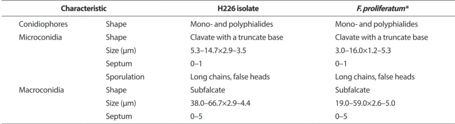
Wilt disease appeared the first in greenhouse-grown safflower (Carthamus tinctorius) in Jeonju, Korea. With the advancement of the disease, the infected plants were withered and died. In order to investigate the causal organism of this symptom disease, fungus was isolated from the infected plants and cultured on potato dextrose agar medium. The fungus showed the white or orange colony color with aerial mycelium. Macroconidia were from falcate to straight, usually 3–5 septate with 38.0–66.7×

2.9–4.4 µm. The fungus was inoculated to a new safflower plant and caused the same wilt. With morphological characters and pathogenicity results, sequence analyses (internal transcribed spacer ribosomal DNA and translation elongation factor 1α) suggested that, the isolated fungus is Fusarium proliferatum. This is the first report of Fusarium wilt disease caused by F. proliferatum on safflower in Korea.

Keywords: Carthamus tinctorius, Fusarium proliferatum, Fusarium wilt, Pathogenicity, Safflower

Research in Plant Disease pISSN 1598-2262, eISSN 2233-9191 www.online-rpd.org

식물병연구

Note

111

(2)

culture, and incubated at 25

o

C. The Isolated pathogen was kept in 20% (v/v) glycerol at –70

o

C for further study.

Morphological characteristic. Fungal isolates were grown on PDA at 25

o

C for 7 days. Morphological character- istics of the isolate were observed under a light microscopy (DM5500B; Leica, Wetzlar, Germany). The isolated fungi H226 produced whitish colony with aerial mycelia on PDA, becom- ing tinged in orange color (Fig. 2A). The mycelial growth of isolated fungi H226 on PDA for 7 days were founded to be optimum at 23

o

C–25

o

C. Microconidia were produced in chains or in false heads (Fig. 2B). Macroconidia were subfal- cate to needlelike, usually 3–5 septate, hyaline, smooth, 38.0–

66.7×2.9–4.4 µm (Fig. 2C). Microconidia were clavate with a truncate base, 0–1 septate, hyaline, smooth, 5.3–14.7×2.9–3.5 µm (Fig. 2D). Conidiophores were cylindrical, often prolif- erated, branched or unbranched, with mono- and poly- phialides. No clamydospores were observed. On the basis of morphological features, the isolated fungus was assumed as F.

proliferatum (Table 1) (Ichikawa and Takayuki, 2000; Nelson et al., 1983).

Pathogenicity assay. Pathogenicity assay was conducted on 3 weeks old safflower plants in green house. The conidial suspension of H226 was prepared from 5-day-old culture

grown on potato dextrose broth and adjusted to 4×10

6

co- nidia/ml. The conidia suspension (10 ml) was inoculated via drenching onto healthy safflowers grown in plastic pots, and then the inoculated plants were placed under green house.

Inoculated plants exhibited same symptom such as discolor- ation and wilting of leaves 1 week after inoculation, but no symptom were shown on the control plant. The fungus was successfully re-isolated from the artificially inoculated plants, fulfilling Koch’s postulates.

Sequence analysis. For further identification, sequences obtained from ribosomal DNA-internal transcribed spacer (rDNA-ITS) region and translation elongation factor 1α (TEF) gene region of the isolated fungus were analyzed. Genomic DNA was extracted from mycelia using cell lysis solution (Qia- gen, Maryland, MD, USA), RNase A solution (Biosesang, Seong- nam, Korea), protein precipitation solution (Qiagen), and Isopropanol (Fisher Scientific Korea Ltd., Seoul, Korea). The region of rDNA-ITS and TEF gene was amplified from genomic DNA using primer pairs ITS1/ITS4 and EF-1/EF-2, respectively (Geiser et al., 2004; Hsuan et al., 2011; White et al., 1990). Each amplified fragment was sequenced and compared with all fungal sequences available in the GenBank database (National Centre for Biotechnology Information; http://blast.ncbi.nlm.

nih.gov/Blast.cgi) and Fusarium ID (http://isolate. fusariumdb.

A

B

C

Fig. 1. Wilt symptoms on safflower grown

in green house. (A) Brown discoloration

and yellowing of leaves. (B) Wilting and

dropping of leaves. (C) Whitish fungal

mass (arrow) in the base of the stem.

(3)

org/blast.php) using BLAST program. With our sequences, reference sequences of Fusarium spp. and related fungi were used for the phylogenetic analysis. A neighbor-joining tree was constructed by the Tajima-Nei parameter model of MEGA 6 (Saitou and Nei, 1987; Tamura et al., 2013).

PCR products (523 bp) of isolate H226 by primer ITS1 and ITS4 were sequenced and deposited GenBank (accession No.

KU218561). This sequence data was compared with other

fungal sequences available in the GenBank database us- ing BLAST search tool. The BLAST search showed that the sequence derived from H226 isolate was 99% similar to the ITS sequence of F. proliferatum (accession Nos. KP407870, GU980584, KJ634671). In addition, PCR products (678 bp) amplified by primer EF-1 and EF-2 were sequenced and de- posited in GenBank (accession No. KU218560). This sequence data showed 99% of similarity to the TEF sequence of F.

Research in Plant Disease Vol. 22 No. 2 113

D

A B

C

Fig. 2. Morphology of Fusarium prolifera­

tum isolated from safflower. (A) Fungal colony on a potato dextrose agar (PDA) after 7 days of incubation at 25

o

C. (B) Chain of microconidia. (C) Macroconidia.

(D) Microconidia of isolated pathogen.

Table 1. Comparison of morphological characteristics between H226 isolate from safflower wilt and Fusarium proliferatum described pre- viously

Characteristic H226 isolate F. proliferatum*

Conidiophores Shape Mono- and polyphialides Mono- and polyphialides

Microconidia Shape Clavate with a truncate base Clavate with a truncate base

Size (µm) 5.3–14.7×2.9–3.5 3.0–16.0×1.2–5.3

Septum 0–1 0–1

Sporulation Long chains, false heads Long chains, false heads

Macroconidia Shape Subfalcate Subfalcate

Size (µm) 38.0–66.7×2.9–4.4 19.0–59.0×2.6–5.0

Septum 0–5 0–5

*Described by Ichikawa and Takayuki (2000) (J. Gen. Plant Pathol. 66: 213–218).

(4)

proliferatum (accession Nos. FJ603514, KP732060, JX174033).

BLAST search using FUSARIUM-ID also showed 99% of similar- ity to TEF sequence of F. proliferatum (accession Nos. FD_01380, FD_01379, FD_01776). Phylogenetic analysis of the ITS and TEF region sequences revealed that H226 isolate belongs to group of F. proliferatum (Fig. 3, 4). Taken together, our results suggest- ed that H226 isolate is F. proliferatum, based on morphological characteristics, sequence analysis and pathogenicity to the host.

To date, there have been no reports of the occurrence of the wilt disease caused by F. proliferatum on safflower in Ko- rea. According to the Systematic Mycology and Microbiology Laboratory Fungus-Host Database, one species of Fusarium, F. oxysporum, was reported on safflower (Farr and Rossman, 2016). To our knowledge, this is the first report of F. prolifera­

tum on safflower in Korea.

Conflicts of Interest

No potential conflict of interest relevant to this article was

reported.

Acknowledgement

This study was supported by a grant (project No. PJ0101180) funded by National Institute of Agricultural Sciences, Rural Development Administration, and by a 2016 Postdoctoral Fel- lowship Program of the National Institute of Agricultural Sci- ences, Rural Development Administration, Republic of Korea.

References

Farr, D. F. and Rossman, A. Y. (2016 onwards). Fungal Databases, Systematic Mycology and Microbiology Laboratory. Version:

1st June 2016. URL http://nt.ars-grin.gov/fungaldatabases/ [13 January 2016].

Geiser, D. M., del Mar Jiménez-Gasco, M., Kang, S., Makalowska, I., Veeraraghavan, N., Ward, T. J., Zhang, N., Kuldau, G. A. and O’donnell, K. 2004. FUSARIUM-ID v. 1.0: A DNA sequence data- base for identifying Fusarium. Eur. J. Plant Pathol. 110: 473-479.

Han, D. S. 1988. Sangyakhak. Dongmyungsa Press, Seoul, Korea. (In Korean)

Hsuan, H. M., Salleh, B. and Zakaria, L. 2011. Molecular identifica- tion of Fusarium species in Gibberella fujikuroi species complex from rice, sugarcane and maize from Peninsular Malaysia. Int. J.

Fig. 3. Phylogenetic analysis of sequence of the internal tran- scribed spacer ribosomal DNA (rDNA) region of the Fusarium proliferatum H226 with closely related strains retrieved from Gen- Bank. The tree was constructed based on the neighbor-joining method with 1,000 replicates. The numbers above the branches represent the bootstrap value. The fungus identified in this study is boldfaced.

Fig. 4. Phylogenetic analysis of sequence of the translation elon-

gation factor 1α region of the Fusarium proliferatum H226 with

closely related strains retrieved from GenBank. The tree was con-

structed based on the neighbor-joining method with 1,000 repli-

cates. The numbers above the branches represent the bootstrap

value. The fungus identified in this study is boldfaced.

(5)

Mol. Sci. 12: 6722-6732.

Ichikawa, K. and Takayuki, A. 2000. New leaf spot disease of Cym­

bidium species caused by Fusarium subglutinans and Fusarium proliferatum. J. Gen. Plant Pathol. 66: 213-218.

Kang, G. H., Chang, E. J. and Choi, S. W. 1999. Antioxidative activity of phenolic compounds in roasted safflower (Carthamus tinc­

torius L.) seeds. J. Food Sci. Nutr. 4: 221-225.

Kim, I. H. 1992. Sinyakboncho. Insandongcheon Press, Seoul, Ko- rea. (In Korean)

Lee, C. B. 1980. Picture Book of Korean Plants. Baekyang Press, Seoul, Korea. (In Korean)

Lee, J. Y., Chang, E. J., Kim, H. J., Park, J. H. and Choi, S. W. 2002.

Antioxidative flavonoids from leaves of Carthamus tinctorius.

Arch. Pharm. Res. 25: 313-319.

Nelson, P. E., Toussoun, T. A. and Marasas, W. F. O. 1983. Fusarium Species: An Illustrated Manual for Identification. Pennsylvania State University Press, University Park, PA, USA.

Saitou, N. and Nei, M. 1987. The neighbor-joining method: a new method for reconstructing phylogenetic trees. Mol. Biol. Evol. 4:

406-425.

Tamura, K., Stecher, G., Peterson, D., Filipski, A. and Kumar, S. 2013.

MEGA6: molecular evolutionary genetics analysis version 6.0.

Mol. Biol. Evol. 30: 2725-2729.

White, T. J., Bruns, T., Lee, S. and Taylor, J. W. 1990. Amplification and direct sequencing of fungal ribosomal RNA genes for phylogenetics. In: PCR Protocols: A Guide to Methods and Ap- plications, eds. by M. A. Innis, D. H. Gelfand, J. J. Sninsky and T. J.

White, pp. 315-322. Academic Press Inc., New York, NY, USA.

Research in Plant Disease Vol. 22 No. 2 115

수치

Fig. 1. Wilt symptoms on safflower grown  in green house. (A) Brown discoloration  and yellowing of leaves
Table 1. Comparison of morphological characteristics between H226 isolate from safflower wilt and Fusarium proliferatum described pre- pre-viously
Fig. 4. Phylogenetic analysis of sequence of the translation elon- elon-gation factor 1α region of the Fusarium proliferatum H226 with  closely related strains retrieved from GenBank

참조

관련 문서

 If your company is already making NSSF contributions under the old NSSF Provident Fund Scheme no new employer registration required BUT must ensure that all existing

The main objective of the Bi Regional Center for SMES Development is oriented towards generating the conditions of cooperation among the countries which

q March 28, 1906: County Magistrate Shim Heung-taek of Uldo-gun[Uldo County] heard this news from the Japanese survey team, he reported to the Korean central government

In a statement to Kuwait News Agency (KUNA) on the sidelines of a meeting of the Arab Parliament's Foreign Affairs Political and National Security

The meeting was attended by Assistant Foreign Minister for GCC Affairs, Ambassador, Nasser Al-Muzayyen, and Deputy Assistant Foreign Minister for the Office of the

“ Sheikh Nasser has a written message from HH the Amir, Sheikh Sabah Al-Ahmad Al-Jaber Al-Sabah to the Chinese President, Chi Gen Beng related to enhancing mutual

On his part, CEO of Express Roads Authority, Saud Al-Naqqi said that the heavy rains of the previous day led to clogging parts of the express

Kuwait will celebrate on Sunday the fourth anniversary of the UN honoring and proclamation of His Highness the Amir, Sheikh Sabah Al-Ahmad Al-Jaber Al-Sabah as