교신저자 : 서 대 원
성균관대학교 의과대학 삼성서울병원 신경과 서울특별시 강남구 일원동 50
TEL) 02-3410-3595, FAX) 02-3410-0052, e-mail) [email protected]
부중심소엽 주변 경막하 전극들에서 기록된 후경골신경 체성감각유발전위
성균관대학교 의과대학 삼성서울병원 신경과
서 대 원
Posterior Tibial Nerve Somatosensory Evoked Potentials Recorded on Subdural Electrodes around Paracentral Lobule
Dae Won Seo, M.D.
Department of Neurology, Sungkyunkwan Univsersity School of Medicine
- Abstract -
Background : Posterior tibial nerve somatosensory evoked potentials (PTSEP) have cortical potentials on primary sensory area of foot around 40 msec. The direct cortical recordings of the cortical potentials shows high voltage posi- tive wave on medial hemisphere, especially on paracentral lobule (PCL). However, it is so difficult to record the potential directly on PCL that the cortical potential of PTSEP is not well understood. We investigated the cortical potential of PTSEP on subdural electrodes. Methods : We recorded cortical potentials to posterior tibial nerve stimu- lation on subdural electrodes which were on medial hemisphere near PCL in 15 intractable neocortical epilepsy patients. The numbers of subdural electrodes were 8 in 10 subjects (1 x 8 array) and 16 in 5 subjects (2x8 arrays).
Seven subjects had three-dimensional imaging fusion (3D-fusion) of MRI and the electrodes using Analyze program.
We investigated the amplitude, latency, polarity, and phase of the waves regarding location. Results : The waves had maximal amplitude on PCL in 4 subjects, precuneus in 1, cingulate gyrus nearest to PCL in 2 among 7 subjects with 3D-fusion. Also the electrodes were located on posterior area of PCL (2 out of 2 subjects with more than two elec- trodes put on PCL in 3D-fusion) and superior area of it (5 out of 5 subjects with 2 x 8 arrays ). All the high (more than 20 uV) amplitude around 40msec had positive polarity in 7 subjects. The phase reversals were detected between the electrodes with the highest amplitude and the just posterior (2 subjects) or anterior (6 subjects) located electrodes.
The just posterior located electrodes had sharper phase reversal than the anterior one. Conclusion : PTSEP might have maximal amplitude of cortical potentials on the more superior and posterior area of PCL. The highest amplitude potential has positivity. The wave with maximal amplitude could have phase reversal of cortical potentials with sur- rounding electrodes, especially shaper with posterior part than with anterior one.
Key Words : Subdural electrode, PTSEP, Paracentral lobule, Medial hemisphere, Phase reversal
서 론
대뇌피질의 감각 영역에 대한 보고로 뇌자극 검사 ( b r a - in stimulation)로의 확인은 P e n f i e l d와 B r o d r e y1에 의해 서 정리되었으며, 그 후 체성감각유발전위 ( s o m a t o s e n- sory evoked potentials:SEP)2 - 4및 경두개 자기장 자극
5 , 6, 기능적 뇌자기공명영상 (functional magnetic reso-
nance imaging:fMRI)7 , 8, 양전자방출단층촬영 ( p o s i t r o n emssion tomography:PET)9에 의해서도 이루어졌다.
특히 뇌자극 검사 또는 S E P는 수술장 내에서 뇌 종양 의 수술중1 0 , 1 1또는 간질 환자에서 경막하 전극하에 장시 간 뇌파를 기록하는 도중1 2 - 1 4뇌 기능을 파악할 수 있겠 다. 이러한 연구들은 감각 영역에 대한 대뇌 피질의 기 능적 국소화를 가능하게 한다2 , 3 , 1 5 - 1 9.
S E P는 흔히 손목에서 정중신경 (median nerve)을 자극하거나 발목에서 후경골신경 (posterior tibial n e r v e )을 자극한 후 대뇌 피질 바로 위 즉 경막하에서 기록하면 각각 일차성 감각영역의 손과 발 부위에서 가 장 큰 진폭을 나타낸다고 볼 수 있다2 5. 또한 경막하 전극 을 이용한 S E P의 연구에 의하면 정중신경 체성감각유발 전위 (median nerve somatosensory evoked poten- tials :MNSEP)의 경우는 중심구의 후벽 부위인 Broadman area 3B에서 주로 이루어져 중심구를 중심 으로 즉 후중심회 (postcentral gyrus) 앞에서 상역전 (phase reversal)이 형성된다고 알려져 있다1 0 , 1 5 , 2 0 - 2 2. 그 러나 후경골신경 체성감각유발전위 (posterior tibial nerve somatosensory evoked potentials:PTSEP)의 경우 일차성 감각피질에서 기록되는 파형은 두피 전극을 C z’에서 F P z를 기준 전극으로 기록할 때 40msec 정도 에서 양극성의 파형으로 나타나는 것으로 알려져 있고 임상에서도 이의 잠복기 진폭 등의 기준을 가지고 이상 여부를 판단한다. 일반적인 일차감각영역인 발목에 해당 하는 부위는 P e n - f i e l d와 B r o d - r e y1에 의한 뇌자극검사 상 결과 겸상막 ( f a l x )과 연한 내측 반구부위 즉 부중심 소엽 (paracentral lobule)에 위치하는 것으로 알려져 있다. 그러나 해부학적으로 피질 정맥(cortical veins)이 상정맥동 (superior sagittal sinus)이 들어가는 부위 주변에 있어 직접적인 경막하 전극을 이용한 기록은 매 우 어려워 이에 대한 연구 역시 부족하다. 또한 중심구가 반구 내측에서 소실되므로 P T S E P의 경우는 부중심소엽 의 발목 감각을 담당하는 부위에서 파형이 이루어진다고 볼 수 있지만 M N S E P와는 구조적으로 다른 중심구가 없는 부중심소엽에서 파형이 이루어지므로 다른 양상의 파형을 이루게 된다고 생각할 수 있다. 특히 M N S E P와 비교할 때 2 0 m s e c에서 음극성(polarity), 및 중심구를 중심으로 한 상역전이 관찰되는지 이에 대한 보고는 아
직 부족하다.
Allison et al2 3에 의하면 경막하전극을 이용하여 기 록한 4례의 증례를 분석하여 부중심소엽의 가장 뒷부분 을 차지하며, 특히 중심구의 내측부위와 의 연장성에 있 는 부위에서 4 0 m s e c에서 양극성의 가장 큰 파형을 형 성하며 구상회에서는 volume conduction에 의해 작은 크기의 파형이 이루어진다고 말하고 있다. 그러나 상역 전이나 주변 부위인 설전부 (precuneus) 나 부중심소 엽의 앞부분에서의 파형에 대한 언급은 없다.
따라서 본 연구에서는 아직 확실히 알려져 있지 않은 P T S E P의 피질 파형을 경막하전극을 이용하여 부중심 소엽 및 그 주변에서 기록하여 분석하고자 하였다.
대상 및 방법
대상군
1 9 9 5년 2월부터 1 9 9년 1 0월까지 삼성의료원 신경과 에서 난치성 간질로 두개강내 전극을 설치한 후 간질 수 술을 시행한 1 3 0례 환자 중 뇌반구 내측 부위 가운데 부중심소엽 주변에 전극이 설치되었고 P T S E P로 파형 이 형성되었던 1 5례를 대상으로 하였다. 남자 7례, 여 자 8례 였으며, 연령은 16 부터 4 3세로 평균연령은 2 2 + 8 . 5세 였다. 신장은 150cm 부터 179cm 였다. 간 질증후군은 전두엽 간질 9례, 두정엽 간질 4례, 측두엽 간질 1례, 다초점성 간질1례 였다. 국소 피질뇌연화증 ( e n c e p h a l o m a l a c i a )이 전두엽에 1례, 두정엽에 3례 있었으며, 핍지교종이 전두엽에 위치한 경우도 1례 있 었고 그 이외의 1 0례는 병변이 관찰되지 않은 환자들이 었다(Table 1).
전극의 설치
경막하전극(PMT, PMT Corporation, MN)은 1례만 2×8 격자 전극을 사용하였고 다른 1 4례는 1×8 선형전 극을 사용하였다. 전극간 거리는 1cm 였다. 4 례는 두 개의 선형전극을 설치하였다. 설치된 전극의 위치는 모두 단순 두개골 촬영을 통해 배열을 확인하였고, 7 례는 두 개강내 전극들이 설치된 상태에서 촬영한 C T와 s p o i l e d gradient echo magnetic resonance images(SPGR M R I )를 삼차원 영상 합치술을 시행하여 내측 피질 위에 전극이 설치된 위치를 확인하였다. 즉 전극이 위치한 부 위가 내측 부위인 상전두회 내측부위 (superior frontal gyrus), 부중심소엽, 설전부 및 대상회 ( c i n g u l a t e gyrus) 중 어디에 위치하는지 확인하였다. 이러한 방법 을 위해서 Analyze (Mayo Clinic, MN) 프로그램을 이 용하였다(Fig. 2).
유발전위 검사
S E P는 비디오-뇌파 집중검사실에서 간질발작을 기록 하면서 각성상태에서 시행하였다. SEP는 Viking VI (Nicolet Instruments, Biomedical Division, Madison, WI) 8채널 기계를 이용하여 기록하였다. 전 기자극은 0.2msec 간의 항전류성 펄스(constant cur- rent pulses)로 자극하였다. 기록전극이 위치한 반대측 후경골신경의 발목부위에서 자극을 가하였고, 자극 강 도는 엄지발가락의 움직임이 관찰되는 역치에서 2 5 % 높은 강도로 자극하였다. 자극의 빈도는 초당 4 . 7회를
주었다. 기록전극은 가장 반구 측면으로 멀리 떨어져 있 으면서 발작파가 나오지 않는 전극을 참고전극으로 하 여 각각의 경막하 전극에서 기록하였다. 필터의 설정은 30Hz. 에서 2 5 0 H z . ( - 3 d B )로 하였으며, 분석시간은 1 0 0 m s e c로 하였으며, 민감도( s e n s i t i v i t y )는 1 - 1 0 u V / d i v .으로 하였다. 1,000 에서 2,000 번을 기록 하여 평균화한 파형을 기록하였다.
파형의 분석
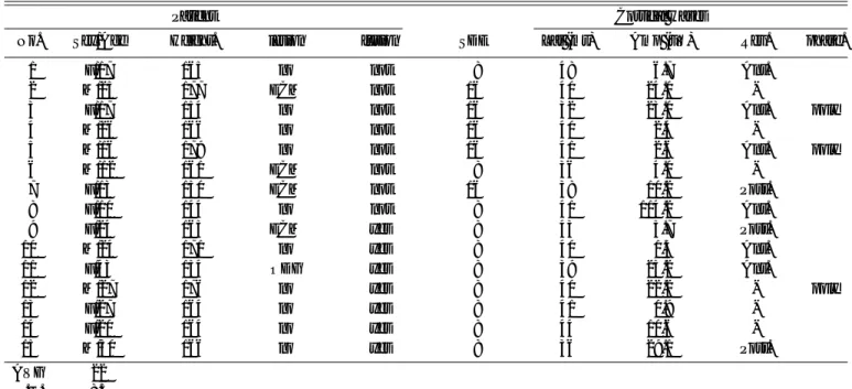
파형은 40msec 주변에서 기록되는 파형을 중심으로 Table 1. Summary of subjects, and recorded subdural electrodes and cortical waves
Patient Cortical waves
No. Sex/Age Height. lesion fusion SDE Lat (ms) Amp (uV) Rev. phase.
01 F/17 165 no not 08 48 6.7 Ant.
02 M/25 177 ECM not 16 40 24.0 -
03 F/17 154 no not 16 32 23.0 Ant. poly
04 M/26 166 no not 16 40 2.4 -
05 M/16 179 no not 16 41 2.6 Ant. poly
06 M/12 161 ECM not 08 36 4.0 -
07 F/13 150 ECM not 16 38 10.2 Post.
08 F/10 144 no not 08 41 114.2 Ant.
09 F/24 165 ECM yes 08 43 5.7 Post.
10 M/24 171 no yes 08 40 1.3 Ant.
11 F/43 154 ODG yes 08 39 25.2 Ant.
12 M/27 176 no yes 08 40 22.2 - poly
13 F/27 164 no yes 08 41 0.9 -
14 F/20 164 no yes 08 44 10.6 -
15 M/30 166 no yes 08 36 29.1 Post.
AVG 22
S.D. 8.5
*No.: number of patient, Lat:latency, Amp:amplitude,
ECM: encephalomalacia, ODG:oligodendroglioma, SDE: number of recorded subdural electrodes Rev. phase reversal of cortical waves. Poly: polyphasic wave
Figure 1. The 3-dimensional fusion images of SPGR MRI and subdural electrodes. (A) The image of patient number 9. The 1x8 subdural strip electrodes were located from medial part of superior frontal gyrus ( ) through paracentral lobule ( ) to precuneus ( ). (B) The image of patient number 14.
The amd electrodes were put on precuneus, on marginal sulcus, on paracentral lobule and on medial part of superior frontal gyrus. The circle numbers in box are the ones on sub - bural strip electrode array.
A B
절대잠복기 및 극대극 진폭 (peak to peak ampli- t u d e )을 측정하였다. 최대의 진폭을 나타내는 전극을 확인하고, 삼차원 영상 합치술을 시행한 7 례에서 그 부 위가 어디에 해당되는지 확인하였고, 관찰되는 파형의 극성 (polarity) 및 모양 ( s h a p e )을 분석하였다. 또한 최대치를 나타내는 전극 주변에 위치한 주변전극에서 기록되는 파형과 비교하였다. 특히 상역전 및 파형 모양 변화를 분석하였다.
결 과
최대치 전극의 위치
삼차원 영상 합치술로 전극의 위치와 상전두회( s u p e- rior frontal gyrus)의 내측 부위, 부중심소엽, 설전부, 및 대상회 (cingulate gyrus) 와의 관계를 볼 때 부중 심소엽에 전극이 지나게 설치된 5 례 중 4례에서는 부 중심소엽에 위치한 전극에서 최대치 진폭을, 1 례에서 는 근접한 설전부에서, 부중심소엽 밑의 대상회를 지나 게 설치된 2례는 모두 부중심소엽 바로 밑의 부위에 위 치한 전극에서 최대치를 나타내었다(Figure 2). 부중 심소엽에 2개 이상의 전극이 설치된 2례에서는 모두 뒤 쪽 전극에서 최대치를 나타내었다. 삼차원 영상합치술 을 시행하지 못하였으나 두개 이상의 선형을 전극 또는 2 x 8 격자 전극을 설치한 5례에서는 모두 상부에 위치 한 전극에서 최대치를 나타내었다.
최대치 전극의 극성 (Table 1)
40msec 부근에서 최대치 진폭을 갖는 파형은 양극성 뿐만 아니라 음극성을 나타내기도 하였다. 그러나 20uV 이상의 국소적으로 큰 진폭을 나타내는 경우는 7 례 였는데 모두 양극성을 나타내었다(Fig. 2A,C).
주변전극과의 상역전 (Table 1)
최대 진폭을 나타내는 전극 주변에서 9례의 경우 상역 전이 관찰되었는데 특히 부중심소엽과 설전부 사이에서 상역전이 관찰된 3례의 경우는 매우 뚜렷한 반면 ( F i g . 2C) 부중심소엽 앞쪽에서 상역전이 관찰된 6례에서는 서서히 변화하는 상역전이 관찰되었다(Fig. 2B).
최대 진폭을 나타내는 전극 앞과 뒤에서 다상성 (polyphasic) 파형이 형성된 경우가 3례 있었다(Fig. 2A).
고 찰
정중신경의 S E P에서 기록되는 첫 피질 파형인 N 2 0 은 일차 감각영역 손부위의 중심구 후벽인 3b 영역에서 처음 발생하는 것으로 알려져 있다. 그러나 후경골신경 의 S E P는 일차 감각영역의 발부위에서 가장 큰 파형을 형성하는 것으로 생각할 수 있다. 그러나 일차감각영역 의 위치가 두정엽 측면에서는 후중심회 ( p o s t c e n t r a l
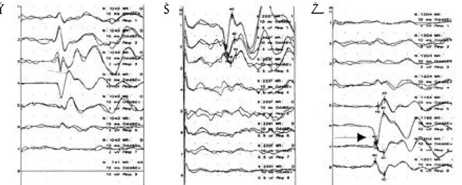
A B C
Figure 2. Cortical waves of PTSEP on subdural electrodes in mesial hemisphere around paracentral lobule. (A) PTSEP of patients number 3. The wave with maximal amplitude was recorded on trace 4 around 32 msec, which had potive polarity. The polyphasic wave also was on trace 3 (anterior to the trace 3 in mesial hemisphere) and negative wave around 30 msec on trace 2. (B) PTSEP of patient number 5. The wave with maximal amplitude was on trace 1 around 38 msec. The phase was slowly reversed on trace 2, and 3 which were located just anterior to trace 1 in medial hemisphere). (C) PTSEP of patient number 6. The wave with maximal amplitude was recorded on trace 6 with positivity. And the electrode showing trace 6 were on paracentral lobule in 3-D fusion. . The phase was sharply reversed on trace 7,8 which were located just posterior trace 6 in medial hemisphere. Arrow indicates phase reversal between trace 6 and 7. The scale of amplitude were described on each trace. The time base of all traces was 10msec. Stimulus delivered at 0 msec. Positive is downward.
g y r u s )에 위치하지만 두정엽 내측면에서는 중심구가 끝나며 전중심회 (precentral gyrus)와 합쳐져서 부중 심소엽 (paracentral lobule)을 형성한다. 일반적으로 다리영역은 Penfield 와 B o l d r e y1에 의한 뇌 자극 검사 상에서는 두정엽 내측면에 위치하는 것으로 알려져 있 다. Allison et al2 3에 의하면 발 감각영역은 후중심구의 말단 부위에 위치하므로 부중심소엽과 만나는 부위에서 가장 잘 나타나는 것으로 보고하였다. 본 연구에서도 삼 차원 영상 합치술로 부중심소엽에 위치한 전극이 두 개 이상을 확인한 2례 모두에서 뒤쪽 부위에 위치한 전극 에서 앞쪽 부위의 전극보다 더욱 높은 진폭을 나타내었 고, 상하로 전극이 위치한 5례의 경우에서 는 상측 부위 에서 기록된 전극에서 더욱 큰 전위를 형성하여 부중심 소엽의 후상부에서 최대의 진폭을 나타내는 것을 알 수 있었다.
최대 진폭을 갖는 파형을 분석해 보면 4 0 m s e c에서 2 0 u v이상의 진폭을 갖는 7례의 파형들은 모두 양극성 을 갖고 있었으며, 그 이외의 낮은 진폭을 갖는 례는 비 록 40msec 에서 음극성을 나타내며 다상성 모양을 보 이기도 하였다. 삼차원 영상 합치술로 확인한 4례 모두 내측 부위 중 하부를 지나 부중심소엽 보다는 그 아래쪽 의 대상회를 지나고 있었다. 그러나 최대의 진폭을 나타 내는 전극들은 바로 부중심소엽의 밑 대상회 부위에 위 치한 전극들이 었다. 특히 최대진폭을 갖는 부위가 양극 성을 갖는다는 점은 피질에서 파형이 형성되는 과정 (cortical electrogenesis)에 대한 가설로 생각해 볼 때 부중심소엽의 구조상 양극성으로 나타날 수 있다고 생 각된다. 즉 감각피질의 심부 제 3 - 5층에 위치한 방추세 포체 근위수상돌기(proximal dendrite) 또는 세포체 에서 저분극(depolarization) 또는 과분극( h y p e r p o- l a r i z a t i o n )이 일어날 경우(sink), 말단수상돌기( a p i- cal dendrite)의 원위부위와 세포외 부분에 전위가 형 성되고(source) 세포체에 근접한 부위와 반대의 극성이 발생하게 되고1 1 , 1 6 , 2 4, 신경세포가 피질에 평행한 면을 이 루고 있다면(radial orientation), 피질의 표재층과 심 부층 및 백질은 반대의 극성을 나타내며 s o u r c e로 작용 하는 피질에서 파형을 기록할 경우는 양극성 파형을 나 타내게 된다.
이러한 점은 P T S E P에서 자극되어 near field로 기 록한 파형이 4 0 m s e c에서 양극성을 나타내며 2 0 u V이 상인 큰 진폭을 나타낼 경우는 부중심소엽의 발과 관련 되는 일차감각영역 또는 그와 아주 근접한 부위에서 기 록된 것으로 생각할 수 있겠다. 아울러 양극성은 2 u v이 상의 큰 진폭을 나타내지 않는다고 해도4 0 m s e c에서 최 대의 진폭을 나타낸 파형의 경우는 다른 전극들 보다 그 부위가 부중심소엽에서 가장 가까운 거리에 위치한 전 극이라고 생각할 수 있겠다.
본 연구에서 관찰된 상역전의 경우 2례에서는 비교적
정확한 상역전을 관찰할 수 있었고 모두 최대치 전극과 그 후방 전극과의 사이에서 형성되었다 즉 부중심소엽과 설전부와의 경계인 대상구 (cingulate sulcus)의 변연 분지 (marginal branch)를 사이에 두고 전극이 위치한 경우로 생각된다 이 중 1례는 삼차원 영상 합치술로 확 인할 수 있었다. 그러나 최대치와 그 전방 전극에서 상역 전이 관찰된 경우 6례 중 3례는 서서히 잠복기가 길어지 면서 상역전이 관찰되었고 1례는 다상의 파형을 나타낸 후 그 다음 전극에서 상역전이 관찰되었다. 즉 부중심소 엽과 상전두회의 경계인 부중심소구(paracentral sul- cus) 로 생각되는 부위에서는 서서히 변화하는 상역전이 관찰되는 경향이 있는 것으로 생각할 수 있다. 이러한 점 은 정중신경 S E P에서 손의 일차 감각영역은 중심구에서 피질이 중심구의 후벽을 형성하여 신경 세포층은 표면과 수직을 이루는 구조로 배열되며(tangential orienta- tion), 이 때 중심구 앞쪽과 뒤쪽으로 반대의 극성을 나 타내는 양극성( d i p o l e )이 형성되어 중심구를 중심으로 상역전이 형성되는 것으로 알려져 있다. PTSEP의 경우 일차적으로 파형이 형성된 부위를 확인할 수 는 없었지 만 일단 형성된 부위 즉 부중심소엽의 뒤쪽 위쪽에서 피 질에서 일차적으로 형성된 전위가 전파한다고 생각할 때 앞쪽 보다는 거리가 가까운 변연구 (marginal sulcus) 에서 분명한 상역전이 형성되기 쉽다고 생각할 수 있다.
요 약
부중심소엽 주변의 경막하 전극들에서 기록한 P T S E P 는 전극의 위치에 따라 다양한 파형을 나타내며, 파형의 최대치는 주로 부중심소엽의 후 상부에 위치한다. 특히 최대값이 주변보다 확연히 차이 나며 1 0 u V이상의 파형 으로 기록될 경우는 4 0 m s e c에서 양극성을 나타낸다. 또 한 주변의 전극과 상역전을 형성할 수 있는데 최대 전극 의 뒤에서 형성될 경우는 더욱 분명한 상역전을 관찰할 수 있다. 따라서 경막하전극을 통한 P T S E P는 내측 반 구의 부중심소엽 주변의 위치를 파악하는데 도움을 줄 수 있다고 생각된다.
참고문헌
01. Penfield W, Boldrey E. Somatic motor and sensory represen- tation in the cerebral cortex of man as studied by electrical stimulation. Brain 1937;60:389-443.
02. Allison T, McCarthy G, Wood CC, et al. Human cortical potentials evoked by stimulation of the median nerve. I.
Cytoarchitectonic areas generating short-latency activity. J Neurophysiol 1989;62(3):694-710.
03. Allison T, McCarthy G, Wood CC, et al. Human cortical
potentials evoked by stimulation of the median nerve. II.
Cytoarchitectonic areas generating long latency activity. J Neurophysiol 1989;62(3):711-722.
04. Chiappa K. Evoked potentials in clinical medicine, 2nd ed.
New York; Raven Press. 1990.
05. Merton PA, Morton HB. Stimulation of the cerebral cortex in the intact human subject. Nature 1980;285:227.
06. Benecke R, Meyer BU, Schonle P, et al. Transcranial magnet- ic stimulation of the human brain: Responses in muscles sup- plied by cranial nerves. Exp Brain Res 1988;71:623-632 07. Wood CC, Cohen D, Cuffin BN, Yarita M, Allison T. Electric
sources in human somatosenory cortex: identification by com- bined magnetic and potentials field recording. S c i e n c e 1 9 8 5 ; 227:1051-1053.
08. Puce Am Condtable RT, Luby ML, et al. Functional magnetic resonance imaging of sensory and motor cortex: comparison with electrophysiologic localization. J Neurosurg 1 9 9 5 ; 8 3 : 2 6 2 - 2 7 0 .
09. Fox PT, Burton H, Raichle ME. Mapping human somatosen- sory cortex with positron emission tomography. J Neurosurg 1987;67:34-43.
10. Kelly DL Jr, Goldring S, O Leary JL. Average evoked soma- tosensory responses from exposed cortex of arm. Arch Neurol 1965;13:1-9.
11. Wood CC, Spencer DD, Allison T, et al. Localization of human sensorimotor cortex during surgery by cortical surface recording of sensory evoked potentials. J Neurosurg 1 9 8 8 ; 6 8 : 9 9 - 1 1 1 . 12. Spencer SS, Spencer DD, Williamson PD, et al. The localizing
value of depth electroencephalography in 32 refractory patients.
Ann Neurol 1 9 8 2 ; 1 2 : 2 4 8 - 2 5 3 .
13. Goldring S, Gregorie EM. Surgical management of epilepsy using epidural recordings to localize the seizure focus. Review of 100 cases. J Neurosurg 1984;60:457-466.
14. Lesser RP, Luders H, Klem G, et al. Extraoperative cortical functional localization in patients with epilepsy. J Clin Neuro- physiol 1987;4:27-53.
15. Luders H, Lesser RP, Hahn J, et al. Cortical somatosensory evoked potentials in response to hand stimulation. J Neurosurg 1 9 8 3 ; 5 8 : 8 8 5 - 8 9 4 .
16. Allison T, McCarthy G, Wood CC, et al. Potentials evoked in human and monkey cerebral cortex by stimulation of the me- dian nerve. Brain 1991;114:2465-2503.
17. Fried, Katz A, McCarthy G, et al. Functional organization of human supplementary motor cortex studied by electrical stim- ulation. J Neurosci 1991;11:3656-3666.
18. Ikeda A, Luders HO, Burgess RC, et al. Movement-related potentials recorded from supplementary motor area and prima- ry motor area. Brain 1992;115:1017-1043.
19. McCarthy G, Allison T, Spencer DD. Localization of the face area of human sensorimotor cortex by intracranial recording for somatosensory evoked potentials J Neruosurgry 1 9 9 3 ; 7 9 : 8 7 4 - 8 8 4 .
20. Jasper H, Lende R, Rasmussen T. Evoked potentials from the exposed somatosensory cortex in man. J Nerv Ment Dis. 1960;
130:526-537.
21. Stohr PE, Goldring S. Origin of somatosensory evoked scalp responses in man. J Neurosurg 1969;31:117-127.
22. Woolsey CN, Erickson TC, Glison WE. Localization in soma- tic sensory and motor areas of human cerebral cortex as deter- mined by direct recording of evoked potentials and electrical stimulation. J Neurosurg 1979;51:476-506.
23. Allison T, McCarthy G, Luby M, Puce M, Spencer DD.
Localization of functional regions of human mesial cortex by somatosensory evoked potential recording and by cortical sti- mulation. EEG and Clin Neurophysiol 1996;100:126-140.
24. Towne AL. On the nature of the primary evoked response.
Exp Neurol 1966;15:113-139.
25. Seo DW, Hong SB. Comparison of perirolandic sensorimotor function using somatosensory evoked potentials and brain stimulation in patients with epilepsy. J Kor Neurol Ass 1999;
17(4):498-504.