• 검색 결과가 없습니다.

Clinical significance of serum galactose-deficient immunoglobulin A1 for detection of recurrent immunoglobulin A nephropathy in kidney transplant recipients

N/A
N/A
Protected

Academic year: 2021

Share "Clinical significance of serum galactose-deficient immunoglobulin A1 for detection of recurrent immunoglobulin A nephropathy in kidney transplant recipients"

Copied!
8
0
0

로드 중.... (전체 텍스트 보기)

전체 글

(1)

Background: Recurrent glomerulonephritis (GN) is a common cause of allograft loss in kidney transplantation (KT), the most frequent of which is immunoglobulin A (IgA) nephropathy (IgAN). Galactose-deficient IgA1 (Gd-IgA1) plays a major role in the pathophysiology of IgAN, but the association between Gd-IgA1 and recurrent IgAN in kidney transplant recipients (KTRs) is uncertain. We aimed to evalu- ate the efficacy of Gd-IgA1 for prediction of recurrent IgAN and graft and patient survival according to Gd-IgA1 level.

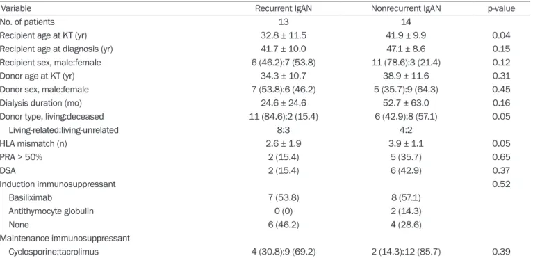
Methods: We enrolled 27 KTRs who underwent allograft biopsy between 2009 and 2016 and measured the serum Gd-IgA1 level of each KTR. We divided the patients into two groups: nonrecurrent IgAN (patients with IgAN prior to KT who were not diagnosed with re- current IgAN) and recurrent IgAN (patients with IgAN prior to KT who were diagnosed with recurrent IgAN).

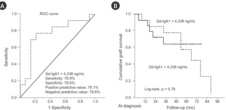
Results: The mean serum Gd-IgA1 level was significantly higher in the recurrent IgAN group than in the nonrecurrent IgAN group (6,419 ± 3,675 ng/mL vs. 3,381 ± 2,844 ng/mL, p = 0.02). The cutoff value of serum Gd-IgA1 in receiver operating characteristic curve analysis was 4,338 ng/mL (area under the curve, 0.76; 95% confidence interval [CI], 0.57–0.95, p = 0.02). Serum Gd-IgA1 lev- el was an independent factor for recurrent IgAN (odds ratio, 17.60; 95% CI, 1.33–233.03, p = 0.03). There was no significant differ- ence in graft or patient survival between the two groups.

Conclusion: Serum Gd-IgA1 can be used as a diagnostic biomarker for recurrent IgAN in KT.

Keywords: Glomerulonephritis, Graft survival, Immunoglobulin A, Kidney transplantation, Survival Kidney Res Clin Pract 2021;40(2):317-324

pISSN: 2211-9132 • eISSN: 2211-9140 https://doi.org/10.23876/j.krcp.20.183

Clinical significance of serum galactose-deficient immunoglobulin A1 for detection of recurrent

immunoglobulin A nephropathy in kidney transplant recipients

Woo Yeong Park

1,2,3

, Yaerim Kim

1,2

, Jin Hyuk Paek

1,2

, Kyubok Jin

1,2

, Seungyeup Han

1,2

1

Division of Nephrology, Department of Internal Medicine, Keimyung University School of Medicine, Daegu, Republic of Korea

2

Keimyung University Kidney Institute, Daegu, Republic of Korea

3

Institute for Cancer Research, Keimyung University, Daegu, Republic of Korea

Received: October 7, 2020; Revised: December 21, 2020; Accepted: December 28, 2020 Editor: Eun Hui Bae, Chonnam National University, Gwangju, Republic of Korea Correspondence: Seungyeup Han

Division of Nephrology, Department of Internal Medicine, Keimyung University School of Medicine, Keimyung University Kidney Institute, 1035 Dalgubeol-daero, Dalseo-gu, Daegu 42601, Republic of Korea. E-mail: [email protected]

ORCID: https://orcid.org/0000-0002-7561-6534 Copyright © 2021 by The Korean Society of Nephrology

This is an Open Access article distributed under the terms of the Creative Commons Attribution Non-Commercial and No Derivatives License (http://

creativecommons.org/licenses/by-nc-nd/4.0/) which permits unrestricted non-commercial use, distribution of the material without any modifications,

and reproduction in any medium, provided the original works properly cited.

(2)

Introduction

One of the most common causes of allograft kidney loss is recurrent glomerulonephritis (GN), and immunoglobulin A (IgA) nephropathy (IgAN) is the most common cause of recurrent GN after kidney transplantation (KT) [1,2]. The pathophysiology of IgAN is the most potent of the “four hits” hypotheses [3], and galactose-deficient IgA1 (Gd-IgA1) plays a major role in the pathophysiology of IgAN [4]. The main mechanism of action of Gd-IgA1 is deposition with an autoimmune IgG complex in the mesangium and endo- thelium, subsequently damaging those cells [4]. The same mechanism can be assumed for recurrent IgAN after KT, but recurrent GN because of immunosuppressive drug adminis- tration does not have the same mechanism of action as IgAN of the general population. Considering that Gd-IgA1 can be measured in the laboratory, it can be used as a diagnostic and prognostic biomarker for IgAN, but it is unclear wheth- er recurrent IgAN after KT is similarly observed based on this clinical pattern and whether de novo IgAN is observed.

However, the association between Gd-IgA1 and occurrence of recurrent IgAN in kidney transplant recipients (KTRs) is uncertain. Therefore, in this study, we aimed to investigate whether Gd-IgA1 is effective as a diagnostic and prognostic marker for recurrent IgAN.

Methods Study design

We enrolled 27 KTRs with stored samples in the Biobank of Keimyung University Dongsan Hospital and who underwent allograft biopsy between 2009 and 2016. There were 17 living donor KTRs (10 living-related donor KTRs and seven liv- ing-unrelated donor KTRs) and 10 deceased donor KTRs. The living-related donor KTRs comprised four parent-to-child and six sibling-to-sibling KTRs; two wife-to-husband KTRs were in- cluded in the living-unrelated donor KTRs. The patients were divided into the nonrecurrent IgAN group (patients with IgAN prior to KT who were not diagnosed with recurrent IgAN after KT [n = 14]) and the recurrent IgAN group (patients with IgAN prior to KT who were diagnosed with recurrent IgAN [n = 13]).

The nonrecurrent IgAN group included KTRs with other recur- rent GN issues, acute and chronic rejection, and calcineurin inhibitor (CNI) toxicity. We evaluated the clinical characteris-

tics of the study population, status of immunosuppression at diagnosis, change in allograft function, severity of proteinuria at diagnosis, and graft and patient survival.

Demographic and clinical data

We investigated the age of donors and recipients at diagno- sis of recurrent IgAN, sex of donors and recipients, KT type, frequency of KT, dialysis type prior to KT, dialysis vintage, causes of end-stage renal disease (ESRD), number of human leukocyte antigen (HLA) mismatches, immunosuppressant for induction and maintenance treatment, previous biop- sy-proven acute rejection (BPAR), severity of proteinuria at diagnosis of recurrent IgAN, panel reactive antibody (PRA) >

50%, and donor-specific antibody positivity. Allograft biopsy was performed when allograft dysfunction (decreased esti- mated glomerular filtration rate [eGFR]) or persistent micro- scopic hematuria and proteinuria were observed. Allograft biopsy was analyzed using the Banff 2013 classification [5].

Allograft function was measured based on eGFR according to the guidelines of the Chronic Kidney Disease Epidemiol- ogy Collaboration within 1 month before diagnosis and at 12 months, 3 years, and 5 years after diagnosis [6]. Proteinuria was measured using the spot urine protein-to-creatinine ra- tio (g/g).

Serum Gd-IgA1 level was measured using the Gd-IgA1 as- say kit (Immuno-Biological Laboratories Co., Ltd., Gunma, Japan) through the enzyme-linked immunosorbent assay [7]. The level of each serum Gd-IgA1 was obtained based on a standard curve with an appropriate regression curve on each plot (four-parameter logistics; optical density, 450 nm).

The assay for Gd-IgA1 was performed in triplicate.

The Institutional Review Board (IRB) of Keimyung Uni- versity Dongsan Hospital approved this study (No. 2017- 02-030). The requirement for informed consent was waived by the IRB because use of patient data for research, except identifying personal information, was explained to all donor families and all recipients before KT. Therefore, as a retro- spective medical record study, this study did not contain any distinguishable personal information, except clinical process and outcome.

Immunosuppression protocols

We administered basiliximab (Simulect, 20 mg on days 0 and

(3)

4; Novartis, Basel, Switzerland) for KTRs with low immunolog- ic risk and antithymocyte globulin (Thymoglobulin, 1.5 mg/

kg on day 0 and 1.0 mg/kg from day 1 to day 3; Genzyme, Cambridge, MA, USA) for KTRs with high immunologic risk as induction immunosuppressants. We administered cyclo- sporine (Sandimmune, 3 mg/kg, twice a day; Novartis AG, Basel, Switzerland) or tacrolimus (Prograf, 0.05 mg/kg, twice a day; Astellas Pharma Inc., Toyama, Japan) as a CNI, pred- nisolone (30 – 20 – 10 – 5 – 0 mg, once a day, on a stepdown regimen), and mycophenolate mofetil (CellCept, 750 or 1,000 mg, twice a day for 1 month after KT and subsequent 500 mg, twice a day; Hoffmann-La Roche Inc., Nutley, NJ, USA) as maintenance immunosuppressants. The treatment pro- tocol for recurrent IgAN was use of an angiotensin receptor blocker at diagnosis; if it elicited no response, then 0.25 mg/

kg per day of oral steroid was used.

Statistical analyses

Continuous variables were analyzed using the Mann-Whit- ney U test, and categorical variables were analyzed using the chi-square or Fisher exact test. Graft and patient survival rates were evaluated using the Kaplan-Meier analysis with log-rank test. Univariate and multivariate analyses with lo- gistic regression analysis were performed to investigate the risk factors for development of recurrent IgAN. The p-values less than 0.05 were considered statistically significant. Statis- tical analysis was performed using the PASW Statistics (ver- sion 18.0; IBM Corp., Armonk, NY, USA).

Results

Baseline characteristics of the study population

The mean follow-up period for all patients was 147.5 ± 83.6 months. Recipient age at KT was significantly lower in the recurrent IgAN group than in the nonrecurrent IgAN group (32.8 ± 11.5 years vs. 41.9 ± 9.9 years, p = 0.04), but there was no significant difference in recipient age at diagnosis.

There was also no significant difference in donor age at KT between the two groups. There were no significant differ- ences in rates of recipient and donor sex, PRA > 50%, use of induction and maintenance immunosuppressants, or previous BPAR between the two groups. The proportion of living donor KT was higher in the recurrent IgAN group than

in the nonrecurrent IgAN group. Moreover, the proportion of deceased donor KT was higher in the nonrecurrent IgAN group than in the recurrent IgAN group. The mean number of HLA mismatches was significantly lower in the recurrent IgAN group than in the nonrecurrent IgAN group (2.6 ± 1.9 vs. 3.9 ± 1.1, p = 0.05). There were no significant differences between the recurrent IgAN and nonrecurrent IgAN groups in the other clinical parameters (Table 1).

Comparison of clinical and laboratory parameters according to recurrent IgA nephropathy

The mean serum Gd-IgA1 level was significantly higher in the recurrent IgAN group than in the nonrecurrent IgAN group (6,419 ± 3,675 ng/mL vs. 3,381 ± 2,844 ng/mL, p = 0.02). The mean time between KT and allograft biopsy was longer in the recurrent IgAN group than in the nonrecurrent IgAN group (108.9 ± 83.3 months vs. 61.7 ± 69.2 months).

The proportion of steroid use was significantly lower in the recurrent IgAN group than in the nonrecurrent IgAN group.

Allograft function between diagnosis and 5 years after diag- nosis and proteinuria at diagnosis did not differ between the two groups (Table 2).

Receiver operating characteristic curve analysis

The cutoff value of serum Gd-IgA1 in receiver operating charac- teristic curve analysis was 4,338 ng/mL (area under the curve, 0.76; 95% confidence interval [CI], 0.57–0.95; p = 0.02). The sensitivity and specificity were 76.9% and 78.6%, respectively.

The positive predictive value and negative predictive value were 76.1% and 78.6%, respectively (Fig. 1A). There was no signifi- cant difference in death-censored graft survival rate after di- agnosis of recurrent IgAN or other diseases according to Gd- IgA1 level (Fig. 1B).

Comparison of death-censored allograft survival and patient survival according to development of recurrent IgA nephropathy and factors related to development of recurrent IgA nephropathy

A total of 14 patients (51.9%; six patients [22.2%] in the re-

current IgAN group and eight patients [29.6%] in the nonre-

current IgAN group) experienced graft failure. The causes

of graft failure were chronic rejection (three [50.0%] and

(4)

Table 1. Comparison of clinical and laboratory parameters according to recurrent IgAN

Variable Recurrent IgAN Nonrecurrent IgAN p-value

No. of patients 13 14

Recipient age at KT (yr) 32.8 ± 11.5 41.9 ± 9.9 0.04

Recipient age at diagnosis (yr) 41.7 ± 10.0 47.1 ± 8.6 0.15

Recipient sex, male:female 6 (46.2):7 (53.8) 11 (78.6):3 (21.4) 0.12

Donor age at KT (yr) 34.3 ± 10.7 38.9 ± 11.6 0.31

Donor sex, male:female 7 (53.8):6 (46.2) 5 (35.7):9 (64.3) 0.45

Dialysis duration (mo) 24.6 ± 24.6 52.7 ± 63.0 0.16

Donor type, living:deceased 11 (84.6):2 (15.4) 6 (42.9):8 (57.1) 0.05

Living-related:living-unrelated 8:3 4:2

HLA mismatch (n) 2.6 ± 1.9 3.9 ± 1.1 0.05

PRA > 50% 2 (15.4) 5 (35.7) 0.65

DSA 2 (15.4) 6 (42.9) 0.37

Induction immunosuppressant 0.52

Basiliximab 7 (53.8) 8 (57.1)

Antithymocyte globulin 0 (0) 2 (14.3)

None 6 (46.2) 4 (28.6)

Maintenance immunosuppressant

Cyclosporine:tacrolimus 4 (30.8):9 (69.2) 2 (14.3):12 (85.7) 0.39

Data are expressed as number only, mean ± standard deviation, or number (%).

BPAR, biopsy-proven acute rejection; DSA, donor-specific antibody; HLA, human leukocyte antigen; IgAN, immunoglobulin A nephropathy; KT, kidney transplantation; PRA, panel reactive antibody.

Table 2. Comparison of clinical outcomes according to recurrent IgAN

Variable Recurrent IgAN (n = 13) Nonrecurrent IgAN (n = 14) p-value

Gd-IgA1 (ng/mL) 6,418 ± 3,675 3,381 ± 2,844 0.02

Time from KT to biopsy (mo) 108.9 ± 83.3 61.7 ± 69.2 0.12

Immunosuppressant at diagnosis

Cyclosporine:tacrolimus 2 (15.4):11 (84.6) 2 (14.3):12 (85.7) >0.99

Mycophenolate mofetil 7 (53.8) 11 (78.6) 0.24

Steroid

Before diagnosis 2 (15.4) 9 (64.3) 0.02

After diagnosis 8 (61.5) 8 (57.1) >0.99

ARB

Before diagnosis 3 (23.1) 3 (21.4) >0.99

After diagnosis 8 (61.5) 5 (35.7) 0.26

Allograft function (CKD-EPI) (mL/min/1.73 m

2

)

Within 1 mo before diagnosis 50.9 ± 25.2 45.9 ± 24.1 0.61

After diagnosis (yr)

1 53.3 ± 28.9 48.9 ± 25.1 0.27

2 48.9 ± 25.1 43.3 ± 22.8 0.58

3 51.1 ± 16.8 35.8 ± 22.8 0.09

4 51.4 ± 23.6 43.9 ± 29.6 0.57

5 44.4 ± 23.5 32.4 ± 23.2 0.35

Proteinuria at diagnosis (g/g) 1.7 ± 1.9 1.0 ± 0.8 0.22

Data are expressed as mean ± standard deviation or number (%).

ARB, angiotensin receptor blocker; CKD-EPI, Chronic Kidney Disease Epidemiology Collaboration; Gd-IgA1, galactose-deficient immunoglobulin A1; IgAN,

immunoglobulin A nephropathy; KT, kidney transplantation.

(5)

six [75.0%] in the recurrent IgAN and nonrecurrent IgAN groups, respectively), recurrent IgAN (three [50.0%] and none in the recurrent IgAN and nonrecurrent IgAN groups, respectively), and patient death with graft function (none and one [12.5%] in the recurrent IgAN and nonrecurrent IgAN groups, respectively) (Table 3). The 10-year graft sur- vival rate was 88.9% in the recurrent IgAN group and 69.6%

in the nonrecurrent IgAN group, but there was no significant difference in overall death-censored graft survival between the two groups (Fig. 2A). There was no significant difference in death-censored graft survival rate after diagnosis of re-

current IgAN or other diseases between the two groups (Fig.

2B). One patient (3.7%) in the nonrecurrent IgAN group died due to cytomegalovirus pneumonia. The 10-year patient survival rate was 100% in the recurrent IgA group and 92.9%

in the nonrecurrent IgAN group, but there was no significant difference in death-censored graft survival between the two groups.

Based on logistic regression analysis, serum Gd-IgA1 level was an independent factor for diagnosis of recurrent IgAN (odds ratio, 17.06; 95% CI, 1.33–233.03; p = 0.03) adjusting for recipient age, sex, living donor KT, dialysis vintage, mainte- Figure 1. The cutoff of Gd-IgA1 level and allograft outcome according to the Gd-IgA1 level. (A) ROC curves for serum Gd-IgA1 level.

ROC AUC for recurrence of IgAN was 0.76 (0.57–0.95) for serum Gd-IgA1 (p = 0.023). (B) Comparison of death-censored graft survival rate after diagnosis of recurrent IgAN according to Gd-IgA1 level.

AUC, area under the curve; Gd-IgA1, galactose-deficient-immunoglobulin A1; IgAN, immunoglobulin A nephropathy; ROC, receiver operating characteristic.

A

Table 3. Comparison of cause of allograft failure and patient death according to recurrent IgAN

Variable Recurrent IgAN (n = 13) Nonrecurrent IgAN (n = 14) p-value

Causes of graft failure 0.08

Chronic rejection 3 (23.1) 6 (42.9)

Recurrent IgAN 3 (23.1) 0 (0)

Others 0 (0) 1 (7.1)

Patient death with a functioning graft 0 (0) 1 (7.1)

Causes of death >0.99

Cytomegalovirus pneumonia 0 (0) 1 (7.1)

Data are expressed as number (%).

IgAN, immunoglobulin A nephropathy.

B

Cumulative graft survival

1.0

0.8

0.6

0.4

0.2

0.0

Follow-up (mo)

At diagnosis 12 24 36 48 60 72 84 96 Gd-IgA1 > 4,338 ng/mL

Gd-IgA1 < 4,338 ng/mL

Log-rank, p = 0.79

1-Specificity

Sensitivity

1.0

0.8

0.6

0.4

0.2

0.0

ROC curve

0.2 0.4 0.6 0.8 1.0

Gd-IgA1 > 4,338 ng/mL Sensitivity: 76.9%

Specificity: 78.6%

Positive predictive value: 76.1%

Negative predictive value: 78.6%

(6)

Figure 2. Allograft outcome of kidney transplant recipients according to the development of recurrent IgAN. Comparison of (A) death- censored overall graft survival rate and (B) death-censored graft survival rate after diagnosis of recurrent IgAN.

IgAN, immunoglobulin A nephropathy; KT, kidney transplantation.

192

A

Cumulative graft survival

1.0 0.8 0.6 0.4 0.2

Follow-up (mo) At KT 0 24 48 72 96 120 144 168

Log-rank, p = 0.26 Recurrent IgAN

Recurrent IgAN 13 13 13 13 13 13 9 8 8 Non-recurrent IgAN 14 13 10 10 10 10 10 9 9

Nonrecurrent IgAN

B

Cumulative graft survival

1.0 0.8 0.6 0.4 0.2

Follow-up (mo)

At diagnosis 0 12 24 36 48 60 72 84 96 Log-rank, p = 0.24

Recurrent IgAN

Recurrent IgAN 13 12 10 10 10 9 9 8 7 Non-recurrent IgAN 14 13 11 9 8 7 6 6 6

Nonrecurrent IgAN

Table 4. Risk factors associated with recurrent IgA nephropathy in KT recipients

Variable Univariate Multivariate

HR 95% CI p-value HR 95% CI p-value

Recipient age at KT 0.92 0.85–1.00 0.05 1.06 0.92–1.24 0.41

Recipient male sex 0.23 0.04–1.25 0.09 0.06 0.01–1.20 0.07

Living donor KT 0.12 0.02–0.86 0.03 13.08 0.94–181.49 0.06

Dialysis vintage 0.99 0.96–1.01 0.20 1.01 0.98–1.04 0.55

Tacrolimus vs. cyclosporine 0.38 0.06–2.52 0.31 8.09 0.09–772.74 0.37

HLA mismatches 0.56 0.30–1.04 0.07 0.69 0.27–1.75 0.44

Time from KT to diagnosis 1.01 1.00–1.02 0.13 1.01 0.98–1.04 0.55

Gd-IgA1 > 4,338 ng/mL 12.22 1.99–75.06 0.007 17.06 1.33–233.03 0.03

Steroid use 0.10 0.02–0.65 0.02 2.09 0.01–556.51 0.80

CI, confidence interval; Gd-IgA1, galactose-deficient-immunoglobulin A1; HLA, human leukocyte antigen; HR, hazard ratio; IgA, immunoglobulin A; KT, kidney transplantation.

nance immunosuppressant, HLA mismatch, time from KT to diagnosis, and steroid use (Table 4).

Discussion

IgAN has been implicated in the most frequent, primary form of GN and is the most common cause of recurrent GN in KTRs. However, the clinical manifestations of recurrent GN include proteinuria and allograft dysfunction such as acute rejection, recurrent GN, or CNI toxicity. To identify the causes of these symptoms, allograft biopsy is currently the

best diagnostic method [8]. However, allograft biopsy is inva- sive, requires hospitalization, and can lead to false diagnosis due to insufficient specimens. Therefore, several studies are being conducted to overcome these shortcomings; in the case of recurrent IgAN, the research has been conducted to identify the mechanism of IgAN in native kidney diseases [9].

In particular, Gd-IgA1, involved in the mechanism of IgAN, has been reported as a diagnostic biomarker [10]. We evalu- ated the usefulness of Gd-IgA1 as a diagnostic biomarker for recurrent IgAN in KTRs.

Temurhan et al. [11] assessed the Gd-IgA1 level of recur-

(7)

rent IgAN in KT, nonrecurrent IgAN in KT, non-transplant IgAN patients, and healthy relatives. They found that, re- gardless of transplantation, Gd-IgA1 level was significantly higher in patients with recurrent IgAN and non-transplant IgAN than in those with nonrecurrent IgAN and healthy relatives (recurrent IgAN, 8,735 ± 10,854 ng/mL; non-trans- plant IgAN, 8,791 ± 8,700 ng/mL; nonrecurrent IgAN, 4,790

± 6,089 ng/mL; healthy relatives, 2,615 ± 1,611 ng/mL). The results of our study are consistent with those of the above- mentioned study. Patients with recurrent IgAN after KT showed significantly higher Gd-IgA1 level than patients with nonrecurrent IgAN after KT.

Because Gd-IgA1 level is specifically elevated in IgAN pa- tients according to pathogenesis of IgAN, IgAN can be pre- dicted by Gd-IgA1 level, although the cause of ESRD is not confirmed through kidney biopsy. In addition, in KTRs, it is possible to indirectly evaluate the status of the allograft kid- ney for presence of acute or chronic rejection or CNI toxicity, although the Gd-IgA1 level is not elevated and the cause of ESRD is not IgAN, which can overcome the disadvantages of allograft biopsy.

In other words, when KTRs with IgAN as a cause of ESRD have allograft dysfunction or new-onset proteinuria or he- maturia, we recommend measuring the Gd-IgA1 level with blood samples. If the Gd-IgA1 level is high, we can consider recurrent IgAN, which will lead us to use an angiotensin re- ceptor blocker or steroid without allograft biopsy.

Interestingly, the proportion of living donor KT was higher in the recurrent IgAN group than in the nonrecurrent IgAN group. Some studies have reported that KT from a living-re- lated donor is the most well-known risk factor for recurrent IgAN, and the incidence of IgAN in renal allografts is higher in living-related donor KT than in deceased donor KT. The results of our study are consistent with those of the above- mentioned studies [12,13]. In our study, multivariate logistic regression analysis showed an odds ratio of living donor KT for occurrence of recurrent IgAN of 13.08 (95% CI, 0.94–

181.49, p = 0.055); therefore, the occurrence of recurrent IgA is associated with living donor KT. However, there was no significant difference in allograft survival of patients with re- current IgAN between living donor KT and deceased donor KT. These observations were not confirmed in other studies [14,15], and there was no speculated reason for the higher recurrence rate of IgAN in living donor KT than in deceased donor KT. Therefore, further large-scale studies are needed.

There is a lack of consensus regarding the role of steroids against occurrence of recurrent IgAN. Allen et al. [16] report- ed that corticosteroids reduced the occurrence of recurrent IgAN in KTR. In our study, the proportion of steroid use at diagnosis was significantly lower in the recurrent IgAN group compared with the nonrecurrent IgAN group, but ste- roid use was not a factor related to development of recurrent IgAN in multivariate analysis. Furthermore, steroid was used when there was no response to angiotensin receptor blocker, and steroid was not effective in most patients with recurrent IgAN.

Our study has some limitations. First, this was a retrospective study. Therefore, since a selection bias can develop, further large-scale prospective studies are needed. Second, we did not assess Gd-IgA1 level regularly after KT because examination for Gd-IgA1 was not commercialized for research purposes.

In other words, we only measured Gd-IgA1 level at the time of allograft biopsy in blood samples stored in the Biobank for evaluation of allograft dysfunction. Third, because the sam- ple size was small, the results of this study might not be rep- resentative. In particular, we cannot compare the difference in Gd-IgA1 level according to the effect of induction immu- nosuppressant because of small sample sizes. However, we did observe significant differences in serum Gd-IgA1 level that might indicate recurrent IgAN in KTRs with allograft dysfunction.

In conclusion, serum Gd-IgA1 can be effective for early detection of recurrent IgAN in KTRs. Therefore, serum Gd- IgA1 can be used as a diagnostic biomarker for recurrent IgAN in KT.

Conflicts of interest

All authors have no conflicts of interest to declare.

Funding

This study was supported by the Basic Science Research Pro-

gram through the National Research Foundation of Korea

(NRF) funded by the Ministry of Education, Science, and Tech-

nology (NRF-2017R1D1A1B03029140) and was also supported

by the First Research Support Project of the National Research

Foundation of Korea (NRF) funded by the Ministry of Science

and ICT in 2018 (NRF-2017R1C1B5076739).

(8)

Authors’ contributions

Conceptualization, Project administration: SH Data curation, Funding acquisition: WYP Investigation: YK, JHP, KJ

Writing–original draft: WYP

Writing–review & editing: All authors

All authors read and approved the final manuscript.

Acknowledgments

The biospecimens and data used for this study were provid- ed by the Keimyung University Dongsan Hospital Biobank a member of the Korea Biobank Network.

ORCID

Woo Yeong Park, https://orcid.org/0000-0003-2662-2898 Yaerim Kim, https://orcid.org/0000-0003-1596-1528 Jin Hyuk Paek, https://orcid.org/0000-0001-8875-1260 Kyubok Jin, https://orcid.org/0000-0002-7836-8863 Seungyeup Han, https://orcid.org/0000-0002-7561-6534 References

1. Wang AY, Lai FM, Yu AW, et al. Recurrent IgA nephropathy in renal transplant allografts. Am J Kidney Dis 2001;38:588–596.

2. An JN, Lee JP, Oh YJ, et al. Incidence of post-transplant glomerulonephri- tis and its impact on graft outcome. Kidney Res Clin Pract 2012;31:219–

226.

3. Chang S, Li XK. The role of immune modulation in pathogenesis of IgA nephropathy. Front Med (Lausanne) 2020;7:92.

4. Floege J. Primary glomerulonephritis: a review of important re- cent discoveries. Kidney Res Clin Pract 2013;32:103–110.

5. Hara S. Banff 2013 update: pearls and pitfalls in transplant renal

pathology. Nephrology (Carlton) 2015;20 Suppl 2:2–8.

6. Inker LA, Eckfeldt J, Levey AS, et al. Expressing the CKD-EPI (Chronic Kidney Disease Epidemiology Collaboration) cystatin C equations for estimating GFR with standardized serum cysta- tin C values. Am J Kidney Dis 2011;58:682–684.

7. Yasutake J, Suzuki Y, Suzuki H, et al. Novel lectin-independent approach to detect galactose-deficient IgA1 in IgA nephropathy.

Nephrol Dial Transplant 2015;30:1315–1321.

8. Al-Awwa IA, Hariharan S, First MR. Importance of allograft bi- opsy in renal transplant recipients: correlation between clinical and histological diagnosis. Am J Kidney Dis 1998;31:S15–S18.

9. Hastings MC, Moldoveanu Z, Suzuki H, et al. Biomarkers in IgA nephropathy: relationship to pathogenetic hits. Expert Opin Med Diagn 2013;7:615–627.

10. Berthelot L, Robert T, Vuiblet V, et al. Recurrent IgA nephropathy is predicted by altered glycosylated IgA, autoantibodies and sol- uble CD89 complexes. Kidney Int 2015;88:815–822.

11. Temurhan S, Akgul SU, Caliskan Y, et al. A novel biomarker for post-transplant recurrent IgA nephropathy. Transplant Proc 2017;49:541–545.

12. Deng R, Dai Y, Zhang H, et al. Higher incidence of renal allograft glomerulonephritis in living-related donor kidney transplantation.

Transplant Proc 2018;50:2421–2425.

13. Han SS, Huh W, Park SK, et al. Impact of recurrent disease and chronic allograft nephropathy on the long-term allograft outcome in patients with IgA nephropathy. Transpl Int 2010;23:169–175.

14. Ponticelli C, Traversi L, Banfi G. Renal transplantation in patients with IgA mesangial glomerulonephritis. Pediatr Transplant 2004;8:334–338.

15. Cosio FG, Cattran DC. Recent advances in our understanding of recurrent primary glomerulonephritis after kidney transplantation.

Kidney Int 2017;91:304–314.

16. Allen PJ, Chadban SJ, Craig JC, et al. Recurrent glomerulonephritis after kidney transplantation: risk factors and allograft outcomes.

Kidney Int 2017;92:461–469.

수치

Table 1.  Comparison of clinical and laboratory parameters according to recurrent IgAN
Table 3. Comparison of cause of allograft failure and patient death according to recurrent IgAN
Table 4. Risk factors associated with recurrent IgA nephropathy in KT recipients

참조

관련 문서

First, there was no significant difference as a result of analyzing the differences in psychological capital, achievement goal orientation, and game roots

There would be difference in a step of maturity of defense mechanism according to a factor of stress by class(elementary school, middle school) and

Fig. After fine grinding of hardness test specimens 29 Fig. Result of tensile strength according to number of welding pass 34 Fig. Result of elongation according

Second, there was no significant difference in the effect of participants' motivation according to age, final educational attainment, motivation to

2) In between-group comparison before and after elastic band exercise, a significant difference was found in the percentage of body fat, fat-free

Fourth, for the sub-factors of personality traits according to weight lifters’ education, while there was a high difference in neuroticism and agreeableness

Third, there was no significant difference in the frequency of second hospital evacuation after moving to the medical room due to experience and

Although there was a trend for a greater decline in quantitative HBsAg titers in the group that achieved HBeAg seroclearance, the difference in the kinetics of qHBsAg