AAH of larger than 2 cm.
Key Words: Lung; Precancerous Conditions; Adenocarcinoma
Address for correspondence: Sung Soo Jung, M.D.
Department of Internal Medicine, Chungnam National
University School of Medicine, 282 Munhwa-ro, Jung-gu,
Daejeon 301-721, Korea
Phone: 82-42-280-8103, Fax: 82-42-257-5753
E-mail: [email protected]
Received: Nov. 20, 2012
Revised: Dec. 4, 2012
Accepted: Feb. 6, 2013
CCIt is identical to the Creative Commons Attribution Non-Commercial
License (http://creativecommons.org/licenses/by-nc/3.0/).
Introduction
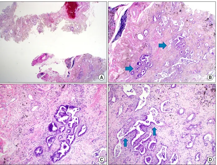
Atypical adenomatous hyperplasia (AAH) is patholog- ically defined as lesion of atypical cells resembling type 2 alveolar cell or cubic cell proliferation usually less than 5 mm in diameter
1. In 1999, World Health Organization reported that AAH is included in precursor lesions of lung cancer along with squamous dysplasial carcinoma in situ , and diffuse idiopathic pulmonary neuroendocrine cell hyperplasia
1. Most studies have not been able to find the direct association of malignant tu- mors and AAH. Instead AAH is explained as a precursor lesion, especially of adenocarcinoma, connecting with the low frequency in noncancerous lung
2. Reports tell that the AAH chest computed tomography (CT) finding shows nodular ground-glass opacity (GGO) pattern
3,4. AAH cases reported until today also report that it is only
scattered in local lesion.
In this paper, we report a 71-year-old female who was diagnosed with lung AAH of larger than 2 cm that was distributed diffusely in the right lobe. She had a history of pulmonary tuberculosis and suffered from dyspnea on exertion showing extensive invasion in the right lung.
Case Report
A 71-year-old female visited the outpatient depart- ment with a 2-week history of coughing with sputum and dyspnea on exertion. She had no other medical his- tory with the exception of pulmonary tuberculosis at the age of 30 years. After administration of anti-tuberculosis medication, she was completely cured.
A chest X-ray performed during the patient's first visit
to the outpatient department showed consolidation in
the right lower lung field and focal consolidation in the
left lower lobe. Sputum acid-fast bacteria (AFB) staining
was negative. On bronchoscopy, the inlet of the right
lower lobe was crushed out, especially the superior seg-
ment of the lower lobe. However, intraluminal lesions
were not found. Based on these results, the patient was
diagnosed with community-acquired pneumonia and
was prescribed clarithromycin for 7 days. She was