Available at http://www.formulastudy.com
HFS
Original Article / 원저
대황과 감초 병용의 항산화 및 간보호효과
이은혜
1,2,#, 백수연
1,#, 김광연
1, 이슬기
1, 김상찬
1, 이형식
2,*, 김영우
1,*1대구한의대학교 한의과대학 MRC 센터, 2의과학대학
Effect of Rheum undulatum Linne extract and Glycyrriza uralensis Fischer extract against arachidonic acid and iron-induced oxidative
stress in HepG2 cell and CCl
4-induced liver injury in mice
Eun Hye Lee
1,2,#, Su Youn Baek
1,#, Kwang-Youn Kim
1, Seul-Gi Lee
1, Sang Chan Kim
1, Hyeong Sik Lee
2,*, Young Woo Kim
1,*1MRC center, Department of Korean Medicine, Daegu Hanny University
2Department of Medical Science, Daegu Hanny Univeristy
ABSTRACT
Objectives : Rheum undulatum Linne and Glycyrriza uralensis Fischer are widely used herbal medicine. In this study, anti-oxidant and liver protective effects of R. undunlatum extract (RUE) and G. uralensis extract (GUE) were investigated in HepG2 cells, respectively. Oxidative stress and liver fibrosis were induced by arachidonic acid (AA) and iron, and CCl4.
Methods : MTT assay was assessed for cell viability, and immunoblotting analysis was performed to detect expression of apoptosis related proteins. In addition, reactive oxygen species (ROS) and mitochondrial dysfunction were measured. In vivo, BALB/c mouse were orally administrated with the aqueous extract of 10 mg/kg RUE and 100 mg/kg GUE for 3 days and then, injected with CCl4 0.5 ml/kg body weight to induce acute liver damage.
Serum ALT level was measured, and histological change was observed in Harris's hematoxylin and eosin stain.
Results : RUE and GUE pre-treatment increased relative cell viability in concentration dependent manner and altered the expression levels of apoptosis-related proteins such as procaspase 3, PARP and Bcl-xL. RUE and GUE also inhibited the mitochondrial dysfunction and excessive reactive oxygen species (ROS) production induced by AA and iron. In addition, RUE and GUE activated liver kinase B1 (LKB1), by increasing phosphorylation. Moreover, RUE and
2016 The Korean Medicine Society For The Herbal Formula Study
ⓒ
This paper is available at http://www.formulastudy.com which permits unrestricted non-commercial use, distribution, and reproduction in any medium, provided the original work is properly cited.
GUE treatment decreased liver injuries induced by CCl4, as evidenced by decreases in histological liver damage as well as serum alanine amino transferase (ALT) level.
Conclusions : These data suggest that RUE and GUE has anti-oxidant and liver protective effects against AA and iron-induced oxidative stress and CCl4-induced liver injury.
Keywords : Rheum undulatum Linne, Glycyrriza uralensis Fischer, Hepatoprotective effect, Oxidative stress, Liver injury.
활성산소종 은
(Reactive Oxygen Species, ROS) 세포의 정상적인 물질대사에서 생성되는 산소의 환 원 대사물로 스트레스, 약물, 환경오염물질, 세균 감염 등의 요인으로 인해 세포 내 대사의 균형이 깨지게 되면 과잉으로 생성된다1,2). 산화적 스트레 스는 활성산소종의 생성과 이를 제거하는 항산화 반응 간의 불균형으로 인해 세포 내의 활성산소종 이 증가하여 DNA나 단백질 지질과 반응하여 손상, 시키는 현상이며 급 만성 간질환을 포함하여 암 동∙ , 맥경화 당뇨 신경퇴화 등 다양한 질병의 요인으로 , , 간주된다3,4). 따라서 세포 내 항산화 기능을 증가시 켜 이러한 질환을 치료할 수 있는 다양한 항산화제 의 탐색과 그 작용기전에 대한 연구가 활발하게 진 행되고 있다5,6).
세포
내 과도하게 증가된 활성산소종은 phospholipase 의 활성화를 통하여 세포막의 인지질과 지방산을 인산화시키면서 아라키돈산(arachidonic acid, AA) 을 유리시킨다6,7). 유리된 AA는 전 염증 매개체로
에 의한 유리 세포질
sphingomyelinase ceramide , Ca2+의 증가 및 미토콘드리아 막 전위를 통해 세포 질로 cytochrome-c를 유리시킴으로서 세포자멸사 를 유도하며 철이 촉매제로 작용하면 세포 손상은 , 가속화된다7). 간세포의 세포자멸사가 지속적으로 발생하게 되면 간의 미세섬유아세포를 섬유화 시킴 으로 간경변을 초래한다 손상 받은 간 조직부위는 . 아교섬유에 의해 정상적으로 수복되지만 세포 외 기질(Extra Cellular Matrix, ECM) 단백질이 축적
되어 섬유화되는 비정상적 수복도 발생하기도 하며 지속적인 간 조직의 손상과 섬유화는 간경화를 초 래한다8). 따라서 간세포에서 , AA와 iron을 이용한 산화적 스트레스 모델은 간질환을 치료할 수 있는 항산화물질을 스크리닝할 수 있는 좋은 세포학적 연구의 모델이 될 수 있다.
대황
(Rheum undulatum Linne)은 아시아에 널 리 분포하는 한약재로 마디풀과(Polygonaceae)에 속하는 다년생 식물이다. 한방에서 복막염, 장염, 담석증 만성설사 황달 및 담석증의 치료에 사용되, , 어 왔으며9,10), 항균 항염 항혈전 등의 다양한 연, , 구가 보고되었다11-13). 특히 대황 추출물 , rhapontigenin 이 대장 세포주인 V79-4 에서 항산화 효과를 보고 된 바 있으며4), 대황에서 추출된 stilbenoids가
세포에서 를 통해 간세포
HepG2 AMPK pathway
보호효과에 대한 연구가 보고된 바 있으나15), 간보 호 효과에 대해서는 연구가 미비한 실정이다.
감초
(Glycyrrhiza uralensis Fischer)는 콩과 에 속하는 다년생 식물로 한국 중국 일
(Fabeceae) , ,
본 등 다양한 곳에서 서식하며 다른 약의 작용을 , 순하게 하는 효과가 있어 한방에서 중요한 한약 재로 쓰인다. 감초는 혈당과 복부지방을 감소시 키는 효능 및 항우울 간보호 효능 항균 효능 등, , 이 보고되었다16-18). 본 연구진은 감초의 활성성분 인 Isoliquiritigenin (IsoLQ)이 사염화탄소 및 사염 화탄소 + buthionine sulfoximine (BSO)로 유도한 산화적 간세포 손상모델에서 간세포를 보호한다는 것을 규명한 바 있다19).
따라서 본 연구에서는 대황 추출물, (R. undulatum
*Corresponding author Young Woo Kim, Department of Korean Medicine, Daegu Haany University, 1, Hanuidae-ro, Gyeongsan-si, : Gyeongsangbuk-do, 38610, Republic of Korea.
Tel +82-53-819-1867, Fax : +82-53-819-1860, e-mail : [email protected]:
*Corresponding author Hyeong Sik Lee, Department of Medical Science, Daegu Haany University, 1, Hanuidae-ro, Gyeongsan-si, : Gyeongsangbuk-do, 38610, Republic of Korea.
Tel +82-53-819-1351, Fax : +82-53-819-1860, e-mail : [email protected]: Received : July 4, 2016 / Revised : August 17, 2016 / Accepted : August 18, 2016
∙
과 감초 추출물
extract, RUE) (G. uralensis extract, 병용처치의 간보호 효능을 와 의 병용
GUE) AA iron
처치를 통해 산화적 스트레스를 유발하는 HepG2 세포 독성 모델과 사염화탄소(carbon tetrachloride, CCl4)에 의해 간독성이 유발된 간경화 동물모델을 활용하여 확인하므로 급 만성 간질환 치료의 후보 ∙ 소재 개발을 위한 가능성을 규명하고자 하였다.
시약 1.
항체
Anti-caspase 3 , anti-poly (ADP-ribose) 항체
polymerase (PARP) , anti-B-cell lymphoma- 항체
extra large (Bcl-xL) , anti-phospho-liver 항체
kinase B1 (LKB1) , HRP-conjugated anti-rabbit 항체
IgG , HRP-conjugated anti-mouse IgG 항체는 Cell Signaling Technology (Beverly,
에서 구매하였으며
MA, USA) arachidonic acid (AA), ferric nitrilotriacetic acid (iron), 3-(4,5- dimethylthiazol-2-yl)-2,5-diphenyl-tetrazoliu m bromide (MTT), Dimethyl sulfoxide (DMSO), rhodamine 123 (Rh123), 2’,7’-Dichlorodihydrofluorescin diacetate (DCFH2-DA), anti- -actin β 항체, carbon tetrachloride (CCl4)는 Sigma-Aldrich
에서 구매하였다
(St. Louis, MO, USA) . Liquid 는
ALT reagent set Pointe Scientific (Canton, 에서 구매하였다
MI, USA) .
추출물 제조 2.
대황 과 감초 은
200g 200g Daewon pharmacy 에서 구매하여 열수추출 후 필터하 (Daegu, Korea)
여 동결건조 하였다 대황의 회수율은 . 11.33% 였으 며 감초의 회수율은 15.61%였다.
세포 배양 및 처치 3.
인간 간암세포주인 세포 세포
HepG2 , SKHep1
및 마우스 간세포주인 AML12 세포는 American 에 Type Culture Collection (Manassas, VA, USA) 서 구입하였고 세포는 10% Fetal Bovine Serum (FBS, Gibco, Grand Island, NY, USA), 1%
이 Normocin (InvivoGen, San Diego, CA, USA) 포함된 Dulbecco’s modified Eagle’s medium
를 사용하여 (DMEM) (Welgene, Daegu, Korea) 3 7 , 5% CO℃ 2 조건에서 배양하였다 세포는 . 12시간 동안 FBS 고갈한 후, RUE와 GUE를 각 실험별 제 시된 농도로 시간 전 처치한 후 1 30 M AAμ 를 12 시간 처치하고 5 M ironμ 을 첨가하여 시간 추가 2 배양하였다.
세포 생존율 측정 4.
대황 및 감초에 의한 세포 생존율을 알아보기 위
해 MTT assay를 실시하였다. HepG2 세포를 에 각 당
48-well plate well 1x10⁵개씩 분주한 뒤
의 로 배양하여 실험에 사용하
80-90% confluency
였다. 12시간 동안 FBS가 첨가되지 않은 배지에서 배양한 후 RUE (3, 10, 30 g/ml)μ 와 GUE (10, 을 처리한 후 시간 전 처리 하였 30, 100 g/ml)μ 1
으며 30 M AA μ 처리 후 12시간 동안 배양하였고
처리 시간 후 를 첨
5 M iron μ 2 MTT (0.1 mg/ml) 가하여 2시간 추가 배양하였다. 배지를 제거하고
의 를 첨가하여
200 ul dimethylsulfoxide (DMSO) 을 용해시키고
formazan crystal ELISA microplate reader (Tecan, Research Triangle Park, NC,
를 사용하여 흡광도에서 세포생존율
USA) 570 nm
을 측정하였다 세포 생존율은 대조군에 대한 백분. 율로 계산하였다.
세포 내 활성산소종 측정 5.
세포 내 활성산소종 생성을 측정하기 위해 DFCH2-DA 염색을 실시하였다. DFCH2-DA는 형광염료로 세포 내 esterase에 의해 비형광 염료로 전환된다 이 비. 형광 염료는 활성산소종에 의해 DCF로 산화되며 형광을 발한다20). HepG2 세포를 Black 96-well
에
plate 2x10⁴개의 세포로 분주하여 80-90%
로 배양하여 실험에 사용하였다 시간
confluency . 12
동안 고갈 후 10 g/ml RUE μ 과 100 g/ml GUEμ 를 시간 전 처치하였다1 . 30 M AA μ 처리 12시간 후 5 M ironμ 에 시간 동안 노출시켰다2 . 10 M μ DCFH2-DA에 30분 동안 염색 시켜 excitation 파 장 460 nm, emission 파장 530 nm에서 활성산소 종을 측정하였다.
분석 6. Western blot
세포를 에
HepG2 6-well plate 1x10⁵개로 분주하
여 처치한 뒤 배지를 제거하고 phosphate buffered 로 회 세척한 후 를 이용하여 saline (PBS) 2 scapper
수거하였다. RIPA buffer (Thermo Scientific, 를 통해 용해시킨 후 Rockford, IL, USA) , 4 , ℃
에서 분 동안 원심분리하여 상층액을 15,000 rpm 30
취하고 BCA kit (Sigma, St. Louis, MO, USA)를 사용하여 단백질 정량하였다 동일한 단백질 . 30 μ
을
g SDS-PAGE (sodium dodecyl sulfate polyacrylamide 에 전기영동하여 분리하고
gel electrophores) nitrocellulose membrane (Thermo Scientific, Rockford, IL, USA) 으로 전이 시킨 후 표적 단백질에 대한 차 항체와 반응 1 시켰다. 2차 항체를 처리 후 chemiluminescent ECL (electrochemiluminescence) kit (Amersham Pharmacia
를 사용하여 표적 단백 Biotech, Piscataway, USA)
질의 발현을 확인하였다.
미토콘드리아 막 전위 측정
7. (MMP)
미토콘드리아 막 전위의 변화를 측정하기 위하여
막 투과성 양이온 형광 염료인 rh123 (Sigma-Aldrich, 를 이용하였다
St. Louis, MO, USA) . 6-well plate 에 배양하여 처치된 HepG2 세포를 0.05 μg/ml
으로 분 동안 염색한 후 을 처리하
rh123 30 , trypsin
여 세포를 취하였다. 1% FBS를 첨가한 PBS에 재부 유하여 샘플 당 2x10⁴개의 세포를 유세포 측정기 (FACSCalibur; BD Bioscience Pharmingen, San Jose, CA, SA)로 측정하였다.
실험동물 8.
체중 정도의 주령 마우스를 오리
18g 5 BALB/c ㈜
엔트 바이오사 (Seong Nam, Korea)로부터 구매 하여 일간 예비 사육하여 환경에 적응 시킨 후 실7 험에 사용하였다. 실험기간 동안 물과 사료(Lab
의 양은 제한 없이 공 Diet 5L79, Lab Diet, USA)
급하였고 온도 25 , ℃ 습도 50~55%에서 12 시간씩 명암주기를 조절하여 사육하였다.
추출물 처치 및 간독성 유발 9.
실험군은 대조군 와
, 10 mg/kg RUE 100 mg/kg 투여군
GUE , CCl4 투여군, RUE 및 CCl4 투여군, 및
GUE CCl4 투여군, RUE와 GUE 및 CCl4 투여군 으로 분류하였다. RUE와 GUE를 일 회1 1 , 3일간 경구투여하였고 마지막 투여 시간 후, 2 , CCl4를 체
중 1kg당 0.5ml씩 복강주사하였다 대조군과 . CCl4
투여군에는 전 실험기간 동안 동일량의 생리식염수 를 경구투여하였다.
혈청 생화학적 검사 10.
실험동물을 로 마취 시킨 후 개복하여 복대
ether
정맥에서 채혈하여 37℃에서 30분 동안 방치한 후, 에서 분간 원심분리하여 얻은 혈청을 5000 x g 20
생화학적 검사에 사용하였다. ALT 측정은 liquid 의 프로토콜에 따라
ALT reagent set reagent 1.2
에 혈청 넣은 후 흡광도 에서 측정
ml 8 l μ 340 nm
하였다. ALT 수치는 IU/L로 표시하였다.
조직병리학적 검사 11.
조직은 적출 즉시 일부분을 에 고정
4% formalin
하였으며, tissue processor를 통해 파라핀을 침투 시킨 후 embedding center를 사용하여 블록을 제 작하였다. Microtome으로 4 m μ 두께의 절편을 제 작하여 Harris’s hematoxylin and Eosin 염색하 고 광학 현미경으로 관찰하였다.
통계처리 12.
모든 실험값은 회 이상 반복 실험한 결과를 기
3
준으로 하였으며 대조군과 각 실험군과의 평균 차 이는 ANOVA로 분석을 실시하여 검정하였다. P-value < 0.05를 유의 수준으로 간주하였으며,
값으로 표기 하였다
mean ± S.E. .
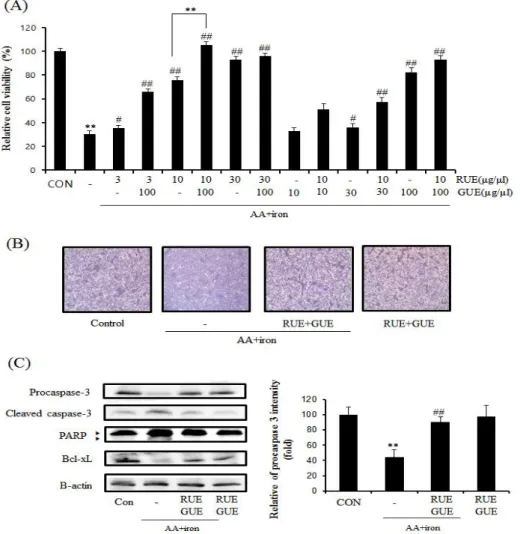
와 의 와 에 의해 유도된 세포자 1. RUE GUE AA iron
멸사에 대한 보호 효과
세포에서 와 에 의해 유도된 세포 HepG2 AA iron
자멸사에서 RUE (3, 10, 30 g/ml)μ 와 GUE (10, 의 효과적인 농도 선정을 위해 30, 100 μg/ml)
를 실시하였다 그 결과
MTT assay . , RUE 10 μ
단독 처리군 보다 와
g/ml 10 g/ml RUEμ 100 μ 의 병용 처치에서 효과적인 세포 보호 효 g/ml GUE
과를 보였다(Figure 1A). 따라서 추후 실험에서는 ,
와 를 병용 처치하
10 g/ml RUEμ 100 g/ml GUEμ
여 진행하였다. AA와 iron에 의해 유도된 산화적 스트레스에 대한 RUE와 GUE의 세포 보호 효능을
광학현미경으로 관찰하였다. AA와 iron을 처리한 군에서는 세포막 손상이 관찰되었고 10 μg/ml
와 전 처리를 통해 세포막의
RUE 100 g/ml GUE μ
손상이 보호되는 것을 확인하였다(Figure 1B). AA 와 iron에 의해 유도된 세포자멸사 억제 효과를
통해 세포자멸사 관련 immunoblotting analysis
단백질의 발현관찰을 통해 확인하였다. AA와 iron 처리군에서는 procaspase 3, PARP, Bcl-xL의 단 백질 발현이 감소하였고 RUE와 GUE 처리군에서는 세포자멸사 관련 단백질의 감소를 억제하였다(Fig. 1).
Fig. 1. The protective effect of R. undulatum extract (RUE) and G. uralensis extract (GUE) on AA and iron-induced apoptosis of HepG2 cells.
(A)Cell viability. HepG2 cells were incubated with 3 to 30 μg/ml RUE and 10 to 100 μg/ml GUE for 1 h and continuously treated with 30 M AA for 12 h, followed by treatment of 5 μ μ M iron for 2 h. Cell viability of RUE and GUE was assessed using MTT assay. Data represents the mean ± S.E. of three experiments.
(B)Cell Morphology. Light microscopic results show the changes of morphology of cells (magnification 200x).
(C)Immunoblot analysis for the proteins associated with apoptosis. Immunoblot analysis of procaspase 3, poly ADP-ribose polymerase (PARP), B-cell lymphoma-extra large (Bcl-xL) and -actin were performed on the lysates of HepG2 cells. HepG2 cells were incubated with 10 β
g/ml RUE and 100 g/ml GUE for 1 h and continuously treated with 30 M AA for 12 h,
μ μ μ
followed by treatment of 5 μM iron for 2 h. The statistical significance of differences between treatments and the vehicle-treated control (**ρ<0.01) or cells treated with RUE and GUE (#ρ<0.05 or ## ρ<0.01) was determined.
와 가 과 활 2. RUE GUE mitochondrial dysfunction
성산소종에 미치는 영향
와 에 의해 유도된 산화적 스트레스는 미 AA iron
토콘드리의 기능 장애를 통하여 세포자멸사를 유도 한다6). RUE와 GUE의 미토콘드리아 기능보호 효과 를 연구하기 위해 HepG2 세포를 rh123 염색하여
로 분석하였다 외부자극으로 flow cell cytometry .
인해 미토콘드리아 막 투과성의 증가로 rh123은 미 토콘드리아 내로 흡수되어 형광을 발한다21). 그 결 과 AA와 iron 처치군은 미토콘드리아 세포막의 손 상시켜 rh123 형광의 감소를 야기하므로 rh123에
대한 낮은 형광 염색 강도의 세포 수 (RN1 가 대조군에 비해 현저히 증가하였다
fraction) .
와 전 처치는 와 에 의해 증가되
RUE GUE AA iron
는 RN1 fraction을 유의성 있게 감소시켰다(Figure 또한 세포 보호효능에 와 에 의해 유
2A). , AA iron
도된 산화적 스트레스의 억제가 매개되는지 관찰하 기 위하여 DCFH2-DA를 이용한 세포 내 활성산소 종 생성에 미치는 영향을 평가하였다. AA와 iron 처치는 세포 내 활성산소종 생성을 대조군에 비해 배 증가시켰으며 와 전 처치에 의해 활
6 RUE GUE
성산소종의 생성을 유의하게 억제되었다(Fig. 2).
Fig. 2. Effect of RUE and GUE on mitochondrial dysfunction and oxidative stress.
(A)Mitochondrial membrane permeability (MMP). HepG2 cells were treated as descried in Figure 1C and stained with 0.05 μg/ml of rhodamine 123 for 1 h, and measured rhodamine 123 intensity by flow cytometry analysis. RN1 fraction indicated cell population.
(B)RN1 fraction. Data represent the mean ± S.E. of three separate experiments. The statistical significance of differences between treatments and vehicle-treated control (**ρ<0.01) or cells treated with AA and iron (##ρ<0.01) was determined.
(C)Cellular ROS production. Cellular ROS production was measured by dichlorofluorescin (DCF) fluorescence. ROS production was analyzed in cells treated with 10 μg/ml RUE and 100 μ g/ml GUE for 1 h and continuously treated as described above. RUE and GUE treatment decreased AA and iron-induced ROS production.
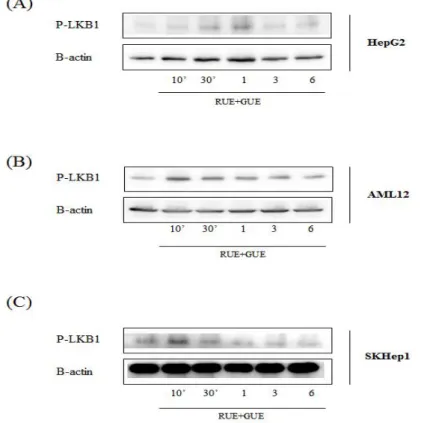
와 가 의 인산화에 미치는 효과 3. RUE GUE LKB-1
와 에 의해 유도된 세포사멸의 보호기전에
AA iron
와 활성화가 매개됨이 보고되었다
AMPK pathway .
인산화를 통하여 활성화된 AMPK는 세포 내 다양한 효소의 활성을 인산화를 통하여 조절하는데, AMPK 의 인산화 상위 kinase인 LKB1이 필요한 것으로 알려져 있다21). 따라서 본 연구에서는 AA와 iron에
의한 산화적 스트레스에 대한 RUE와 GUE의 보호 효과에 LKB1의 활성화가 매개되는지 여부를 알아 보기 위해 immunoblotting analysis를 시행하였 다. RUE 10 g/mlμ 과 GUE 100 g/mlμ 을 10분 ~
시간 동안 처치한 결과 의 인산화가
6 LKB1 HepG2
세포에서는 시간에서 증가하였으며1 , AML12 세포 와 SKHep1 세포에서는 10분에서 증가하였다(Fig. 3).
Fig. 3. The effect of RUE and GUE on the phosphorylation of LKB1.
Immunoblot analysis of p-LKB1 and -actin were performed in lysates of HepG2 (A), AML 12 β (B) and SKHep 1 (C) cells that had been treated with 10 g/ml RUE and 100 g/ml GUE for μ μ the indicated time periods.
와 가
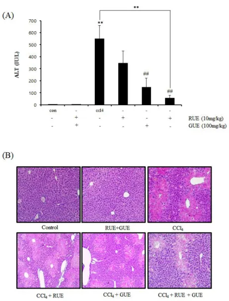
4. RUE GUE CCl4에 의해 유도된 간 손상 모델 에 미치는 영향
동물실험에서 와 가
RUE GUE CCl4로 유도된 급성 간 손상에 대한 간보호에 미치는 영향을 알아보기 위해 혈청 내 ALT를 측정하였다. CCl4투여 군에서 혈청 ALT는 대조군(4.18 IU/L)에 비하여 549.90
로 현저히 증가하였으며 단독
IU/L RUE 10 mg/kg
처치군은 347.36 IU/L, GUE 100 mg/kg 단독 처 치군은 146.90 IU/L, RUE와 GUE 병용 처치군에
서는 56.36 IU/L로 매우 낮은 ALT 수치를 확인하 였다(Figure 4A). RUE와 GUE의 간보호 효과를 조 직학적으로 확인해 보기 위하여 H&E 염색을 진행 하였다. CCl4를 처치하지 않은 정상 대조군은 정상 적인 간의 조직학적 구조를 보였으며 CCl4 투여군 에서는 세포괴사 및 세포질 내에 많은 액포와 중심 정맥 주변으로 세포질의 호산성과 염증세포의 침윤 이 증가되었다. RUE나 GUE의 단독 전처군에 비해
와 병용 처치군은
RUE GUE CCl4 처치군 간 조직에 비해서 완화된 양상을 보였다(Fig. 4).
Fig. 4. The effect of RUE and GUE on CCl4-induced liver toxicity in mice.
(A)Serum level of ALT. Mice were administrated RUE and GUE and then injected CCl4 as described in the Materials and Methods. Serum level of ALT was assayed by semi-automated blood chemistry analyzer. Data represent the mean ± S.E. Statistical significance of differences between treatments and vehicle-treated control (** <0.01) or cells treated with ρ CCl4 (## <0.01) was determined. ρ
(B)Histological analysis. Liver tissue sections were stained with Harris hematoxylin and eosin (magnification 200x).
세포의 대사과정에서 발생되는 활성산소종은 유
전자의 발현 세포증식 조절 등의 매개체로서 작용, 을 한다 그러나 과도한 활성산소종의 생성은 산화. , 적 스트레스를 일으켜 세포의 손상을 유발한다22).
임상적으로도 산화적 스트레스는 염증 노화 종양 , , 등과 같은 질환 및 세포사멸과 연관된다23). 인체에 서 물질대사가 가장 활발한 장기인 간은 대사체들 로 인해 산화적 스트레스의 영향을 많이 받으며 간, 조직 내 과도한 산화적 스트레스는 세포 내의 효소, 신호분자 유전자의 발현을 교란하여 염증과 간섬유,
화를 유발하므로 급 만성간질환의 주요 원인으로 ∙ 알려져 있다24). 그러므로 산화적 스트레스로 인한 , 독성을 억제할 수 있는 물질의 발굴을 통해 간보호 후보소재를 찾고자 하는 연구가 활발히 이루어지고 있다.
대황은 항균 항염 항혈전 등 다양한 효능이 입증, , 된 바 있다9-11, 13). 대황의 성분으로 안트라퀴논계 화합물인 chrysophanol, emodin, aloe-emodin, 및 스틸벤 rhein, physcion, sennoside A, B, C, D, E
계 화합물인 rhaponticin, piceid, deoxyrhaponticin 등 이 알려져 있다25). 특히 대황의 성분인 piceatannol는 의 신호를 통한 의 발현 증가로 항산화
Nrf2 HO-1
효과를 나타내는 것으로 보고되고 있다26). 감초는 혈당과 복부지방 감소, 항염증, 항알러지, 항산화 효과 등 다양한 효능이 보고되었다16-18, 27). 아직까 지 산화적 스트레스에 의해 유도된 간손상에 미치 는 대황과 감초의 병용 효과에 대해 보고 된 바 없 다 따라서 본 연구에서는 대황과 감초의 병용 처치. 가 HepG2세포에서 AA와 iron에 의해 유도된 산화 적 스트레스와 CCl4에 의해 유도된 간경변 모델 실 험동물에 미치는 영향을 규명하였다.
산화적 스트레스를 통해 유도한 세포 독성에 대
한 RUE와 GUE의 보호효능을 MTT assay를 통해 알아보았다 그 결과 . RUE와 GUE는 AA와 iron에 의해 유도된 세포 독성을 농도 의존적으로 억제함 을 확인하였으며, RUE 와 GUE 병용 처치가 각각 의 단독처치보다 더 높은 세포 생존율을 나타냄을 확인하였다 이와 더불어 . RUE와 GUE의 세포 보호 효능을 세포자멸사 관련 단백질인 procaspase 3,
의 발현 변화를
PARP, Bcl-xL immonoblotting
로 확인하였다 와 처치군에서는
analysis . AA iron
단백질의 발현이 감 procaspase 3, PARP, Bcl-xL
소하였고, RUE와 GUE의 전 처치가 이를 유효하게 억제하는 것을 확인하였다. Caspase 3는 세포 내에 서 불활성화 형태인 procaspase 3 상태로 존재하지 만 활성산소종 등의 자극에 반응하여 분해되면서 활성화 된다28). PARP 역시 활성산소종의 자극에 의해 분해되어 활성화 된다29). 또한 세포자멸사 방 어인자의 하나인 Bcl-xL은 미토콘드리아의 외막에 존재하며 cytochrome-c의 유리를 제어하여 세포자 멸사를 억제하는 기능을 담당하는 것으로 알려져 있다30,31). 따라서 RUE와 GUE의 병용 처치가 AA와
에 의해 유도된 세포자멸사에 세포 보호효과를 iron
보인다고 여겨진다 또한. , RUE와 GUE의 병용처치 는 AA와 iron에 의해 유도된 활성산소종 생성을 효 과적으로 억제하였다. 이러한 결과를 종합하면
와 의 병용처치는 산화적 스트레스에 의해 RUE GUE
유발된 간세포의 자멸사에 대해 활성산소종 생성억 제와 항산화 효과를 통해 세포보호작용을 나타내는 것으로 사료된다.
미토콘드리아는 외부 자극에 반응하여 여러 유기
물질에 저장된 에너지를 산화적 인산화 과정을 통 하여 ATP를 생산하는 세포 생존에 관여하는 주요 세포 내 소기관이다 따라서 과도한 산화적 스트레. , 스는 세포 내 활성산소종의 축적을 야기하여 미토, 콘드리아의 막 전위 변화를 통한 기능 장애를 유발 하므로 미토콘드리아의 세포자멸사 회로를 활성화 시킨다32). 본 연구에서 유세포 분석기 분석을 통해 미토콘드리아 막 전위 변화를 측정한 결과, AA와 의 처치는 미토콘드리아 막 전위의 저하를 일 iron
으켰으며, RUE와 GUE 전 처치가 AA와 iron에 의 해 유도된 미토콘드리아 막 전위의 저하를 통계적 으로 유의하게 억제하였다 본 연구를 통해 . RUE와
의 와 으로 유도된 미토콘드리아의 기 GUE AA iron
능 장애에 대한 보호 효과를 확인하였다.
은 종양 억제 인자로 알려져 있으며 세포의 LKB1
에너지 고갈로 인한 세포자멸사에 대한 보호 효능 이 보고되었다33). LKB1은 AMPK upstream kinase 중 하나로 에너지 대사 조절 인자인 AMPK의 활성 화를 촉진시키며, 간에서는 LKB1의 아세틸화가
의 세포 내 위치와 활성에 관여하는 것으로 LKB1
나타났다34). 따라서, immunoblotting analysis를 통해 간세포주인 HepG2 세포, AML12 세포,
세포에서 와 가 의 활성화에
SKHep1 RUE GUE LKB1
미치는 영향을 알아보았다. HepG2 세포에서는 시1 간, AML12 세포와 SKHep1 세포에 10분에서 LKB1 의 활성화를 보였다. 그러므로 RUE와 GUE가
의 활성화를 통한 간세포 보호 효능이 있을 LKB1
것으로 여겨진다. HepG2 세포에서 RUE와 GUE의 산화적 스트레스에 의한 효능을 토대로 CCl4에 의 해 유도된 간경변 마우스 모델에서 RUE와 GUE의 효능을 실험하였다. In vivo 모델에서 사용된 대표 적인 간독성 유발물질인 CCl4는 체내에서 산소와 반응해 간독성을 일으키는 CCl3을 만들며, 이들은
세포막의 인지질을 공격하여 지질의 과산화를 유발 한다 이러한 지질의 과산화는 미토콘드리아나 . DNA, 세포막을 포함하는 주요 세포구조물에 유독한 영향 을 미쳐 간세포의 괴사를 일으킨다 이러한 활성산. 소종 생성 및 세포사멸은 알코올성 및 비알콜성 지 방간염 혈색소증 및 간독성 등의 여러 간질환에 기, 여한다35). RUE와 GUE는 CCl4로 유도한 간독성 모 델에서 혈청학적 지표인 ALT의 증가를 개선하였고, 조직학적 지표인 간세포의 변형 및 염증세포의 침 윤을 억제하였다 이러한 효과는 . RUE와 GUE의 병 용 처치가 각 추출물의 단독 처리보다 효과적으로 나타났다 따라서. , RUE와 GUE는 in vivo 모델에 서 CCl4에 의해 유도된 산화적 스트레스에 의한 간 조직 내 손상 및 이로 인한 염증을 억제를 통해 효 율적인 간보호 효과가 있음을 확인하였다.
본 연구에서는 와 에 의해 산화적 스트레
AA iron
스가 유도된 HepG2 세포를 활용한 in vitro 모델 과 CCl4에 의해 급성 간손상이 유발된 마우스의 in vivo 모델에서 활성산소종에 의한 간독성에 대한 와 의 항산화 및 간보호 효과를 평가하였 RUE GUE
다. In vitro 모델에서는 MTT assay를 통하여 세 포 보호효과 및 immunoblotting analysis로 세포 자멸사 단백질 발현을 관찰함으로써 세포자멸사 억 제 효능을 확인하였다 또한 유세포 분석을 실시하. , 여 미토콘드리아 보호효과를 입증하였고 HepG2 세 포, AML12 세포, SKHep1 세포 세 종류의 간세포 주에서 LKB1의 인산화를 확인하였다. In vivo 모델 에서도 RUE와 GUE의 병용 처치가 혈청 중 ALT 수치를 감소시키고 간조직 내 손상 및 염증을 억제, 하는 것으로 확인되었다.
이러한 결과는 대황과 감초의 병용처치가
in
vitro와 in vivo 모델에서 산화적 손상에 대해 세포 보호 효능과 간보호 효과가 있음을 입증한 것으로 대황과 감초는 항산화 효과와 더불어 간을 보호하 는 유효한 약물 후보이다 따라서 대황과 감초의 병. 용처치 효과의 명확한 세포 보호효과 기전에 대한 지속적인 연구가 필요할 것으로 생각된다.
This work was supported by the National Research Foundation of Korea (NRF) grant funded by the Korea government [MSIP] (No.
2012R1A5A2A42671316) and the NRF grant funded by the Korea government [MSIP] (No.
2014R1A2A2A01007375), and also by the Grant K16830 awarded to Korea Institute of Oriental Medicine (KIOM) from Korea Ministry of Education, Science and Technology (MEST).
1. Halliwell B, Reactive oxygen species in living systems: source, biochemistry, and role in human disease. The American journal of medicine, 1991;91(3):S14-S22.
2. Lee J, Koo N, Min D, Reactive oxygen species, aging, and antioxidative nutraceuticals. Comprehensive reviews in food science and food safety, 2004;3(1):21-33.
3. Choi SH, Kim YW, Kim SG, AMPK-mediated GSK3beta inhibition by isoliquiritigenin contributes to protecting mitochondria against iron-catalyzed oxidative stress. Biochem Pharmacol, 2010;79(9):1352-62.
4. Okuda M, Li K, Beard MR, Showalter LA, Scholle F, Lemon SM, et al., Mitochondrial injury, oxidative stress, and antioxidant gene expression are induced by hepatitis C virus core protein. Gastroenterology, 2002;122(2):
366-75.
5. Kim YW, Kim YM, Yang YM, Kim TH, Hwang SJ, Lee JR, et al., Inhibition of SREBP-1c-mediated hepatic steatosis and oxidative stress by sauchinone, an AMPK- activating lignan in Saururus chinensis.
Free radical biology & medicine, 2010;48(4):
567-78.
6. Ko HL, Jegal KH, Song SY, Kim NE, Kang J, Byun SH, et al., Water Extract of Rosa laevigata Michx. Protects Hepatocytes from Arachidonic Acid and Iron-mediated Oxidative Stress. The Korea Journal of Herbology, 2015;30(6):7-15.
7. Balboa MA, Balsinde J, Oxidative stress and arachidonic acid mobilization. Biochimica et biophysica acta, 2006;1761(4):385-91.
8. Bataller R, Brenner DA, Liver fibrosis. The Journal of clinical investigation, 2005;115(2):
209-18.
9. Park S-M, Lee G-W, Cho Y-H, Effect of Rheum undulatum extract on antioxidant activity and activity of matrix metalloproteinase-1 in human skin fibroblasts. Journal of Life Science, 2008;18(12):1700-4.
10. Matsuda H, Tewtrakul S, M orikaw a T, Yoshikawa M, Anti-allergic activity of stilbenes from Korean rhubarb (Rheum undulatum L.): structure requirements for inhibition of antigen-induced degranulation and their effects on the release of TNF-α and IL-4 in RBL-2H3 cells. Bioorganic &
medicinal chemistry, 2004;12(18):4871-6.
11. Song J-H, Yang T-C, Chang K-W, Han S-K, Yi H-K, Jeon J-G, In vitro anti- cariogenic activity of dichloromethane
fraction fromRheum undulatum L. root. Archives of pharmacal research, 2006;29(6):490-6.
12. Hw ang IY, Jeong CS, A Study on the Antigastritic Effects of Rheum Species Extracts and Their Active Components. Journal of Food Hygiene and Safety, 2013;28(4):330-6.
13. Yang W-K, Sung Y-Y, Cheon M-S, Yoon T-S, Kim H-K, Comparative Study of Extracts from Rhubarb on Anti-Thrombotic and Anti-Platelet Activity. The Korea Journal of Herbology, 2010;25(4):1-6.
14. Dong Gz, Lee Yi, Jeong JH, Zhao HY, Jeon R, Lee HJ, et al., Stilbenoids from
Rheum undulatum Protect Hepatocytes Against Oxidative Stress Through AMPK Activation. Phytotherapy Research, 2015;29(10):
1605-9.
15. Zhang R, Kang KA, Piao MJ, Lee KH, Jang HS, Park MJ, et al., Rhapontigenin from Rheum undulatum protects against oxidative-stress-induced cell damage through antioxidant activity. Journal of Toxicology and Environmental Health, Part A, 2007;70(13):
1155-66.
16. Jeon JH, Park D, Shin S, Kang D-H, Moon S-H, Kim Y-B, Effect of green tea and licorice extracts on cyclophosphamide teratogenicity. Reproductive Toxicology, 2007;24(1):75.
17. Fiore C, Eisenhut M, Krausse R, Ragazzi E, Pellati D, Armanini D, et al., Antiviral effects of Glycyrrhiza species. Phytotherapy research, 2008;22(2):141-8.
18. Dong GZ, Lee YI, Jeong JH, Zhao HY, Jeon R, Lee HJ, et al., Stilbenoids from Rheum undulatum Protect Hepatocytes Against Oxidative Stress Through AMPK Activation. Phytotherapy research : PTR, 2015;29(10):1605-9.
19. Kim YW, Ki SH, Lee JR, Lee SJ, Kim CW, Kim SC, et al., Liquiritigenin, an aglycone of liquiritin in Glycyrrhizae radix, prevents acute liver injuries in rats induced by acetaminophen with or without buthionine sulfoximine. Chemico-biological interactions, 2006;161(2):125-38.
20. Rastogi RP, Singh SP, Häder D-P, Sinha RP, Detection of reactive oxygen species (ROS) by the oxidant-sensing probe 2 , ′ 7 -dichlorodihydrofluorescein ′ diacetate in the cyanobacterium Anabaena variabilis PCC 7937. Biochemical and biophysical research communications, 2010;397(3):603-7.
21. Pereira CF, de Oliveira CR, Oxidative glutamate toxicity involves mitochondrial
dysfunction and perturbation of intracellular Ca 2+ homeostasis. Neuroscience research, 2000;37(3):227-36.
22. Lizcano JM, Göransson O, Toth R, Deak M, Morrice NA, Boudeau J, et al., LKB1 is a master kinase that activates 13 kinases of the AMPK subfamily, including MARK/PAR‐
1. The EMBO journal, 2004;23(4):833-43.
23. Orrenius S, Gogvadze V, Zhivotovsky B, Mitochondrial oxidative stress: implications for cell death. Annu. Rev. Pharmacol.
Toxicol., 2007;47:143-83.
24. Parola M, Robino G, Oxidative stress-related molecules and liver fibrosis. Journal of hepatology, 2001;35(2):297-306.
25. Jang Y-S, Chu VM, Lee K-J, Seo E-Y, Kim D-H, Kang J-S, Effects of the decoction water on the extraction of the bioactive compounds from rhubarb. Analytical Science and Technology, 2011;24(1):38-44.
26. Lee H-H, Park S-A, Almazari I, Kim E-H, Na H-K, Surh Y-J, Piceatannol induces heme oxygenase-1 expression in human mammary epithelial cells through activation of ARE-driven Nrf2 signaling. Archives of biochemistry and biophysics, 2010;501(1):
142-50
27. Yoon T-S, Cheon M-S, Kim S-J, Lee A, Moon B-C, Chun J-M, et al., Evaluation of solvent extraction on the anti-inflammatory efficacy of Glycyrrhiza uralensis. Korean Journal of Medicinal Crop Science, 2010;18(1):
28-33.
28. Cai L, Li W, Wang G, Guo L, Jiang Y, Kang YJ, Hyperglycemia-induced apoptosis in mouse myocardium mitochondrial cytochrome c mediated caspase-3 activation pathway– . Diabetes, 2002;51(6):1938-48.
29. Kang Y-H, Yi M-J, Kim M-J, Park M-T, Bae S, Kang C-M, et al., Caspase-Independent Cell Death by Arsenic Trioxide in Human Cervical Cancer Cells Reactive Oxygen
Species-Mediated Poly (ADP-ribose) Polymerase-1 Activation Signals Apoptosis-Inducing Factor Release from Mitochondria. Cancer research, 2004;64(24):8960-7.
30. Hsu Y-T, Wolter KG, Youle RJ, Cytosol- to-membrane redistribution of Bax and
Bcl-XL during apoptosis. Proceedings of the National Academy of Sciences, 1997;94(8):
3668-72.
31. Kharbanda S, Pandey P, Schofield L, Israels S, Roncinske R, Yoshida K, et al., Role for Bcl-xL as an inhibitor of cytosolic cytochrome C accumulation in DNA damage-induced apoptosis. Proceedings of the National Academy of Sciences, 1997;94(13):
6939-42.
32. Bonnard C, Durand A, Peyrol S, Chanseaume E, Chauvin M-A, Morio B, et al., Mitochondrial dysfunction results from oxidative stress in the skeletal muscle of diet-induced insulin-resistant mice. The Journal of clinical investigation, 2008;118(2): 789-800.
33. Corradetti M N, Inoki K, Bardeesy N, DePinho RA, Guan K-L, Regulation of the TSC pathway by LKB1: evidence of a molecular link between tuberous sclerosis complex and Peutz-Jeghers syndrome. Genes & development, 2004;18(13):1533-8.
34. Suchankova G, Nelson LE, Gerhart-Hines Z, Kelly M, Gauthier M-S, Saha AK, et al., Concurrent regulation of AMP-activated protein kinase and SIRT1 in mammalian cells. Biochemical and biophysical research communications, 2009;378(4):836-41.
35. Bisht S, Khan MA, Bekhit M, Bai H, Cornish T, Mizuma M, et al., A polymeric nanoparticle formulation of curcumin (NanoCurc )™ ameliorates CCl4-induced hepatic injury and fibrosis through reduction of pro-inflammatory cytokines and stellate cell activation. Laboratory investigation, 2011;91(9):1383-95.