209
and Safety
Available online at http://www.foodhygiene.or.kr
Brazilin Inhibits of TPA-induced MMP-9 Expression Via the Suppression of NF-κB Activation in MCF-7 Human Breast Carcinoma Cells
Byeong-Soo Kim*
Department of Companion and Laboratory Animal Science, Kongju National University, Yesan 340-702, Korea (Received June 24, 2010/Revised July 7, 2010/Accepted July 11, 2010)
ABSTRACT
- Metastasis is the primary cause of from breast cancer mortality. Cell migration and invasion play important roles in neoplastic metastasis. Matrix metalloproteinase-9 (MMP-9), which degrades the extracellular matrix (ECM), plays an important role in cancer cell invasion. NF-κB is transcription factor important in the regula- tion of MMP-9, as the promoter of MMP-9 gene contains binding sites for NF-κB. Brazilin, an active component of sappan wood (Caesalpinia sappan), decreases TPA-induced MMP-9 expression and invasion in MCF-7 cells. Also, brazilin suppressed NF-κB activation in TPA-treated MCF-7 cells. Taken together, we demonstrated that the inhibi- tion of TPA-induced MMP-9 expression and cell invasion by brazilin is mediated by the suppression of the NF-κB pathway in MCF-7 cells. This result suggest brazilin provide a potential therapeutic approach for the treatment of breast cancer.Key words: Brazilin , MMP-9, metastasis, invasion, NF-κB, MCF-7
Introduction
Brazilin (7, 11b-dihydrobenz[b]indeno[1,2-d]pyran-3,6a,9, 10 (6H)-tetrol), the major component of Caesalpinia sappan L., is a natural red pigment. It is usually used for histological staining1,2). Previous studies, brazilin was demonstrated various biological effects including anti-hepatotoxicity3), anti-platelet activity4), inhibition of protein kinase C and insulin receptor kinase5), induction of immunological tolerance6,7), and anti-inflammatory activities8-10). Furthermore, recent studies revealed that brazilin involve regulation of transcription factors NF-κB and AP-19).
Breast cancer is one of the leading causes of malignancy related death in woman11). Most of breast cancer death cases are caused by distant metastasis from the primary tumor site.
Despite successful treatment of the primary malignancy, relapse and subsequent metastatic spread can still occur at other areas of the body through the bloodstream or lymphatic channels. This leads to local, regional or distant metastasis, including bone, lung, liver, kidney, thyroid and brain12). Invasion and metastasis are the fundamental properties and major causes of morbidity and mortality in breast cancer patients. These processes require degradation of the extracellular matrix
(ECM), which provides biochemical and mechanical barriers to cell movement in cancer cells13). ECM consists of type IV collagen, laminin, heparan sulfate proteoglycan, nidogen and fibronectin14). ECM degradation requires extracellular proteinases, of which the matrix metalloproteinases (MMPs) have been shown to play a critical role in breast cancer.
MMPs are a family of zinc- and calcium-dependent endo- peptidases, consisting of four subclasses based on substrate, including collagenases,gelatinases, stromelysins and membrane- associated MMPs. MMP-9 is reported to be a key enzyme for degrading type IV collagen, which is a major component of the basement membrane. Elevated MMP-9 levels are functionally linked to elevated metastasis in many tumors, including brain15), prostate16), bladder17) and breast18,19). Several mechanisms regu- late MMP-9 activity, including gene transcription, proenzyme activation, and endogenous inhibitors such as tissue inhibitors of metalloproteinases (TIMPs). A variety of stimuli, including cytokines and phorbol ester, can stimulate MMP-9 synthesis and secretion during various pathological processes such as tumor invasion, atherosclerosis, inflammation, and rheumatoid arthritis. MMP-2, on the other hand, is usually expressed constitutively19,20). Cytokine and TPA treatments can induce MMP-9 expression via activation of transcription factors such as nuclear factor-κB (NF-κB). NF-κB is transcription factor important in the regulation of MMP-9, as the promoter of MMP-9 gene contains binding sites for NF-κB21). Therefore, it was hypothesized that brazilin may have anticancer properties inhibiting cell invasion.
In this study, brazilin was examined for its potential on
*Correspondence to: Byeong-Soo Kim, Department of Companion and Laboratory Animal Science, Kongju National University, Yesan 340-702, Korea
Tel: 82-41-330-1523; Fax: 82-41-330-1529 Email : [email protected]
TPA induced cell invasion and MMP-9 expression in MCF- 7 cells with related molecular mechanisms. Our results demonstrated that brazilin suppresses TPA-induced MMP-9 expression by blocking the NF-κB signaling pathways and the suppression of MMP-9 expression is correlated well with its inhibition of cell invasion.
Materials and Methods
Cells and Material
MCF-7 cells were obtained from the American Type Culture Collection (Manassas, VA). Cells were cultured in DMEM supplemented with 10% fetal bovine serum (FBS) and 1% antibiotics at 37oC in a 5% CO2 incubator. Brazilin was purchased from Sigma (St. Louis, MO. USA) and dissolved in dimethyl sulfoxide (DMSO). 12-O-tetradecanoyl- phorbol-13-acetate (TPA) and 3-(4,5-dimethyl-thiazol-2-yl)- 2,5-diphenyltetrazolium bromide (MTT), DMSO and anti-β- actin antibodies were obtained from Sigma (St. Louis, MO.
USA). Primary antibodies for MMP-9, p50, p65, PCNA, and Horseradish peroxidase (HRP)-conjugated IgG were pur- chased from Santa Cruz Biotechnology (SantaCruz, CA.
USA). [c32-P]ATP was obtained from Amersham (Bucking- hamshire, UK). High glucose-containing Dulbecco’s modified Eagle’s medium (DMEM), FBS and phosphate-buffered saline (PBS) were obtained from Gibco-BRL (Gaithersburg, ME. USA).
Determination of cell viability
The effect of brazilin on MCF-7 cell viability was deter- mined using an MTT assay. Briefly, cells were seeded to 3× l04cells/well, allowed to attach. After 24 h, cells were treated with various brazilin concentrations (10, 20, 50, 100 and 200µM). After incubation for 24 h, cells were washed with PBS, MTT (0.5 mg/ml PBS) was added to each well and the plates were incubated at 37oC for 30 min. Formazan crystals were dissolved with DMSO (100µl/well) and detected at 570 nm using a microplate reader (Model 3550, BIO- RAD, Richmond, CA, USA).
Western blot analysis
MCF-7 cells (5× 10⁵) were pre-treated with brazilin (20µM and 50 µM) for 1 h and then incubated with TPA for 24 h. Cells were lysed with ice-cold M-PER® Mammalian Protein Extraction Reagent (Pierce Biotechnology, Rockford, IL). The protein concentration in the lysate was determined using the Bradford method22). Samples (20µg) were separated by SDS-PAGE with 10% acrylamide, and transferred to hybond™-PVDF membranes using a Western blot apparatus.
The PVDF membranes were blocked with 2% bovine serum albumin or 5% skim milk, and then incubated overnight with
1µg/ml primary antibodies for MMP-9, β-actin, p50, p65, or PCNA. HRP-conjugated IgG was used as a secondary antibody. Protein expression levels were determined by signal analysis using an image analyzer (Fuji-Film, Japan).
Gelatin Zymography assay
Conditioned media were collected after 24 h stimulation, mixed with non-reducing sample buffer, and electrophoresed in a polyacrylamide gel containing 0.1% (w/v) gelatin. The gel was washed at room temperature for 30 min with 2.5%
Triton X-100 solution, and subsequently incubated at 37oC for 16 h in 5 mM CaCl2, 0.02% Brij, and 50 mM Tris–HCl (pH 7.5). The gel was stained for 30 min with 0.25% (w/v) Coomassie brilliant blue in 40% (v/v) methanol/7% (v/v) acetic acid and photographed on an image analyzer (Fuji-Film, Japan). Proteolysis was imaged as a white zone in a dark blue field. Densitometric analysis was performed using Multi Gauge Image Analysis software (Fuji-Film, Japan).
Quantitative real-time PCR assay
Total RNA was extracted from cells using a FastPureTM RNA Kit (TaKaRa, Shiga, Japan). The RNA concentration and purity were determined by absorbance at 260/280 nm.
cDNA was synthesized from 1µg total RNA using a PrimeScriptTM RT reagent Kit (TaKaRa, Shiga, Japan). MMP- 9 and GAPDH mRNA expression were determined by real- time PCR using the ABI PRISM 7900 sequence detection system and SYBR® Green (Applied Biosystems, Foster City, CA, USA). The primers were: MMP-9 (NM 004994) sense, CCTGGAGACCTGAGAACCAATCT; antisense, CCACC- CGAGTGTAACCATAGC and GAPDH (NM 002046) sense, ATGGAAATCCCATCACCATCTT; antisense, CGCCCCA- CTTGATTTTGG. To control for variation in mRNA concen- tration, all results were normalized to the housekeeping gene, GAPDH. Relative quantitation was performed using the comparative ∆∆Ct method according to the manufacturer’s instructions.
Preparation of nuclear extract
MCF-7 cells (2× 10⁶) were treated with brazilin in the presence or absence of TPA for 4 h. Cells were immediately washed twice, scraped into 1.5 ml of ice-cold PBS (pH 7.5), and pelleted at 1,500 g for 3 min. Cytoplasmic and nuclear extracts were prepared from cells using the NE-PER® Nuclear and Cytoplasmic Extraction Reagents (Pierce Biotechnology, Rockford, IL).
Electrophoretic mobility shift assay (EMSA)
NF-κB activation was assayed with a gel mobility shift assay using nuclear extracts. An oligonucleotide containing the κ-chain (κB, 5’-CCGGTTAACAGAGGGGGCTTTC-
CGAG-3’) binding site was synthesized and used as a probe for the gel retardation assay. The two complementary strands were annealed and labeled with [α-32P]dCTP. Labeled oligonucleotides (10,000 cpm), 10µg of nuclear extracts, and binding buffer (10 mM Tris-HCl, pH 7.6, 500 mM KCl, 10 mM EDTA, 50% glycerol, 100 ng poly (dI·dC), 1 mM DTT) were then incubated for 30 min at room temperature in a final volume of 20µl. The reaction mixtures were analyzed by electrophoresis on 4% polyacrylamide gels in 0.5X Tris- borate buffer. The gels were dried and examined by autora- diography. Specific binding was controlled by competition with a 50-fold excess of cold κB oligonucleotide.
Invasion assay
The invasion assay was carried out in 24-well chambers (8µm pore size) coated with 20 µl matrigel diluted DMEM.
The matrigel coating was re-hydrated in 0.5 ml DMEM for 30 min immediately before the experiments. 2× 10⁵cells were added to the upper chamber with chemoattractant in the bottom well. Conditioned medium (0.5 ml) was added to the lower compartment of the invasion chamber. The chambers were incubated for 24 h. After incubation, cells on the upper side of the chamber were removed using cotton swabs, and cells that had migrated were fixed and stained with Toluidine blue solution. Invading cells were counted in five random areas of the membrane using a light microscope. Analyzed data are the means ± SE from three individual experiments performed in triplicate.
Statistical analysis. Statistical data analysis was performed using ANOVA and Duncan’s test. Differences with a p <
0.05 were considered statistically significant.
Results
Effect of brazilin on of MCF-7 cell viability
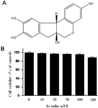
We first investigated the cytotoxicity of brazilin (Fig. 1A) on MCF-7 cells, the cells were seeded into 96-well culture plates at a density of 1× 10⁵cells/well. Effect of brazilin on MCF-7 cellular toxicity was analyzed using the MTT assay. Treatment of MCF-7 cells with indicated concentrations of brazilin for 24 h did not show significant cytotoxicity (Fig. 1B).
Brazilin inhibits TPA-induced MMP-9 expression in MCF-7 cells
To investigate the effect of brazilin on TPA-induced MMP- 9 expression, we performed western blot analysis, real-time PCR, and zymography in MCF-7 cells. Western blot analysis revealed that brazilin treatment in MCF-7 cells blocked the up-regulation of TPA-induced MMP-9 protein expression (Fig. 2A). Real-time PCR revealed that TPA increased the MMP-9 level in MCF-7 cells, and that brazilin blocked TPA-
induced MMP-9 up-regulation in a dose-dependent manner (Fig. 2B). To determine the effect on TPA-induced MMP-9 secretion by brazilin, we performed zymography; MCF-7 cell treatment with TPA resulted in increased MMP-9 secretion.
Brazilin significantly diminished TPA-induced MMP-9 secretion (Fig. 2C). These results indicate that brazilin is a potent inhibitor of TPA-induced MMP-9 expression in MCF-7 cells.
Brazilin suppresses TPA-induced NF-κB DNA binding activity in MCF-7 cells
To determine the mechanism of brazilin -mediated inhibition of MMP-9 expression, the effect of brazilin on TPA-induced activation of NF-κB was evaluated using EMSA. As shown in Fig. 3, TPA increased substantially NF-κB binding activity.
Pre-treatment with brazilin inhibited TPA-stimulated NF-κB binding activity. Brazilin itself had no effect on NF-κB binding activity. These results suggest that brazilin specifically blocks NF-κB activation in MCF-7 cells.
Brazilin inhibits TPA-induced invasion of MCF-7cells It has been reported that the up-regulation of MMP-9 expression contributes to invasion of cancer cells23,24). An in vitro invasion assay was used to investigate the inhibitory effects of brazilin on the invasive potency of breast carcinoma
Fig. 1. Structure of brazilin and effects of brazilin on cell viability of MCF-7 cell. Chemical structure of brazilin (A). To cytotoxicity test of brazilin, Cells were cultured in 96-well plates until 70%
confluence and various concentrations of brazilin were added to cells for 24h. MTT assay was used to detect the viability of the cells (B). The optical density value of control was regarded as 100%.
Data points are the mean ± SE of three independent experiments.
MCF-7 cells. TPA treatment increased MCF-7 cell invasion when compared with untreated control cells, as determined by a Matrigel invasion assay. Brazilin inhibited the TPA- induced MCF-7 cell invasion (Fig. 4).
Discussion
Breast cancer is the main cause of death from cancer in women globally. It is the second leading cause of woman death in the United States. Metastasis is the primary cause of breast cancer mortality. Tumor metastasis is a multistep process by which a subset of individual cancers disseminate from a primary tumor to distant secondary organs or tissues in a complex process that includes cell proliferation, ECM degradation, cell migration, and tumor growth at metastatic sites19,25). MMP-9 has been regarded as major critical molecules in processing tumor invasion and metastasis. MMP-9 activation has been shown to be especially associated with tumor progression and invasion, including mammary tumors27). In previous reports, inflammatory cytokines, growth factors, or phorbol esters stimulated MMP-9 by activating different intracellular-signaling pathways in breast cancer cells28-30).
The inhibitory effects on expression are important for the development of a therapeutic experimental model of tumor metastasis.
NF-κB is transcription factor important in regulating MMP- 9, as the MMP-9 gene promoter contains binding sites for transcription factors21). NF-κB comprises a family of inducible transcription factors which regulate host inflammatory and immune responses31). Diverse signal transduction cascades Fig. 3. Brazilin blocks TPA-induced NF-κB activation in MCF-7 cells. Cells were treated with brazilin in the presence of TPA.
Following 3 h of incubation, nuclear extracts were prepared as described in Materials and Methods. NF-κB DNA binding was analyzed by electrophoretic mobility shift analysis as described in Materials and Methods.
Fig. 4. Effect of brazilin on TPA-induced Matrigel invasion in MCF-7 cells. Cells were seeded onto the upper chamber and drugs placed in the well. After a 24 h incubation, cells on the bottom of filter were fixed, stained, and counted. 1, Control; 2, TPA alone (100 nM); 3, TPA with brazilin (50µM). Each value represents the mean ± SEM of three independent experiments. *p < 0.01 vs. TPA.
Fig. 2. Brazilin inhibits TPA-induced MMP-9 expression in MCF- 7 cells. MCF-7 cells in monolayer were treated with the indicated brazilin concentrations in the presence of TPA for 24 h. Cell lysates were analyzed by Western blot with anti-MMP-9. The blot was reprobed with anti-β-actin to confirm equal loading (A). Con- ditioned medium was prepared and used for gelatin zymography (B). MMP-9 mRNA levels were analyzed by real-time PCR and GAPDH was used as an internal control (C). Each value repre- sents the mean ± SEM of three independent experiments. *p < 0.01 vs. TPA.
mediate NF-κB pathway stimulation31). NF-κB elements are centrally involved in MMP-9 gene induction by TPA25,32). Our results show that brazilin inhibited MMP-9 expression by suppression of NF-κB in breast carcinoma cells.
In this study, we have for the first time provided evidence that brazilin inhibits TPA-induced expression of MMP-9 in breast carcinoma cells. Furthermore, we also demonstrated the molecular mechanism of brazilin. Our results also showed that brazilin blocked TPA-induced NF-κB activation. Recent studies have clearly implicated multiple targets of brazilin action. Recent several studies focused on heme oxygenase-1 activation by brazilin, but many studies have been growing interest in effect of anti-inflammatory by brazilin9,10,33). Bae et al. demonstrated that NF-κB is molecular target in brazilin treated cells9). These results indicate that brazilin can affect proliferation signals and apoptotic signals via modulation of NF-κB activity.
In conclusion, our results have demonstrated that brazilin is a potent inhibitor of TPA-induced MMP-9 expression and strongly blocks the ability of NF-κB signalling pathway in breast carcinoma cells. This is the first study showing brazilin suppress TPA-stimulated cancer cell invasion by inhibiting MMP-9 expression. Thus, brazilin may be a potential candidate to prevent breast tumor invasion and metastasis in vivo.
Acknowledgements
This work was supported by the National Research Founda- tion of Korea (NRF) grant funded by the Korea Government (MEST) (No. 2010-0001355).
References
1. Puchtler, H. and Sweat, F.: On the mechanism of sequence iron-hematein stains. Histochemie, 4, 197-208 (1964).
2. Puchtler, H., Meloan, S.N. and Waldrop, F.S.: Application of current chemical concepts to metal-hematein and -brazilein stains. Histochemistry, 85, 353-364 (1986).
3. Moon, C.K., Park, K.S., Kim, S.G., Won, H.S. and Chung, J.H.: Brazilin protects cultured rat hepatocytes from BrCCl3- induced toxicity. Drug Chem Toxicol, 15, 81-91 (1992).
4. Hwang, G.S., Kim, J.Y., Chang, T.S., Jeon, S.D., So, D.S. and Moon, C.K.: Effects of Brazilin on the phospholipase A2 activity and changes of intracellular free calcium concentra- tion in rat platelets. Arch Pharm Res, 21, 774-778 (1998).
5. Kim, S.G., Kim, Y.M., Khil, L.Y., Jeon, S.D., So, D.S., Moon, C.H. and Moon, C.K.: Brazilin inhibits activities of protein kinase C and insulin receptor serine kinase in rat liver. Arch Pharm Res, 21, 140-146 (1998).
6. Choi, S.Y. and Moon, C.K.: Effects of brazilin on the altered immune functions in the early phase of halothane intoxica- tion of C57BL/6 mice. Planta Med, 63, 400-404 (1997).
7. Mok, M.S., Jeon, S.D., Yang, K.M., So, D.S. and Moon, C.K.: Effects of Brazilin on induction of immunological tol- erance by sheep red blood cells in C57BL/6 female mice.
Arch Pharm Res, 21, 769-773 (1998).
8. Hikino, H., Taguchi, T., Fujimura, H. and Hiramatsu, Y.: Anti- inflammatory principles of Caesalpinia sappan wood and of Haematoxylon campechianum wood. Planta Med, 31, 214-220 (1977).
9. Bae, I.K., Min, H.Y., Han, A.R., Seo, E.K. and Lee, S.K.: Sup- pression of lipopolysaccharide-induced expression of inducible nitric oxide synthase by brazilin in RAW 264.7 macrophage cells. Eur J Pharmacol, 513, 237-242 (2005).
10. Sasaki, Y., Hosokawa, T., Nagai, M. and Nagumo, S.: In vitro study for inhibition of NO production about constituents of Sappan Lignum. Biol Pharm Bull, 30, 193-196 (2007).
11. Jemal, A., Murray, T., Ward, E., Samuels, A., Tiwari, R.C., Ghafoor, A., Feuer, E.J. and Thun, M.J.: Cancer statistics, 2005. CA Cancer J Clin, 55, 10-30 (2005).
12. Friedel, G., Pastorino, U., Ginsberg, R.J., Goldstraw, P., Johnston, M., Pass, H., Putnam, J.B. and Toomes, H.: Results of lung metastasectomy from breast cancer: prognostic criteria on the basis of 467 cases of the International Registry of Lung Metastases. Eur J Cardiothorac Surg, 22, 335-344 (2002).
13. Woessner, J.F., Jr.: Matrix metalloproteinases and their inhib- itors in connective tissue remodeling. FASEB J, 5, 2145-2154 (1991).
14. Nakajima, M., Welch, D.R., Belloni, P.N. and Nicolson, G.L.: Degradation of basement membrane type IV collagen and lung subendothelial matrix by rat mammary adenocarci- noma cell clones of differing metastatic potentials. Cancer Res, 47, 4869-4876 (1987).
15. Saito, N., Hatori, T., Murata, N., Zhang, Z.A., Ishikawa, F., Nonaka, H., Iwabuchi, S. and Samejima, H.: A double three- step theory of brain metastasis in mice: the role of the pia mater and matrix metalloproteinases. Neuropathol Appl Neu- robiol, 33, 288-298 (2007).
16. Castellano, G., Malaponte, G., Mazzarino, M.C., Figini, M., Marchese, F., Gangemi, P., Travali, S., Stivala, F., Canevari, S. and Libra, M.: Activation of the osteopontin/matrix metal- loproteinase-9 pathway correlates with prostate cancer pro- gression. Clin Cancer Res, 14, 7470-7480 (2008).
17. Kanayama, H.: Matrix metalloproteinases and bladder can- cer. J Med Invest, 48, 31-43 (2001).
18. Lin, C.W., Hou, W.C., Shen, S.C., Juan, S.H., Ko, C.H., Wang, L.M. and Chen, Y.C.: Quercetin inhibition of tumor invasion via suppressing PKC delta/ERK/AP-1-dependent matrix met- alloproteinase-9 activation in breast carcinoma cells. Carcino- genesis, 29, 1807-1815 (2008).
19. Lee, S.O., Jeong, Y.J., Kim, M., Kim, C.H. and Lee, I.S.:
Suppression of PMA-induced tumor cell invasion by capilla- risin via the inhibition of NF-kappaB-dependent MMP-9 expres- sion. Biochem Biophys Res Commun, 366, 1019-1024 (2008).
20. Nabeshima, K., Inoue, T., Shimao, Y. and Sameshima, T.:
Matrix metalloproteinases in tumor invasion: role for cell migration. Pathol Int, 52, 255-264 (2002).
21. Eberhardt, W., Huwiler, A., Beck, K.F., Walpen, S. and Pfeilschifter, J.: Amplification of IL-1 beta-induced matrix met- alloproteinase-9 expression by superoxide in rat glomerular mesangial cells is mediated by increased activities of NF- kappa B and activating protein-1 and involves activation of the mitogen-activated protein kinase pathways. J Immunol, 165, 5788-5797 (2000).
22. Bradford, M.M.: A rapid and sensitive method for the quanti- tation of microgram quantities of protein utilizing the principle of protein-dye binding. Anal Biochem, 72, 248-254 (1976).
23. Chambers, A.F. and Matrisian, L.M.: Changing views of the role of matrix metalloproteinases in metastasis. J Natl Can- cer Inst, 89, 1260-1270 (1997).
24. Stetler-Stevenson, W.G., Hewitt, R. and Corcoran, M.: Matrix metalloproteinases and tumor invasion: from correlation and causality to the clinic. Semin Cancer Biol, 7, 147-154 (1996).
25. Chung, T.W., Moon, S.K., Chang, Y.C., Ko, J.H., Lee, Y.C., Cho, G., Kim, S.H., Kim, J.G. and Kim, C.H.: Novel and therapeutic effect of caffeic acid and caffeic acid phenyl ester on hepatocar- cinoma cells: complete regression of hepatoma growth and metastasis by dual mechanism. FASEB J, 18, 1670-1681 (2004).
26. Scorilas, A., Karameris, A., Arnogiannaki, N., Ardavanis, A., Bassilopoulos, P., Trangas, T. and Talieri, M.: Overex- pression of matrix-metalloproteinase-9 in human breast can- cer: a potential favourable indicator in node-negative patients.
Br J Cancer, 84, 1488-1496 (2001).
27. Cho, H.J., Kang, J.H., Kwak, J.Y., Lee, T.S., Lee, I.S., Park,
N.G., Nakajima, H., Magae, J. and Chang, Y.C.: Ascofuranone suppresses PMA-mediated matrix metalloproteinase-9 gene activation through the Ras/Raf/MEK/ERK- and Ap1-depen- dent mechanisms. Carcinogenesis, 28, 1104-1110 (2007).
28. Kajanne, R., Miettinen, P., Mehlem, A., Leivonen, S.K., Bir- rer, M., Foschi, M., Kahari, V.M. and Leppa, S.: EGF-R reg- ulates MMP function in fibroblasts through MAPK and AP- 1 pathways. J Cell Physiol, 212, 489-497 (2007).
29. Srivastava, A.K., Qin, X., Wedhas, N., Arnush, M., Linkhart, T.A., Chadwick, R.B. and Kumar, A.: Tumor necrosis factor- alpha augments matrix metalloproteinase-9 production in skel- etal muscle cells through the activation of transforming growth factor-beta-activated kinase 1 (TAK1)-dependent sig- naling pathway. J Biol Chem, 282, 35113-35124 (2007).
30. Yamamoto, Y. and Gaynor, R.B.: Therapeutic potential of inhibition of the NF-kappaB pathway in the treatment of inflammation and cancer. J Clin Invest, 107, 135-142 (2001).
31. Hong, S., Park, K.K., Magae, J., Ando, K., Lee, T.S., Kwon, T.K., Kwak, J.Y., Kim, C.H. and Chang, Y.C.: Ascochlorin inhibits matrix metalloproteinase-9 expression by suppress- ing activator protein-1-mediated gene expression through the ERK1/2 signaling pathway: inhibitory effects of ascochlorin on the invasion of renal carcinoma cells. J Biol Chem, 280, 25202-25209 (2005).
32. Washiyama, M., Sasaki, Y., Hosokawa, T. and Nagumo, S.:
Anti-inflammatory constituents of Sappan Lignum. Biol Pharm Bull, 32, 941-944 (2009).