• 검색 결과가 없습니다.

Fractured Surface Morphology and Mechanical Properties of Ni-Cr Based Alloys with Mo Content for Dental Applications

N/A
N/A
Protected

Academic year: 2021

Share "Fractured Surface Morphology and Mechanical Properties of Ni-Cr Based Alloys with Mo Content for Dental Applications"

Copied!
5
0
0

로드 중.... (전체 텍스트 보기)

전체 글

(1)

한국표면공학회지 J. Korean Inst. Surf. Eng.

Vol. 49, No. 3, 2016.

http://dx.doi.org/10.5695/JKISE.2016.49.3.260

<연구논문>

ISSN 1225-8024(Print) ISSN 2288-8403(Online)

Fractured Surface Morphology and Mechanical Properties of Ni-Cr Based Alloys with Mo Content for Dental Applications

Hyun-Soo Kim

a

, Mee-Kyoung Son

b

, Han-Cheol Choe

c*

a

College of Dentistry, Chosun University, Gwangju 61452, Korea

b

Department of Prosthodontics, College of Dentistry, Chosun University, Gwangju 61452, Korea

c

Department of Dental Materials, Research Center of Nano-Interface Activation for Biomaterials, Chosun University, Gwangju 61452, Korea

(Received June 23, 2016 ; accepted June 29, 2016)

Abstract

In this study, fractured surface morphology and mechanical properties of Ni-Cr-Mo alloys with various contents of Mo for dental material use have been evaluated by mechanical test. The alloys used were Ni- 13Cr-xMo alloys with Mo contents of 4, 6, 8, and 10 wt.%, prepared by using a vacuum arc-melting furnace.

Ni-13Cr-xMo alloys were used for mechanical test without heat treatment. The phase and microstructure of alloys using an X-ray diffraction (XRD) and optical microscopy (OM) were evaluated. To examine the mechanical properties of alloys according to microstructure changes, the tensile test and the hardness test were carried out using tensile tester. To understand the mechanism of Mo addition to Ni-Cr alloy on mechanical property, the morphology and fractured surfaces of alloys were investigated by field-emission scanning electron microscope (FE-SEM). As a result, 79Ni-13Cr-8Mo alloy was verified that the tensile strength and the hardness were better than others. Varying Mo content, the changes of microstructures of alloys were identified by OM and SEM and that of 79Ni-13Cr-8Mo alloy was proved fabricated well. Microstructures of alloys were changed depending on Mo content ratio. It has been observed that 8% alloy had the most suitable mechanical property for dental alloy.

Keywords : Ni-Cr-Mo alloy, Molybdenum, Mechanical properties, Dental materials

1. Introduction

For many years, clinicians have used high noble- content alloys for dental materials. These alloys possess compatible biological properties and good corrosion resistance and physical strength [1]. However, their dental applications became restricted because of the increasing cost of gold and following the global financial crisis. Consequently, non-precious material alloys, such as nickel–chromium (Ni–Cr), are now commonly used for the dental materials [2]. As stated above, the essential properties of a dental cast

alloys are biocompatibility and mechanical strength and corrosion resistance. Among several alloys, Ni based alloys were widely used in dental skeletal structures and implant fixtures and prosthodontic restoration. The advantages of these alloys are low cost, acceptable mechanical and tribological properties and matching thermal expansion coefficient with the ceramics of metal-ceramic restorations [3-4]. In commercial Ni based alloys, the compositions of Cr and Mo usually range from 11% to 25% and from 0 to 10% (mass fraction), respectively [5]. In this study, the alloys used were Ni-13Cr-xMo, and Ni-13Cr-xMo alloys with Mo contents of 4, 6, 8, and 10 wt.%

The commercial nickel-base alloys are very multipurpose and relatively inexpensive. They possess a good mechanical properties and high corrosion resis- tance [6]. The remarkable properties of Ni–Cr alloys are due to their complex composition. Basically,

* Corresponding Author :Han-Cheol Choe

Department of Dental Materials, Research Center of Nano- Interface Activation for Biomaterials, Chosun University Tel : +82-62-230-6896 ; Fax: +82-62-230-6885

E-mail : hcchoe@chosun.ac.kr

(2)

these alloys are composed of Ni (68 - 80%) and Cr (11.9 - 26.3%), but alloying with other elements is required to ensure the achievement of mechanical and corrosion resistance, castability, and porcelain bonding. Iron, aluminum, molybdenum, silicon, copper, titanium, gallium, cobalt, tin, magnesium, manganese, and beryllium are added to Ni–Cr alloys in the range of 0.1 - 14% [7]. Especially, resistance of crevice corrosion is increased by addition of Mo in Ni-Cr base alloys[8]. The objective of this study was to investigate the microstructure, mechanical properties, and fractured surface status with tensile test specimens of Ni–Cr-xMo dental alloys.

2. Experiment Details

The alloys used were Ni-13Cr-xMo, and Ni-13Cr- xMo alloys with Mo contents of 4, 6, 8, and 10 wt.%

were melted by using a vacuum arc-melting furnace.

Ni-13Cr-xMo alloys were used for mechanical test without heat treatment. The obtained cylindrical bars were 10 mm in diameter and 3 mm in height (The bars were prepared in disc form by cutting, 10mm in diameter and 3mm in thickness). They were polished in grits of emery paper (#200 to 1500) and finally wet polished using with aluminium dioxide to produce mirror surfaces. After polishing process, the surfaces were washed with propyl alcohol to prevent any pollution. The polished samples were dip into an acid mixture of reagent (20 mL water, 6 mL hydrofluoric acid, 6 mL nitric acid) to obtain the surface pattern and the reaction was stopped with 70% alcohol. After drying, the microstructural patterns were examined by optical microscope at X50, X100 magnifications and scanning electron microscope (SEM) at X300, X600 magnifications.

X-ray diffraction (XRD) analysis was performed on same disc used for SEM using Cu target with a voltage of 40 kV and a current of 30 mA.

Vickers hardness test were also carried out on the disc specimens with a micro hardness tester. The mean values were obtained by means of 10 indentations with distance 200 μm between each of them with a load of 1000 g for 10 s. Data were analyzed using one-way ANOVA followed by Tukey test. The significance level was set at 5% (α = 0.05).

Smooth plate specimens which were cross sectioning of 0.75 mm × 1.5 mm × 6 mm dimensions for tensile test were prepared. The geometry of tensile test specimens is shown in Fig. 1 The specimen were wet polished using a #1500 emery

paper to obtaining shiny surface. Tensile tests were performed on the specimens using an Instron-type machine with a crosshead speed of 8.33 × 10

−6

m/s in air. Data were analyzed using one-way ANOVA followed by Tukey test. The significance level was set at 5% (α = 0.05).

After tensile test, the fracture surfaces of specimens were examined by scanning electron microscope (SEM) to observe fractured surface patterns of them.

3. Results and Discussion

Ni-Cr-Mo alloy is a nickel based superalloy, a non- precious metallic alloy which can be used at high temperatures, available on the market for medical and dental use. These small quantities of Cr and Mo could alter the surface hardness, bulk composition, microstructure, and composition of passive films over the alloy surface [9-10].

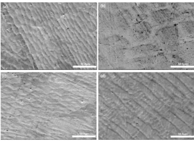
Figure 2 shows the microstructure of surface in Mo ratio 4%, 6%, 8%, and 10% (wt.%), respectively.

Although patterns of surface of alloy are different a little, the surfaces are characterized by a solid- solution arrays in dendritic disposition (primary phase) and interdendritic patterns (second phase) regularly distributed. With ascending Mo ratio from 4% to 10%, dendritic and interdendritic patterns are

Fig. 1. Dimensions of specimen for tensile test.

Fig. 2. Optical micrographs of Ni-Cr-Mo alloys. (a) Ni-

13Cr-4Mo, (b) Ni-13Cr-6Mo, (c) Ni-13Cr-8Mo, (d) Ni-

13Cr-10Mo.

(3)

more dense and 10 Mo% specimen shows grain boundaries. The differences may be come from differences of the solidification mode with changes of amount of alloying components [8]. The pre- dominant structure is dendritic patterns and it is known that the dendrites have concentration of present metals, difference of metals concentration in the interdendritic spaces. This is due to solidification process, where dendrites solidify firstly and have a resistence to different chemical and electrochemical attack [11].

Figure 3 shows the SEM images of Ni-Cr-Mo alloys with two phase microstructures, consisted of primary (dendritic) phase and secondary (interdendritic) phase generally. They also show smaller precipitation

particles on surfaces and the particles are probably constituted of Cr and Mo carbides, mainly Mo. The secondary(interdendritic) phase, an inter-metallic phase, is responsible for the increased-temperature resistance of the material and its strength to creep deformation. The amount of secondary phase depends on the chemical ratio and temperature [12].

Figure 4 shows XRD spectra of alloys. The only strong austenite (γ-Ni) peak can be detected. When Mo adding to Ni-Cr base alloy, the austenite (γ-Ni) is only remain broadly in room temperature and γ’( Ni

2

Cr) is absent. Thus, austenite is only detected in appearance although other intermetallic compounds are existed in very small amount from the isothermal diagram of tertiary Ni-Cr-Mo in room temperature (25

o

C) as shown in Fig. 5.

Fig. 3. FE-SEM morphologies of Ni-Cr-Mo alloys. (a) Ni-13Cr-4Mo, (b) Ni-13Cr-6Mo, (c) Ni-13Cr-8Mo, (d)

Ni-13Cr-10Mo. Fig. 4. XRD peaks of Ni-Cr-Mo alloys.

Fig. 5. Isothermal diagrams of Ni-Cr-Mo ternary alloy.

(4)

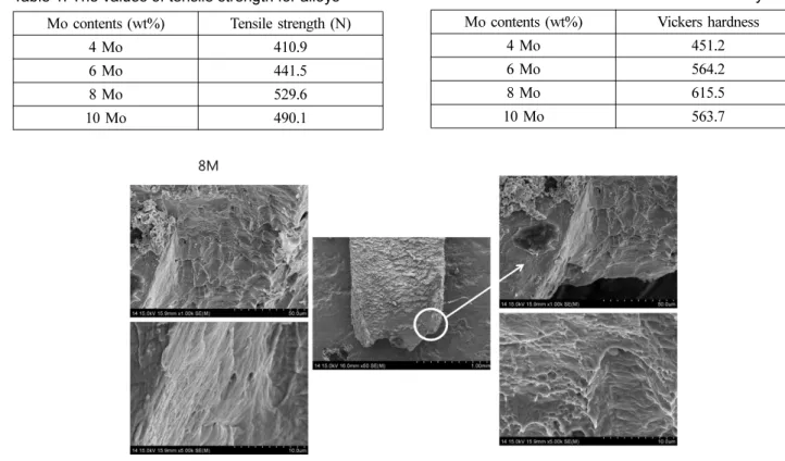
Figure 6 shows the tensile test results of Ni-Cr-Mo alloy with Mo ratio (wt.%) changing from 4% to 10%. The tensile strength is gradually increased to 8% and decreased in 10% as shown in Table 1.

Consequently, the tensile strength is greatest in 8%

Mo ratio among them. In this experiment, tensile tests were only performed but compression test and horizontal shearing test were not tested. Besides tensile strength, it is necessary to examine mechanical properties such as compression and resistance in following research.

Figure 7 shows the results of hardness test of alloys. With Mo ratio (wt.%) changing from 4% to 10%, the Vickers hardness index was gradually

increased to 8% and decreased in 10% as shown Table 2. This results show that the hardness of alloy is also greatest in 8% Mo ratio. The elements (Cr and Mo) are solid-solution strengtheners both in primary and secondary phase as a dendritic brute fusion microstructure with precipitates dispersed in the entire matrix, that can be observed through of a columnar dendritic growth and the eutectoid element formation, which shows extensive solid solubility of chromium in nickel, and as a result of binary alloys hardened [13-15].

Figure 8 shows the fractured surface patterns of alloy. There are dimple patterns which are remar- kable in ductile fracture and faucet patterns which Fig. 6. The results of tensile test for alloys. Fig. 7. The results of hardness test for alloys.

Fig. 8. FE-SEM micrographs of fractured surface of Ni-13Cr-8Mo alloy after tensile test.

Table 1. The values of tensile strength for alloys Mo contents (wt%) Tensile strength (N)

4 Mo 410.9

6 Mo 441.5

8 Mo 529.6

10 Mo 490.1

Table 2. The values of Vickers hardness for alloys Mo contents (wt%) Vickers hardness

4 Mo 451.2

6 Mo 564.2

8 Mo 615.5

10 Mo 563.7

(5)

are noticeable in brittle fracture are also shown in fractured surface. Fracture was probably started in ductile pattern region and ended in brittle fracture region. Among alloys, it seemed that elongation length of fracture fragment is more longer and dimple patterns are shown more in the 8% Mo (wt.%) alloy than others. Viewed in these characters, it is reasonable that the tensile displacement and tensile strengths are greatest in alloy of 8% Mo ratio.

As a limitation of this study, although the dental materials is used in processing forms which is suitable to oral condition, the alloys used in this study were tested in forms of plates and discs . Thus, to obtain practical data, specimens of dental use form should be tested in further investigation.

4. Conclusion

The surfaces of alloys are characterized by a solid- solution arrays in dendritic disposition (primary phase) and interdendritic patterns (second phase) regularly distributed. The tensile strength and Vickers hardness were greatest in alloy of 8% Mo ratio among them. There are dimple patterns which are remarkable in ductile fracture and faucet patterns which are noticeable in brittle fracture are also shown in fractured surface. Fracture was probably started in ductile pattern region and ended in brittle fracture region.

Acknowledgments

This study was supported by research funds from Education and Cultural Foundation of College of Dentistry, Chosun University, 2015.

References

[1] R. P. O’connor, J. R. Mackert, M. L. Myers, E.

E. Parry, Castability, opaque masking, and porcelain bonding of 17 porcelain-fused-to-metal alloys, J.

Prosthet. Dent. 75 (1996) 367-374.

[2] K. F. Leinfelder, An evaluation of casting alloys used for restorative procedures, J. Am. Dent. Ass.

128 (1997) 37-45.

[3] J. C. Wataha, N. L. O'Dell NL, B. B. Singh, M.

Ghazi, G. M. Whitford, P. E. Lockwood, Relating nickel-induced tissue inflammation to nickel release in vivo, J Biomed Mater Res. 58 (2001) 537-544.

[4] R. M. Joias, R. N. Tango, J. E. Junho de Araujo, M. A. Junho de Araujo, Ferreira Anzaloni Saavedra Gde S, T.J. Paes-Junior, E.T. Kimpara, Shear bond strength of a ceramic to Co-Cr alloys, J Prosthet Dent. 99 (2008) 54-59.

[5] C. M. Wylie, R. M. Shelton, G. J. Fleming, A.

J. Davenport, Corrosion of nickel-based dental casting alloys, Dent Mater. 23 (2007) 714-723.

[6] J. Qiu, W. Q. Yu, F. Q. Zhang, R. J. Smales, Y.

L. Zhang, C. H. Lu, Corrosion behaviour and surface analysis of a Co-Cr and two Ni-Cr dental alloys before and after simulated porcelain firing, Eur J Oral Sci. 119 (2011) 93-101.

[7] A. El-Bediwi, M. Saad, A. A. El-Fallal & T. El- Helaly, Electrochemical corrosion behavior, Vickers microhardness, and microstructure of Co–Cr and Ni–Cr dental alloys, Radiation Effects & Defects in Solids 164 (2009) 712-718.

[8] D. C. Choi, Microstructure and mechanical properties of Ni-Cr-Mo based dental cast iron for porcelain- fused-to-metal firing, J. Kor. Foundry Society, 27 (2007) 120-125.

[9] J. D. Bumgardner, L.C. Lucas, Cellular response to metallic ions released from nickel-chromium dental alloys, J. Dent. Res. 74 (1995) 1521-1527.

[10] H. H. Huang, Surface characterization of passive film on NiCr-based dental casting alloys, Biomaterials, 24 (2003) 1575-1582.

[11] F. Faot, W. J. Siva, R. C. Matheus, R. Garcia, A. A. Cury, Microstructural characterization of Ni- Cr-Mo-Ti and Ti-6Al-4V alloys used in prosthetic abutments, Rev. odonto. ciênc. 24 (2009) 401-405.

[12] J. R. Davis, Nickel, cobalt, and their alloys, Materials Park, ASM International (2000).

[13] A. J. Lewis, The mechanism of tensile failure in a nickel-base casting alloy, J. Dent. Res, 56 (1977) 631-634.

[14] A. J. Lewis, The metallography of a nickel base casting alloy, Aust Dent J. 20 (1975) 298-303.

[15] G. R. Baran, The metallurgy of Ni-Cr alloys for

fixed prosthodontics, J. Prosthet. Dent, 50 (1983)

639-650.

수치

Figure 2 shows the microstructure of surface in Mo ratio 4%, 6%, 8%, and 10% (wt.%), respectively.
Figure 4 shows XRD spectra of alloys. The only strong austenite (γ-Ni) peak can be detected
Figure 8 shows the fractured surface patterns of alloy. There are dimple patterns which are  remar-kable in ductile fracture and faucet patterns whichFig

참조

관련 문서

Surface characteristics of hydroxyapatite coated Ti-40Ta-xNb alloys by plasma electrolytic oxidation and sputtering

The mechanical properties of the Ir-based films by the change of the alloy element content show that the hardness value is decreased with Re contents

In this study, we investigated the surface characteristics of hydroxyapatite film on the micro-pore structured Ti-35Ta-xNb alloys by

Control of Mechanical Properties by Proper Heat Treatment in Iron-Carbon

Consequently, Zr-Cu binary alloys have the potential to be used as biomaterials with nullifying magnetic properties for magnetic resonance imaging diagnosis and

Various studies have been performed to improve properties of material, electrical and mechanical properties of railway train according to the increase of the speed in the

however, such scaffolds could have poor mechanical strengths. 19) For these reasons, multi-pore sized scaffolds have been announced to increase the mechanical

: Development of Microstructure and Alteration of Mechanical Properties.. 4.6 The homogeneous nucleation rate as a function of undercooling ∆T. ∆T N is the critical