PGHN
Original Article
Primary Sclerosing Cholangitis with Inflammatory Bowel Disease in Korean Children
Jisun Yoon, Seak Hee Oh, Hyun Jin Kim, Sang Hyoung Park, Byong Duk Ye*, Suk-Kyun Yang*, and Kyung Mo Kim
Department of Pediatrics, Asan Medical Center Children’s Hospital, *Department of Internal Medicine, Asan Medical Center, University of Ulsan College of Medicine, Seoul, Korea
Purpose: Primary sclerosing cholangitis (PSC) is a rare condition that can be associated with inflammatory bowel disease (IBD). The aim of this study was to evaluate PSC and its association with IBD in children.
Methods: We retrospectively enrolled 13 pediatric patients (<18 years) with PSC treated at Asan Medical Center between June 1989 and December 2013. Clinical findings and long-term outcomes were investigated. During the same period, the incidence of PSC among IBD patients was evaluated among 600 Crohn disease (CD) and 210 ulcerative colitis (UC) patients.
Results: All 13 study patients diagnosed with PSC also presented with IBD. Eleven boys and two girls with a median age of 15.0 years old (9.0-17.8 years) were included. The cumulative incidence of PSC for UC was 5.7% (12 of 210) and 0.2% for CD (1 of 600), respectively. PSC occurred during follow-up for IBD for five patients (38.5%) whereas, IBD developed during follow-up for PSC for two patients (15.4%), and was diagnosed during the initial work-up for PSC for 6 patients (46.2%). For the 77.3 month median follow-up period, 9/13 patients (69.2%), neither the clinical symptoms nor blood test results worsened. Two cases (15.4%) developed liver cirrhosis and underwent liver transplantation. Among 13 PSC patients with IBD, two (15.4%) developed colorectal cancer, and no one developed cholangiocarcinoma.
Conclusion: All patients with PSC in this study had associated IBD. The incidence of PSC was not rare compared to reports in adults. PSC should be considered during the management of IBD and vice versa in children.
Key Words: Sclerosing cholangitis, Inflammatory bowel diseases, Ulcerative colitis, Crohn disease, Colorectal neoplasms, Liver transplantation
Received:September 8, 2015, Revised:October 27, 2015, Accepted:November 14, 2015
Corresponding author: Kyung Mo Kim, Department of Pediatrics, Asan Medical Center Children’s Hospital, University of Ulsan College of Medicine, 88 Olympic-ro 43-gil, Songpa-gu, Seoul 05505, Korea. Tel: +82-2-3010-3380, Fax: +82-2-473-3725, E-mail: [email protected] Copyright ⓒ 2015 by The Korean Society of Pediatric Gastroenterology, Hepatology and Nutrition
This is an openaccess article distributed under the terms of the Creative Commons Attribution NonCommercial License (http://creativecommons.org/licenses/by-nc/4.0/) which permits unrestricted noncommercial use, distribution, and reproduction in any medium, provided the original work is properly cited.
INTRODUCTION
Primary sclerosing cholangitis (PSC) is a chronic hepatobiliary disease with findings that include a beaded pattern of dilatation, stricture, and occlusion in the intrahepatic and extrahepatic bile ducts on cholangiography [1-3]. The clinical progress of pa- tients with PSC is variable. In some cases, after un- dergoing biliary liver cirrhosis due to the fibrosis and stricture of the intrahepatic and extrahepatic bile ducts, as well as portal hypertension, and hepatic failure, patients with PSC may require liver trans- plantation [1,4,5]. In addition, PSC is closely related to the onset of cholangiocarcinoma or colon cancer, thereby requiring regular follow-up of the patient’s clinical progress [6-8].
Cases of comorbid PSC and inflammatory bowel disease (IBD) and the comorbidity levels in adults are variable, as reported in various countries. In a study conducted with adult American PSC patients in 1989, IBD comorbidity was 71% [7]. In a cohort study conducted with 604 PSC patients between 1970 and 1998 in Sweden, IBD comorbidity was 80%
[6]. According to a report in 2011 in Japan, PSC and IBD comorbidity in adults was 68.9%, indicating that comorbidity is also high in Asia [9]. Cases of PSC and IBD comorbidity reported in children are rare com- pared to those reported in adults. Kaplan et al. [10]
reported the PSC prevalence rate among adults as 1.11 cases per 100,000 person-years, but the PSC prevalence rate among children was only 0.23 cases per 100,000 person-years. However, in a 2001 study that examined PSC and IBD comorbidity in children, the comorbidity of IBD among patients <18 years of age was high, at 83% [11].
Among the studies reported in South Korea to date—although cases of children diagnosed with PSC have been reported [12-14]—no study has yet looked at the process of diagnosing children with PSC, at IBD comorbidity, or at treatment and clinical progress. The present study analyzes the character- istics of pediatric patients diagnosed with PSC at the Asan Medical Center, whether or not the condition was associated with IBD. The study analyzed clinical
and histological characteristics, clinical progress and treatment, and liver cirrhosis and colon cancer in- cidence rates during the follow-up period.
MATERIALS AND METHODS
Subjects
This study was a retrospective case series of 13 pa- tients with PSC under 18 years of age diagnosed at the Asan Medical Center (Seoul, Korea) between June 1989 and December 2013. The cumulative in- cidence of PSC among IBD patients was calculated according to the number of IBD patients who had been diagnosed with ulcerative colitis (UC; n=210) or Crohn disease (CD; n=600) in the same period.
The study was approved by the institutional review board of the Asan Medical Center.
Diagnosis of PSC
Patients were diagnosed with PSC according to a laboratory test, liver histology, endoscopic retro- grade cholangiopancreatography (ERCP) findings, and magnetic resonance cholangiopancreatography (MRCP) findings. PSC was suspected in those cases where the alkaline phosphatase (ALP) or γ-gluta- myl transferase (GGT) increased to more than 50%
above the normal values for their age groups [15].
Liver histology was defined by the presence/absence of bile duct damage, onion-skinned periductal fib- rosis, inflammation, portal edema or fibrosis, ducto- penia, ductular proliferation, or cholestasis [1]. With regard to imaging characteristics, patients were di- agnosed with PSC when the ERCP or MRCP in- dicated multifocal strictures, focal dilatation, or beading of the biliary tree [16]. Patients whose bio- chemical and histological tests indicated PSC but whose imaging tests showed normal biliary tree findings were diagnosed with small duct PSC [17].
Patients with sclerosing cholangitis due to secondary causes such as surgery, trauma, cancer, or infection were excluded from the study. A complete blood count, serum aspartate aminotransferase (AST), ala- nine aminotransferase (ALT), ALP, GGT, total and direct bilirubin, albumin, prothrombin and activated
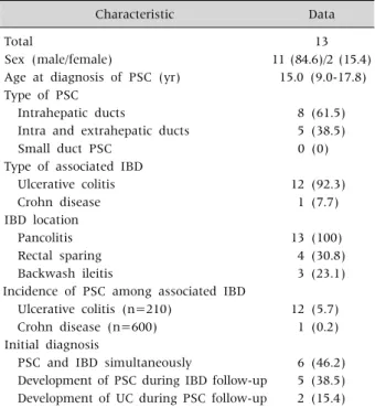
Table 1. Study Patient Characteristics
Characteristic Data
Total 13
Sex (male/female) 11 (84.6)/2 (15.4)
Age at diagnosis of PSC (yr) 15.0 (9.0-17.8) Type of PSC
Intrahepatic ducts 8 (61.5)
Intra and extrahepatic ducts 5 (38.5)
Small duct PSC 0 (0)
Type of associated IBD
Ulcerative colitis 12 (92.3)
Crohn disease 1 (7.7)
IBD location
Pancolitis 13 (100)
Rectal sparing 4 (30.8)
Backwash ileitis 3 (23.1)
Incidence of PSC among associated IBD
Ulcerative colitis (n=210) 12 (5.7)
Crohn disease (n=600) 1 (0.2)
Initial diagnosis
PSC and IBD simultaneously 6 (46.2) Development of PSC during IBD follow-up 5 (38.5) Development of UC during PSC follow-up 2 (15.4) Data are expressed as number only, number (%), or median (range).
PSC: primary sclerosing cholangitis, IBD: inflammatory bowel disease, UC: ulcerative colitis.
partial thromboplastin time, IgG, antinuclear anti- body, anti-smooth muscle antibody, and antineu- trophil cytoplasmic antibody (ANCA) tests were con- ducted at the time of diagnosis through blood tests.
Special blood tests and urinalyses were carried out to exclude other immunological diseases such as acute viral hepatitis, and Wilson’s disease. In cases where patients were diagnosed at other hospitals and transferred to our center, the results of biochemical, histological, and imaging tests at other hospitals were reviewed again at the study center. In cases where PSC was suspected in patients who were al- ready being followed due to UC or CD, an ERCP or MRCP was carried out.
Diagnosis of IBD
Once a patient was diagnosed with PSC, a colono- scopy was conducted for IBD screening both in cases where the patient complained of gastrointestinal symptoms and in cases where there were no symp- toms. A definitive IBD diagnosis was made according to the patient’s medical history, physical examina- tion, and laboratory, radiologic, endoscopic, and his- tologic criteria described elsewhere [18].
Follow-up and treatment
During the median follow-up period of 77.3 months, the patients visited the outpatient clinic pe- riodically to enable their AST, ALT, ALP, GGT, and bi- lirubin values to be checked. All patients diagnosed with PSC took ursodeoxycholic acid (UDCA) conti- nuously. Patients diagnosed with IBD took sulfasa- lazine or 5-aminosalicylates (5-ASA), and steroids, immunosuppressants, or infliximab were admini- stered when any of the patients showed acute ex- acerbation.
RESULTS
Patient population and clinical presentation Among the 13 patients that participated in the present study, 11 were male and 2 were female. Their median age when diagnosed was 15.0 years (range, 9.0-17.8 years). All 13 study patients diagnosed with
PSC also presented with IBD. Twelve (92.3%) had UC, and one (7.7%) had CD as the first presentation, and PSC was diagnosed during follow-up for five pa- tients (38.5%); whereas, for two patients (15.4%), PSC was the first presentation and IBD was diag- nosed later during follow-up. For six patients (46.2%), PSC presented first, and IBD was diagnosed subsequently at the time of their PSC diagnosis dur- ing their initial work-up for PSC (Table 1).
Incidence of PSC in IBD patients
All 13 patients suffered from IBD. Twelve patients associated with UC and one patient associated with CD. Therefore, the cumulative incidence rates of co- morbid PSC were 5.7% for UC (12 of 210) and 0.2%
for CD (1 of 600), respectively.
Clinical features at diagnosis
Common clinical symptoms at the time of diag- nosis were jaundice, itching, and abdominal pain in
Fig. 1. (A) Endoscopic retrograde cholangiopancreatography (ERCP) showing focal dilatation and narrowing of the intrahepatic and extrahepatic bile ducts. (B) ERCP showing focal dilatation and narrowing of the intrahepatic bile ducts. (C) Magnetic resonance cholangiopancreatography revealing irregular dilatation and narrowing and beaded appearance of the intrahepatic bile duct.
Table 2.Clinical Presentation and Laboratory Data at Diagnosis Signs and symptoms at presentation Data
Jaundice 4 (30.8)
Pruritus 4 (30.8)
Abdominal pain 4 (30.8)
Asymptomatic 4 (30.8)
Hepatomegaly 2 (15.4)
Laboratory data
AST (U/L) 144.9±215.8
ALT (U/L) 88.8±112.5
Total bilirubin (mg/dL) 5.1±7.7 Direct bilirubin (mg/dL) 4.1±7.7
GGT (U/L) 138.0±105.3
ALP (U/L) 286.5±219.7
Data are expressed as number (%) or mean±standard deviation.
AST: aspartate aminotransferase, ALT: alanine aminotransferase, GGT: γ-glutamyl transferase, ALP: alkaline phosphatase.
four cases (30.8%), as indicated in Table 2. In the lab- oratory results, 53.8% (n=7) of the study patients showed increased AST and ALT levels, with values of 144.9±215.8 U/L (normal range, 15-40 U/L) and 88.8±112.5 U/L (normal range, 5-45 U/L), respec- tively. Their total and direct bilirubin levels were 5.1±7.7 mg/dL and 4.1±7.7 mg/dL, respectively.
GGT was measured in 12 patients, and all patients showed elevated GGT values over a range of 138.0±
105.3 U/L (normal range, 5-25 U/L). When compared to normal values by age group (the normal range in a 10- to 15-year-old male is 116-483 U/L and in an
age-matched female is 93-386 U/L; in a 16- to 19- year-old male is 58-237 U/L and in an age-matched female is 45-116 U/L), two children (15.4%) showed increased ALP with values in the range of 287.5±219.7 U/L. The ratio of patients with total bi- lirubin in the normal range was 38.5% (n=5) in tests conducted when the ERCP or MRCP results in- dicated PSC.
Cholangiographic features
Nine patients (69.2%) underwent an ERCP, three (23.1%) underwent both an ERCP and a MRCP, and one (7.7%) underwent a MRCP alone. All 13 patients showed multifocal strictures, focal dilatation, or beading of the biliary tree on cholangiography, as ex- pected in a PSC diagnosis (Fig. 1).
Among biliary involvements identified through cholangiography, cases with abnormal findings in the intrahepatic duct were the most frequent, affect- ing eight patients (61.5%). Involvement of both in- trahepatic and extrahepatic ducts was evident in five cases (38.5%). Results indicating a PSC diagnosis were identified in all 13 patients undergoing an ERCP and a MRCP. In the present study, therefore, no pa- tient was diagnosed with small duct PSC (Table 1).
Histological features
In the present study series, two patients (15.4%)
Fig. 2. Microscopic findings showing onion-skin type periductal fibrosis and mild lymphocytic infiltration around the inter- lobular bile ducts (H&E stain, ×100).
underwent a liver biopsy. According to the biopsy re- sults, both patients showed onion-skinned periduc- tal fibrosis and inflammation, portal fibrosis, ducto- penia, ductular proliferation, and cholestasis con- ditions—criteria matching a PSC diagnosis (Fig. 2).
Treatment, clinical outcomes, and long-term survival of PSC patients with IBD
During 77.3 months of median follow-up (range, 25.5-285.2 months), all patients began to take UDCA and 5-ASA (n=13). Some also took systemic steroids (n=6) depending on their IBD activity. During the most recent follow-up period, a remission was main- tained in 11 patients (84.6%).
Among the 13 study patients, 2 (15.4%) under- went liver transplantation. The first patient was si- multaneously diagnosed with the intrahepatic type of PSC and UC at 15 years of age. The diseases pro- gressed into liver cirrhosis at 17 years of age, and the patient underwent a liver transplantation four years later at 19 years of age. The other patient was diag- nosed with PSC at 17 years of age while being fol- lowed-up after an UC diagnosis. This patient under- went a liver transplantation four years later at 21 years of age. We have continued to follow this pa-
tient, so far without any particular problem. As for UC progress, both are taking 5-ASA and have main- tained their remission states. No one developed chol- angiocarcinoma in our study.
Colon cancer developed in two male patients (15.4%). One was diagnosed with UC, PSC, and co- lon cancer at the ages of 10, 26, and 30 years old.
Following a proctocolectomy, the patient is disease- free during 3 years of follow-up. The other patient was diagnosed with UC, PSC, and colon cancer at the ages of 15, 16 and 28 years old with mortality.
During the long-term follow-up of the other pa- tients, nine patients neither underwent liver trans- plantation nor were diagnosed with colon cancer.
One was first diagnosed with both the intrahepatic and extrahepatic types of PSC, and then with UC at 14 years of age. This patient died during the ambula- tory follow-up period for unknown reasons. We con- tinue to follow the remaining eight patients (61.5%) as outpatients while they are being continuously treated.
In conclusion, a Kaplan Meier survival analysis was carried out, based on the clinical progress of the patients with both PSC and IBD (n=13), who were followed up for long periods of time. On the basis of follow-up for mean 12.1 years (median, 11.4 years), the incidence ratio of major medical complications such as liver transplantation, cancer, and death was 0.38 (5/13), and the mean time of occurrence was 14.0 years.
DISCUSSION
The present study introduces the largest number of cases of children with PSC-IBD in a domestic sin- gle-center study. Major outcomes in our current study series included analysis of both the natural course of the disease and IBD comorbidity, and the incidence of major medical complications such as liv- er transplantation, colon cancer and death through long-term follow-up of patients under 18 years of age when diagnosed with PSC. Because all the cases with complications involved patients with PSC-UC, our present findings demonstrated that the relevant
facts were recognized and the need for periodic fol- low-up was clarified.
With regard to other studies conducted with PSC-IBD patients, when 47 patients with PSC were analyzed through a retrospective medical record re- view, the mean age at the time of diagnosis was 11 years, the IBD comorbidity was 59%, the rate of pan- colitis findings through colonoscopy was 24%, and the rate of rectal sparing findings was 24% [11]. In another retrospective medical record review study conducted with 52 PSC patients, IBD comorbidity was high, at 84%. Of the relevant patients, 89% were diagnosed with UC and 11% were diagnosed with CD, indicating that PSC-UC was predominant. Dur- ing follow-up, 80% of colonoscopy findings indicated pancolitis; whereas, 26.7% showed rectal sparing [11]. A large cohort study conducted with 1,649 IBD patients in the early 2000s reported PSC incidence rates five years after IBD diagnosis as 0.5% (95% con- fidence interval [CI] 0.2-13%) in CD and 2.7% (95%
CI 1.5-4.8%) in UC, indicating that UC was more of- ten associated with PSC than CD [19].
Among the current study patients with PSC, IBD comorbidity was 100% and UC was predominant, with a comorbidity rate of 92.3%. The colonoscopy findings for all patients indicated pancolitis, and 30.8% showed rectal sparing conditions. This sug- gests higher IBD prevalence rates than other reports, although the rates from colonoscopy findings are considered similar to those of other reports. In addi- tion, during the same period, among IBD patients under 18 years of age and followed at this center, PSC occurred in 5.7% of UC patients and 0.2% of CD pa- tients, indicating that PSC comorbidity was higher among UC patients.
Our current patients were diagnosed with PSC when their serum ALP or GGT increased to at least 50% above the normal maximum values for their age group and where the ERCP or MRCP results in- dicated multifocal strictures, focal dilatation, or the beading of the biliary tree [15]. All of the patients in- cluded in this study also satisfied the criteria for a PSC diagnosis because their GGT in biochemical tests increased to at least 50% above the normal max-
imum values for their age group. However, only 15.4% of these cases showed an increased ALP. In a long-term (20-year) cohort study carried out by Feldstein et al. [4] to observe the long-term prog- nosis of patients with PSC, 25% of the patients diag- nosed with PSC through cholangiography had nor- mal ALP values. Because serum ALP values identify total ALP values, this suggests that when hepatic ALP isoenzyme values are measured in children, the values may be higher than the normal standard value. This variation is present because children have large personal differences depending on growth and display large ranges of normal standard values. In addition, the standard values vary with age. As a re- sult, GGT has been suggested to be a more accurate indicator when diagnosing PSC in children [4].
Pursuant to the diagnostic recommendations of the American College of Gastroenterology (ACG) [1], a liver biopsy was not conducted when a patient met the criteria for a PSC diagnosis as a result of ERCP or MRCP findings, but was considered in cases where small duct PSC was suspected. However, all 13 pa- tients had findings that matched a PSC diagnosis.
According to the results of long-term follow-up of PSC-IBD patients in our present study, 15.4% under- went liver transplantation due to hepatobiliary cirr- hosis. In the 2003 annual report of Studies of Pediatric Liver Transplantation (SPLIT) in the United States, the ratio of child cases where patients underwent liv- er transplantation due to PSC was 3.5% [20]. In 2009, a study by Miloh et al. [15] showed that 19% of pa- tients with PSC underwent liver transplantation due to repetitive cholangitis or uncompensated liver cir- rhosis, and the incidence rate in that study was high- er than previous reports. However, the authors of that study stated that there might have been more subjects in their hospital than in other hospitals be- cause that institution specialized in liver treatment.
To date, no treatment that can prevent the pro- gression of liver cirrhosis has been discovered. How- ever, since many complications of end-stage liver dis- ease can be effectively treated, PSC should be diag- nosed early, and the use of hepatotoxic drugs should be avoided. Liver cirrhosis should also be diagnosed
early so that complications can be actively treated, and biliary tract cancer screening tests should also be conducted periodically [5].
Colon cancer incidence rates in PSC-UC patients are reported to be higher than in patients with UC only. The cumulative incidence rates of colon cancer in PSC-UC patients reported in Sweden were 9% at 10 years, 31% at 20 years, and 50% at 30 years; where- as, rates in UC patients were 2%, 5%, and 10%, re- spectively, indicating significant differences between the two conditions [21]. This was also the case in a meta-analysis reported in 2002 in the United States (odds ratio, 4.79; 95% CI, 3.58-6.41) [19]. As a result, adult PSC-UC patients are currently recommended to undergo a colonoscopy every year [7,22,23]. Al- though the occurrence of IBD in pediatric patients is not an independent risk factor for colon cancer, the need for colon cancer screening tests in children has also been emphasized recently. The cumulative rate of occurrence of colon cancer among child patients diagnosed with extensive UC before 15 years of age is 40%, and disease durations are more important than the chronological ages of IBD pediatric patients [23].
Although there are currently no guidelines for peri- odic examinations for the early diagnosis of colon cancer in children with UC and PSC, colon cancer clearly should be diagnosed and treated early. Colon cancer occurred in 15.4% of PSC-IBD children in the present study, all of whom were UC patients. PSC co- morbidity accounted for 5.7% of patients receiving follow-up due to UC in the same period.
A limitation of the present study stems from the fact that the results are technical reports prepared by a single tertiary institution. They cannot, therefore, be statistically verified and there are difficulties in generalizing the prevalence rate and progress of PSC-IBD in children in South Korea. However, these findings are important in that no previous study has reported 13 cases of children with PSC-IBD and be- cause they included long-term follow-up results.
In conclusion, PSC in children is often associated with IBD. UC comorbidity is particularly high. There- fore, if a patient shows increases in serum GGT or ALP, or where findings in ERCP or MRCP tests in-
dicate PSC, the patient should be evaluated for possi- ble IBD. In the case of patients receiving follow-up due to IBD, the existence of PSC should be checked by serological tests conducted during the follow-up, and the results should be evaluated. Finally, the fre- quent occurrence of liver cirrhosis and colon cancer in PSC-IBD patients should be recognized and such conditions should be intensively monitored.
REFERENCES
1. Lindor KD, Kowdley KV, Harrison ME; American College of Gastroenterology. ACG Clinical Guideline:
primary sclerosing cholangitis. Am J Gastroenterol 2015;110:646-59; quiz 660.
2. Vergani D, Mieli-Vergani G. Autoimmune hepatitis and PSC connection. Clin Liver Dis 2008;12:187-202, x.
3. Schrumpf E, Boberg KM, Karlsen TH. Primary scleros- ing cholangitis - the Norwegian experience. Scand J Gastroenterol 2015;50:781-96.
4. Feldstein AE, Perrault J, El-Youssif M, Lindor KD, Freese DK, Angulo P. Primary sclerosing cholangitis in children: a long-term follow-up study. Hepatology 2003;38:210-7.
5. Deneau M, Jensen MK, Holmen J, Williams MS, Book LS, Guthery SL. Primary sclerosing cholangitis, auto- immune hepatitis, and overlap in Utah children: epi- demiology and natural history. Hepatology 2013;58:
1392-400.
6. Lindström L, Lapidus A, Ost A, Bergquist A. Increased risk of colorectal cancer and dysplasia in patients with Crohn's colitis and primary sclerosing cholangitis. Dis Colon Rectum 2011;54:1392-7.
7. Broomé U, Bergquist A. Primary sclerosing cholangitis, inflammatory bowel disease, and colon cancer. Semin Liver Dis 2006;26:31-41.
8. Kornfeld D, Ekbom A, Ihre T. Is there an excess risk for colorectal cancer in patients with ulcerative colitis and concomitant primary sclerosing cholangitis? A pop- ulation based study. Gut 1997;41:522-5.
9. Sano H, Nakazawa T, Ando T, Hayashi K, Naitoh I, Okumura F, et al. Clinical characteristics of inflam- matory bowel disease associated with primary scleros- ing cholangitis. J Hepatobiliary Pancreat Sci 2011;
18:154-61.
10. Kaplan GG, Laupland KB, Butzner D, Urbanski SJ, Lee SS. The burden of large and small duct primary scleros- ing cholangitis in adults and children: a popula- tion-based analysis. Am J Gastroenterol 2007;102:
1042-9.
11. Faubion WA Jr, Loftus EV, Sandborn WJ, Freese DK, Perrault J. Pediatric "PSC-IBD": a descriptive report of associated inflammatory bowel disease among pedia- tric patients with psc. J Pediatr Gastroenterol Nutr 2001;33:296-300.
12. Jung JA, Kwak IK, Lee HR, Jang SH, Kim KM, Yoo ES.
A case of overlap syndrome with auoimmune hepatitis and cholangiopathy in a child. Korean J Pediatr Gast- roenterol Nutr 2006;9:269-75.
13. Kim JS, Choe YH, Kim CJ, Kim WS, Kim IO, Seo JK.
A case of primary sclerosing cholangitis with ulcerative colitis. J Korean Pediatr Soc 1996;39:1448-54.
14. Hong J, Song MK, Ko JS, Kang GH, Kim WS, Seo JK.
Autoimmune hepatitis-primary sclerosing cholangitis overlap syndrome in a 10-year-old girl with ulcerative colitis. Korean J Pediatr 2009;52:504-7.
15. Miloh T, Arnon R, Shneider B, Suchy F, Kerkar N. A ret- rospective single-center review of primary sclerosing cholangitis in children. Clin Gastroenterol Hepatol 2009;7:239-45.
16. MacCarty RL, LaRusso NF, Wiesner RH, Ludwig J.
Primary sclerosing cholangitis: findings on cholangiog- raphy and pancreatography. Radiology 1983;149:39- 44.
17. Björnsson E, Boberg KM, Cullen S, Fleming K, Clausen OP, Fausa O, et al. Patients with small duct primary sclerosing cholangitis have a favourable long term prognosis. Gut 2002;51:731-5.
18. Levine A, Griffiths A, Markowitz J, Wilson DC, Turner D, Russell RK, et al. Pediatric modification of the Montreal classification for inflammatory bowel dis- ease: the Paris classification. Inflamm Bowel Dis 2011;17:1314-21.
19. Soetikno RM, Lin OS, Heidenreich PA, Young HS, Blackstone MO. Increased risk of colorectal neoplasia in patients with primary sclerosing cholangitis and ul- cerative colitis: a meta-analysis. Gastrointest Endosc 2002;56:48-54.
20. McDiarmid SV, Anand R; SPLIT Research Group.
Studies of Pediatric Liver Transplantation (SPLIT): a summary of the 2003 Annual Report. Clin Transpl 2003:119-30.
21. Broomé U, Löfberg R, Veress B, Eriksson LS. Primary sclerosing cholangitis and ulcerative colitis: evidence for increased neoplastic potential. Hepatology 1995;
22:1404-8.
22. Khaderi SA, Sussman NL. Screening for malignancy in primary sclerosing cholangitis (PSC). Curr Gastroen- terol Rep 2015;17:17.
23. Farraye FA, Odze RD, Eaden J, Itzkowitz SH, McCabe RP, Dassopoulos T, et al; AGA Institute Medical Posi- tion Panel on Diagnosis and Management of Colorectal Neoplasia in Inflammatory Bowel Disease. AGA medi- cal position statement on the diagnosis and manage- ment of colorectal neoplasia in inflammatory bowel disease. Gastroenterology 2010;138:738-45.