대한임상병리사회지
:제 19 권 제
1호
1987혈청 단백 과
LD H isoenzyme측정 시
agarose
gel법 과
celluloseacetate법 과의 비 교
연세의료원 임상병리과 박 영숙, 김 태숙
The Comparison of Beckman Paragon
Agarose Gel Technique to Cellulose Acetate Technique in Serum Protein and LD H Isoenzyme Fractiona tion
Y oung Sook Pàrk, Tae Sook Kirn
Yonsei University, College 01 Medicine, Severance Hosþital
= Abstract=
Electrophoresis for protein fractionation has been a valuable seperation and quantitation technique in a clinical laboratory. A newly automated agarose electrophoresis system (Paragon@) supplied by Beckman Co. has been introduced to our hospital. We analyzed serum protein electrophoresis and LDH isoenzyme fractionation by using a conventional cellulose acetate electrophoresis system paralleling with this new agarose electrophoresis system.
And the results were as follows ;
1 . The correlations of serum electrophoresis between a conventional cellulose acetate system and a agarose gel electrophoresis system are good in the major fractionation (y = 0.72, 0.73). But, the relations of LDH a'1, a'2, and
β
are not so good (y= -0.48, 0.59, -0.11).2 . The correlations of LDH isoenzyme fractionations are good in LDH1 and LDH2 fractions
(y = 0.98, 0.98) but not so good in fractions of LDH3, 4,5 (y = 0.43, 0.44, 0.33).
3. The results of precisions which expressed as CV (%) are better in agarose system than cellulose acetate system.
From the above results, we confirmed that agarose system is better in the view point of resolution and sensitivity. And the Paragon system is suitable for a clinical laboratory especially in its simple procedure and reproducibility.
I .
서 론 검사실에서 널리 쓰이고 있다.
ZoneEP는 지지체로
cellulose accetate, acrylamide,
agarose를 사용한 1937년 Tiselius가 액체 지지체를 이용한
mov-다. 전기영동의 원리는 전기장에서 하전된 입자들이
ing-boundary electrophoresis
(EP) 를 개발한 후 점 각기 다론 이동성을 갖는다는 사실에 기초하여 차 지지체, 완충액 등이 개선된
zoneEP 법이 임상 protein을 분리한다 1)
-122-
이때 protein의 이동속도는
1)분자의
net elec- tric charge 2)분자의
sizey. shape 3)지 지 체 의 특성
4) temperature of operation등에 의 해 달라 진 다. 본 검 사실에 서 는
serumprotein과
LDHisoenzyme을 측 정 하는데 지 지 체 로서
celluloseacetate를 사용하고 있었으나
agarosegel을 사용하 는 Beckman의
paragon EPsystem 이 새 로 도 입 되 었 다
. Agarose gelEP는
serum protein, he- moglobin variants, LDH isoenzymes, lipopro- tein fractions등을 분석 하는데 널 리 이 용되 고 있 다
. Agarosegel은 protein에 대 한 affinity 가 적 고 자체의 수명도가 있어 gel을
drying후
den-sitometer로 scanning하여 좋은 결과를 얻을 수 있 다
2)특히
paragon EPsystem에 서 는 최 근 개 발된
template application
방법 을 사용하므로
surfaceartifact를 피 할 수 있 다
.지 지 체 로
agarosegel을 사용할 경우 resolution과 sensitivity 가 좋다는 것은 이미 보고되고 있다 3) 본 검사실에서는
Beck-man paragon EPsystem을 사용하여
serum pro - tein,LDH
isoenzyme을 측정하는데 기존의
cel-luloseacetate 법과의 상관성, 정밀도를 비교 실험하였다.
11.
실험재료 및 실험방법
1 .
실험재료
Beckman 1. D . Zone normal protein serum
control로 정밀도 실험하였다. 검사를 의뢰한 10 명 의 환자검체로 상관성 실험을 하였다. 그 중 정상검 체와 비정상 검체를 각기 택하여 정밀도 실험을 하 였으며 이 때
serum proteinelectrophoresis의 경 우 검체는 buffer로
1:2희석하여 사용하였다(단
agarose
gel 을 사용한 경 우)
.2.
시약 및 기구
1 ) Cellulose
acetate법
Gelman sepraphore III, Helena trisbarbital- sodium barbital buffer (pH 8.6 - 9.0) , super z applicator (twin wire applicator), sepra clear II
(40% aqueous N-methyl, pyrrolidone), 5 % acetic acid. Helena ponceau S, LD vis isoenzyme reagent (Helena)
2) Agarose
gel법
Paragon SPE Kit (SPE gel, barbital buffer pH 8.6, Blue stain, template, template blotter, gel
blotter), glacial acetic acid, methanol, paragon LDH isoenzyme Kit (LD gel, barbital buffer pH 8.2, LD substrate containing Lithium L- Lactate, NAD, NBT, PMS), template, tem- plate blotter, gel blotter, press dryer assem- bly, drying blotter)
3.
실험방법
1 ) Cellulose
acetate법
Cellulose acetate
strip을
pH 8.6 barbital buffer에 서 prewetting시 켜
5분 후 과도한 수분을 제 거 하 고
super Zapplicator로 검 체 를 apply 한다
. 350V18분간 전기영동 후
ponceau S염색시약에서 5 분 간 염색 후
5 % acetic acid용액으로 탈색한다.
sepra clear II
용액 에
5분간 담꾀
slide위 에 부착 시킨 후
900Coven에서 말려서 densitometer로
scanning
한다.
LD H isoenzyme
경우에는 위와 같은 방법으로
350V
20분간 전 기 영 동 후
cellulose acetatestrip을
slide
위 에 부 착시 킨 뒤
LD vis isoenzyme reagent를 까하여 다시 strip을 덮고 slide를 덮어 빛을 차단 시 킨 뒤
moisturechamber에 넣 어
3TC incubator에서 말색시켜 densitometer로 600mm에서
scan- ning한다.
2) Agarose
gel법
Agarose
gel을
gelblotter로 blotting한 후
template application
한다
.Template의 slot에 apply한 sample 이
5분 동안
diffusion되 게 한 후
template
blotter로 과도한 혈 청 을 blot한 후
100volt에서 25분간 전기 영동한다. Gel을
acid-alcohol용액에 3 분 담군 뒤 dryer에서 완전히 말린 뒤
bluestain"하여
acetic acid용액 으로 탈색 하고 다시
dry하여
scanning한다 LDH
isoenzyme경우엔
sample apply
후
100volt에 서 20분간 전 기 영 동 후
incubation
box에
LDsubstrate로 포화시 킨
gelblotter를
gel위 에 덮 어
450C'30분간
incubation한 다. Gel을
5 % acetic acid용액에
1분간 담근 뒤
press
dry하고 dryer에서 말린 뒤 scan한다.
111.
실험결과 및 고찰
Serum protein electrophoresis (SPE)
경 우 두 방
법의 상관성 실험결과는
Table1 과 같았다.
LDH isoenzymeelectrophoresis경 우 두 방법 의 상관성
실험결과는
Table2 와 같았다.
SPE경우 주요 분
m ωTable 1. Correlation between Beckman paragon(agarose gel) and cellulose acetate for SPE.
Gamma (Mg/dL) Beckman n
짜 -빼
맨
-B a -
앙
- pu
Alpha 2 (mg/dL) -
Cellulose Beckman Alpha 1 (mg/dL)
Cellulose Beckman Albumin (mg/dL)
Cellulose Beckman
Cellulose I
2 3 4 5 6 7 8 9
0.77 1.52 1.63 1.85 1.87 1.49 0.99 1.19 0.82 l.51
l.96 l.37 l.89 1.81 l.69 l.23 l.42 0.89 0.56
0.69 0.66 0.83 0.83 0.62 0.63 0.68 0.61 Cellulose
0.57 0.66 l.51 0.73 0.66 0.82 0.97 0.79 0.73 0.69
0.98 l.11 l.08 1.26 1.07 0.86 0.88 0.93 0.31
0.34 0.67 0.74 0.71 0.52 0.70 0.59 0.51 0.34
0.36 0.36 0.35 0.38 0.24 0.25 0.23 0.24 0.15
0.17 0.21 0.23 0.35 0.21 0.37 0.24 0.16 2.62
2.70 3.37 3.70 2.74 4.20 3.45 4.29 3.62 3.56
3.28 3.34 4.20 3.36 4.35 2.93 4.26 3.91 No.
r = 0.72 r= -0.48 r=0.59 r= -0.11 r=0.73 Albumin y=0.88x+0.16
Alpha 1 y= -0.13x+0.36 Alpha 2 y = 0.62x + 0.63 Beta y= -0.04x-0.71 Gamma y = 0.99x + 0.02
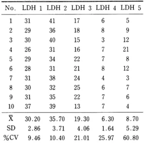
acetate for LDH No. LDH 1 (IU/L) LDH 2 (IU/L) LDH 3 (IU/L) LDH 4 (IUfL) LDH 5 (IU/L)
Cellulose Beckman Cellulose Beckman Cellulose Beckman Cellulose Beckman Cellulose Beckman
34 27 50 58 27 20 10 11 7 11
2 126 146 94 95 40 24 13 15 25 16
3 255 294 195 181 72 28 14 18 18 34
4 123 86 92 72 26 33 7 19 10 47
5 79 112 61 67 41 15 15 8 18 12
6 49 43 49 43 30 28 7 12 8 14
7 63 41 73 69 42 39 13 23 16 34
8 39 35 40 39 22 20 7 8 4 10
9 144 145 76 89 29 14 8 8 12 8
10 115 115 108 99 56 43 18 20 16 37
11 49 45 66 40 25 5 9 5 9
12 40 35 70 68 33 27 6 13 6 13
13 263 300 263 300 72 32 14 13 11 13
14 41 30 41 30 37 24 11 19 11 19
Correlation between Beckman paragon(agarose gel) and cellulose Table 2
LDH 1 Y = 1.18x - 16.26 r = 0.98 LDH 2 y=1.08x-5.49 r=0.98 LDH 3 y=0.23x+ 17.44 r=0.43 LDH 4 y=0.55x+8.14 r=0.44 LDH 5 y=0.70+ 11.41 r=0.33
이 정확하고 resolution 이 좋기 때문에 minor fraction 인
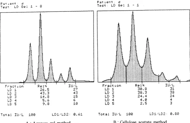
LDH- 4, LDH- 5의 분리가 훨씬 예 민하게 나타남을 알 수 있다. 또한 agarose gel plate상의 성상에서도 각 band간의 분리가 명확하였 다 (Fig.
2).N ormal protein EP
control을 사용하여 두 방법 에 대한 정밀도 실험을 한 결과는
table 3, Table4 와 같다. Agarose gel을 사용한 경우 전 분획에서
celluloseacetate법에서 보다 좋은
%,cv값을 얻
-124-획 인
albumin,y-globulin의 상관성은 높으나
lrb lr2,β-globulin의 상관성이 적은 것으로 미루어
agarosegel 법의 경우 분획의 sensitivity가 증가하
여 minor fraction에 보다 많은 영 향을 미 친 다고 생
각할 수 있 다.
LD H isoenzyme경 우도
majorfraction인 LDH
1, LDH 2 의 상관성은 좋으나
minorfraction의 상관성 이 적 게 나타났다
.특히
LDH isoenzyme경우 Fig.
1에서 보는 바와 같이
Densitogram에 서
celluloseacetate법 보 다
baseline‘
- .J..
Pa "t ~ent. d
T 흔.s: t:
LD Gel :1.Pa': l.:-t',1:. d
T t?드':
L[) GE'l 1 - 8‘」「l?」ξ) -잉
/ ’
--4
4--b 4-
”니
T{
F!''''
글 =τ
;’그 n
I r\ 서
LL‘
£
LD’ 3
L[:i 츄
LD 5
딩 .8 。
LD1 ’LD2
:1.딩딩
Total IU/L
0. 쉰 i
L[;'1/LD‘.-.
:L 띤딩
T ct.:: 1
工꾀 /L
B : Cellulose acetate method
Fig. 1 .Comparison of LDH isoenzyme densitogram between cellulose acetate and agarose gel method
A : Agarose gel method
리
(t)띠
Gl
n
빼
o• p
히
(t)띠
Gm ,
때
a
p.
----.--...._- ...
-
---- --
-‘ ---
. ‘ -
10
@ Fig. 2. LDH isoenzyme electrophoretic patterns obtained by agarose gel electrophoresis
m 냉
9 8
4 5 6 7
BECKM'AN 3
2
@
9
4 5 6 7 8
BECKMAN
3 2
agarose gel
방법 의 정 밀도가 높았다. 이 상의 실험 결과로 미루어
agarosegel을 지지체로 사용할 경우 resolution 이 좋고 재현성이 좋은 결과를 얻을 수 있 다고 할 수 있다 4) 특히
paragon E. P.system은 각 각 의 chemistry 마 다
gel의 구 성 성 분 과
bufferTable 5. Within-run precision (cellulose acetate, SPE, normal patient serum)
였다. 한편 정상과 비정상인 경우의 환자검체를 각 기
1명씩 택하여 정밀도를 실험하여
table 5, table 6의 결 과를 얻 었 다
. Agarosegel을 사용한 결 과는
table 7, table 8과 같다
. SPE경 우 두 방 법 중
agarose gel방법의 정밀도가 높았으며,
LDHisoenzyme에 대해서도 두 방법간의 정밀도 실험한 결 과
(table 9, table 10, table 11, table 12)N o. AlbuminAlpha 1 Alpha 2 Beta 2 Gamma 1.58 1.50 1.62 1.61 1.67 1.69 1. 75 1.82 1.68 1.71 1.90 1.93 0.61 0.64 0.69 0.77 0.62 0.72 0.69 0.57 0.70 0.66 0.86 0.88 0.42
0.56 0.48 0.46 0.45 0.48 0.43 0.35 0.43 0.41 0.50 0.50 0.20 0.27 0.23 0.22 0.18 0.31 0.18 0.19 0.21 0.19 0.19 0.21 4.08
3.92 3.88 3.83 3.98 3.70 3.85 3.97 3.88 3.94 3.44 3.38 1 2 3 4 5 6 7 8 9
m n u
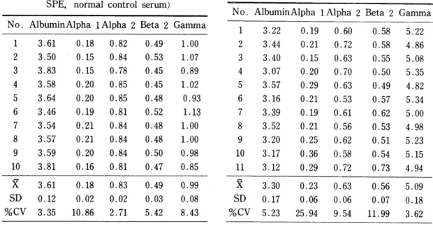
Table 3. Within-run precision (cellulose acetate, SPE, normal control serum)
N o. AlbuminAlpha 1 Alpha 2 Beta 2 Gamma 0.95 0.95 0.76 1.01 1.12 1.09 0.84 0.87 1.01 1.06 0.58
0.58 0.53 0.60 0.65 0.57 0.49 0.51 0.48 0.53 0.47 0.47 0.51 0.54 0.46 0.41 0.49 0.48 0.48 0.45 0.18 0.18 0.16 0.21 0.16 0.15 0.16 0.15 0.12 0.14 3.92
3.92 4.14 3.73 3.71 3.87 4.11 4.09 4.01 3.93
1i
nι
qu
A시1 ζ니 ζU
7l n6
9
10 1.71
0.13 7.50 0.70 0.10 13.61 0.45
0.06 12.81 0.21
0.04 18.23 3.82
0.21 5.58
-x m α
‘‘
0.97 % 0.12 11.95 0.55
0.05 9.65 0.48 0.03 7.13 0.16 0.02 15.34 3.94
0.15 3.77
-x m
쨌
Table 6. Within-run precision (cellulose acetate, SPE, abnormal patient serum) Table 4. Within-run precision (agarose gel,
SPE, normal control serum)
N o. AlbuminAlpha 1 Alpha 2 Beta 2 Gamma 5.22 4.86 5.08 5.35 4.82 5.34 5.00 4.98 5.23 5.15 4.94 0.58 0.58 0.55 0.50 0.49 0.57 0.62 0.53 0.51 0.54 0.73 0.60
0.72 0.63 0.70 0.63 0.53 0.61 0.56 0.62 0.58 0.72 0.19 0.21 0.15 0.20 0.29 0.21 0.19 0.21 0.25 0.36 0.29 3.22
3.44 3.40 3.07 3.57 3.16 3.39 3.52 3.20 3.17 3.12 1 2 3 4 5 6 7 8 9
m n
No. AlbuminAlpha 1Alpha 2 Beta 2 Gamma 1.00 1.07 0.89 1.02 0.93 1.13 1.00 1.00 0.98 0.85 0.49
0.53 0.45 0.45 0.48 0.52 0.48 0.48 0.50 0.47 0.82
0.84 0.78 0.85 0.85 0.81 0.84 0.84 0.84 0.81 0.18 0.15 0.15 0.20 0.20 0.19 0.21 0.21 0.20 0.16 3.61
3.50 3.83 3.58 3.64 3.46 3.54 3.57 3.59 3.81 1 2 3 4 5 6 7 8 9
m
5.09 0.18 3.62 0.56 0.07 11.99 0.63
0.06 9.54 0.23 0.06 25.94 3.30
0.17 5.23
-x m
U f
% 0.99
0.08 8.43 0.49
0.03 5.42 0.83
0.02 2.71 0.18 0.02 10.86 3.61
0.12 3.35
-x m
w
‘‘
%
-126-
3)
appraise 의 특 성 은 수 있도록 되 어 있으며
(Fig.table
13과 같다
.Table 9. Within-run precision (cellulose acetate, LDH isoenzyme, normal patient serum)
system 이 독특-하으로 분 2] 능이 좋다.
또 한
paragon gel은
isoenzyme측 정 에 서 Linearity 가 광범위하여 낮은 농도의 겸체에 대해서 도 예민하게 측정된다. 각 실험에 사용한
Beckman appraisedensitometer는
calibrationstandard 가 있 어 서 densitometer의
detection상태 를
check할
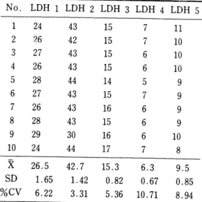
LDH 1 LDH 2 LDH 3 LDH 4 LDH 5
5 9 U
긴
8
m
3 7 6 4
”b
QU
q나 껴i
”i QU
A4i
”b
”I
꺼t
No.
1 2 3 4 5 6 7 8 9
m
Table 7. Within-run precision (agarose gel, SPE, normal normal serum) No. AlbuminAlpha 1Alpha 2 Beta 2 Gamma
8.70 5.29 60.80 6.30
1.64 25.97 19.30
4.06 21.01 35.70
3.71 10.40 30.20
2.86 9.46
-x m α
‘‘
% 2.01
1.95 2.01
1.96
1.96
1.97
1.97
1.89
1.96 2.01 2.01
Table 10. Within-run precision (cellulose acetate, LDH isoenzyme, abnormal patient serum)
1.97 0.04
1.95 0.63
0.01
1. 79 0.71
0.02 2.12 0.27
0.02 7.80 3.33 0.05
1.40
-x m
U 5
%
LDH 1 LDH 2 LDH 3 LDH 4 LDH 5 301 312 281 312 361 381 304 306 264 271 103
80 88 107 75 86 78 107 113 116
%
nm
띠갱 따냈 대ω Xω 딘 닮 띠ω 때
끼J
AA1
A41
qJ
1i j4 1i
po
”o pn
ζU
CU
”I FD
A*
9J
?l A*
껴l
CU
얘 앙
%
”%
낌 m삶
mJ
%
%
n
No.
1 2 3 4 5 6 7 8 9
m
Table 8. Within-run precision (agarose gel, SPE, abnormal patient serum)
4.54 4.81 4.70 4.47 4.54 4.75 4.63 4.63 4.53 4.56 4.65 0.60
0.50 0.62 0.56 0.60 0.51 0.56 0.45 0.54 0.56 0.59 0.39
0.35 0.36 0.37 0.39 0.30 0.27 0.32 0.28 0.32 0.32 3.11
3.01 3.03 3.26 3.11 3.37 3.23 3.42 3.37 3.27 3.14
No. AlbuminAlpha 1 Alpha 2 Beta 2 Gamma
1.17
1.12
1.10
1.15
1.17 0.96
1.11
1i
?ι
qJ
j4 Fhu
pU
”i
n
mm
πω mω 껑 이 μ 낌 야ω 껑
mu 14
Gu
nU
1i i4
1i nxu
oι
Ru
ng j4
qJ
j4
qu
qJ
qu
nJ
qtu
qJ
qJ
이 M m깅 m애 Mω mω %ω
”M
m애 와 ”이
0.64 0.64 0.62 0.62 0.62 0.64 0.62 0.65 0.63 0.64 0.64 0.73
0.70 0.71 0.69 0.71 0.68 ‘ 0.69 0.72 0.71 0.72 0.73 0.28
0.23 0.23 0.28 0.27 0.27 0.28 0.28 0.28 0.29 0.28 3.25
3.36 3.33 3.35 3.34 3.34 3.38 3.37 3.32 3.24 3.25
1 2 3 4 5 6 7 8 9
m n
309.3 36.90 11.93 95.3
15.49 16.25 53.5
7.47 13.97 58.8
14.5 24.66 53
11.93 22.51
-x m
0.98 %
1.08
1.09
1.11
8 9
m n
4.62 0.10 2.22 0.55
0.05 9.18
1.09 0.07 6.26 0.33 0.04 12.47 3.21
0.14 4.39
-x m α
‘‘
%
Table 12. Within-run precision(agarose gel, LDH isoenzyme, abnormal patient serum) Table 11. Within-run precision(agarose gel, LDH
isoenzyme, normal oatient serum)
LDH 1 LDH 2 LDH 3 LDH 4 LDH 5 410 400 412 407 409 422 429 401 412 438
mω mι
%
η
mω
%
없
%m
w
던
m깅 앓 이 U 이 U
E
M 깅 %ω mω
E
Mm
πω ”이 싫 %냉 야에 Mω m깅 야ω 싫 %ω
%ω m 깅 %ω %ω Mμ
%“
%ω %ω %ω mω
No.
1 2 3 4 5 6 7 8 9
m
1 2 3 4 5 6 7 8 9
m
LDH 1 LDH 2 LDH 3 LDH 4 LDH 5
n m m m
9 9 9 9
m
8
7·7·GU
CU
다u
7l Ru
”o
PO
7·
πω πω πω πω
M
πω mω πω
”m
n
염 껑
엄쩌 μ%
쇄 섭섭 mω μ 입
%섭 깨 낀 %ω %ω 낀 %ω %ω m정 잃
No.
414 12.1
2.92 67.9
5.20 7.65 29.6
2.46 8.30 33.1
3.00 9.06 25.3
2.79 11.03
-x m
U 5
% 9.5
0.85 8.94 6.3
0.67 10.71 15.3
0.82 5.36 42.7
1.42 3.31 26.5
1.65 6.22
-x m
U
‘‘
%
굉 -13
APPRAISE
DensitoMεter
CalibrationCheζk
f세』드 ζ, t-b 글 nζ e Calibration 8-13F.:e
l'~
6:3.3
ε‘ ε.1
7.S 1.6 13.5 Fraction
i 3 4 s
F.:e 1. ÀbSOt-bance
ε.97
2.4'~2.131 1. 51 1.134 0.50
빠
ε 1i
3 4 5 -b
’ E
Fig. 3. Calibration check of Appraise Densitometer
Chem., 27: 1944-1945(1981).
4 . Hicks, E.J. and Hughes, B.J., Chem., 21, 1072(1975).
5 .
김진규외
4명,
Cobas Bio자동화학 분석기의 평가, 대한임상병리와 정도관리학회,
Vo1.6, No.1, 1984.Clin.
-128- Chemistry
Clin.
참고문헌
1 . Tietz, Textbook of Saunders, 98-109.
2. J eppsson, J. 0 . , Laurell, C. B. et al. Clin.
Chem., 25: 629-638, 1979.
3 . P. Bourdoux, C. Bhanders, et al.
Clinical