INTRODUCTION
Delayed heart rate (HR) recovery after exercise is a function of vagal reactivation, and it is a predictor of overall mortality (1, 2) and adverse cardiovascular events (3) in the general population who has no prior evidence of clinical cardiovas- cular disease (CVD). Its predictive value for adverse events has also been shown in patients with ischemic heart disease (4-6), or with diabetes (7). Parasympathetic tone dominates the resting state, while exercise arouses sympathetic activa- tion with the withdrawal of vagal tone. After exercise, HR recovers to the level of normal by parasympathetic activation followed by sympathetic withdrawal (8, 9). Therefore, a de- layed HR recovery after exercise implies a dysfunction of vagal reactivation. Sympathetic overactivity is known to be asso- ciated with hyperinsulinemia or insulin resistance, which underlies the metabolic syndrome (10, 11). There have been studies indicating that autonomic dysfunction, involving both sympathetic and parasympathetic activity, is associated with obesity (12), insulin resistance (13), type 2 diabetes (14), and brain hemorrhage (15). Attenuated parasympathetic activi- ty has recently been seen in the first-degree relatives or off- spring of type 2 diabetes patients (16, 17). However, less is known about the relationship between metabolic syndrome and impaired vagal tone.
Therefore, the aim of this study is to investigate the asso- ciation between metabolic syndrome and HR recovery, which reflects post-exercise parasympathetic reactivation.
MATERIALS AND METHODS Study subjects
The subjects for the study (n=1,434, M:F=978:456, mean age 51±9 yr) were volunteers for a routine health check-up in the health promotion center, Samsung Medical Center, Seoul, Korea. The subject’s past medical histories were obta- ined through a structured questionnaire. Those who had his- tory of stroke, established heart disease, and/or known dia- betes or who were on medication were excluded from the analysis. Twelve percent of the subjects were either hyper- tensive or on antihypertensive medication and were included in the study. Written consents were obtained from all subjects and the institutional review board approved our study.
Anthromorphometry and laboratory tests
Blood sampling for serum lipid profile and glucose was obtained after at least 14 hr of fasting. Metabolic syndrome
Jidong Sung, Yoon-Ho Choi, Jeong Bae Park*
Center for Health Promotion, Samsung Medical Center and Department of Medicine/Cardiology*, Cheil General Hospital, Sungkyunkwan University School of Medicine, Seoul, Korea
Address for correspondence Jeong Bae Park, M.D.
Department of Medicine/Cardiology, Cheil General Hospital, Sungkyunkwan University School of Medicine, 1-19 Mookjung-dong, Jung-gu, Seoul 100-380, Korea Tel : +82.2-2000-7249, Fax : +82.2-2000-7152 E-mail : [email protected]
621
Metabolic Syndrome Is Associated with Delayed Heart Rate Recovery after Exercise
Heart rate (HR) recovery after exercise is a function of vagal reactivation, and its impairment is a predictor of overall mortality and adverse cardiovascular events.
While metabolic syndrome is associated with sympathetic overactivity, little is known about the relationship between metabolic syndrome and HR recovery. A symptom- limited exercise stress test in healthy subjects (n=1,434) was used to evaluate HR recovery. Metabolic syndrome was defined according to the National Cholesterol Education Program’s Adult Treatment Panel III (NCEP ATP-III) criteria. Seventeen percent of subjects had ≥≥3 criteria for metabolic syndrome. HR recovery was lower in men than women and in smokers than nonsmokers. The subject with metabolic syndrome (vs. without) showed lower HR recovery (10.3±±11.6 vs. 13.6±±9.7 per minute) and higher resting HR (64.3±±10.3 vs. 61.6±±9.1 per minute). HR recovery correlated inversely to age (r=-0.25, p<0.0001), but not to resting HR or maximal oxygen uptake. Delayed HR recovery was associated with metabolic syndrome after an adjustment for age, sex, resting HR and smoking (p<0.01). Metabolic syn- drome is associated with impaired vagal reactivation. Adverse cardiovascular out- comes associated with metabolic syndrome may be mediated by the failure of vagal reactivation in addition to sympathetic overactivity.
Key Words : Heart Rate; Heart Rate Recovery; Metabolic Syndrome X; Vagal Reactivation
Received : 7 September 2005 Accepted : 13 December 2005
was defined according to the Adult Treatment Panel III (ATP- III) criteria (18), except that body mass index (BMI) (>25 kg/m2) was substituted for waist circumference (19). The subjects were diagnosed as having metabolic syndrome if 3 or more of the 5 criteria were met; 1) BMI >25 kg/m2, 2) triglycerides≥150 mg/dL, 3) HDL cholesterol <40 mg/dL in men, <50 mg/dL in women, 4) blood pressure ≥130/≥85 mmHg, 5) fasting blood glucose ≥110 mg/dL.
Cardiopulmonary function test
A maximal exercise stress test was done in all subjects using the modified Bruce protocol. Blood pressure, HR and the Borg scale of rating of perceived exertion (RPE) (20) were measured at 2 min of each stages of the exercise. Exercise was stopped when the subject demanded cessation of the tread- mill due to exhaustion, or if the heart rate achieved was more than 90% of estimated maximal HR (220-age), or if the RPE was more than 17 or the respiratory exchange ratio was more than 1.15. During the recovery phase, the subjects continued to walk for 30 sec at the speed of 1.2 mph and then they sat down for 5 min with continued medical monitoring. HR recovery was calculated as the decrease of HR per minute between the peak exercise period and 3 min post-exercise ((HRat peak-HR3 min post-exercise)/3). Those subjects who showed signs suggestive of myocardial ischemia were excluded from the analysis. Positive myocardial ischemia was defined as J- point and ST80 (defined as the point that is 80 msec from the J point) depression of 0.1 mV or more and/or an ST-seg- ment slope within the range of ±1 mV/sec in 3 consecutive beats. Tightly sealing breathing mask connected to an airflow sensor was used. Respiratory gas analysis was done by dynamic breath-by-breath measurement using JAEGER system (VI- ASYS Healthcare, Hoecherg, Germany). Various respiratory parameters including minute ventilation, oxygen uptake and carbon dioxide output were measured with a sampling inter- val of 8 sec to determine the maximal oxygen uptake.
Statistical analysis
All values were expressed as a mean±standard deviation.
Comparisons of means between the 2 groups were done using a Student t-test. Bivariate correlations between variables were evaluated by Spearman correlation. A multiple regression model was constructed with HR recovery as the dependent variable, and independent variables were those variables show- ing significant association with HR recovery upon uni- and bivariate analysis. Both stepwise regression and empirical exploration were used for the final model selection. Criteria for stepwise regression were: 0.25 of probability to enter and 0.10 of probability to leave. Interactions between the inde- pendent variables were explored and insignificant interaction terms were removed from the final model. A p value <0.05 was considered statistically significant. SAS for Windows v.
8.01 (Cary, NC, U.S.A.) was used for the analysis.
RESULTS Clinical characteristics
Demographic and laboratory characteristics of the popu- lation are shown in Table 1. The majority of the subjects had a sedentary lifestyle, with only 15% doing regular exer- cise more than 4 times per week. Twelve percent of the sub- jects reported that they were either hypertensive or on anti- hypertensive medications. Seventeen percent of the subjects were found to have metabolic syndrome, and their clinical and basic laboratory characteristics are shown in Table 1. Of the subjects with metabolic syndrome, 12.8% had 3, 4.4%
had 4 and less than 1% had all 5 components of metabolic syndrome. As is shown in Table 1, these variables display significant difference between those with and without meta- bolic syndrome in the expected direction, except that the proportion of smokers was not different between the 2 groups.
Their heart rates at rest, at the peak exertion time and at 3 min post-exercise were 62±9, 151±16 and 112±29 per minute, respectively.
Predictors of HR recovery by univariate analysis
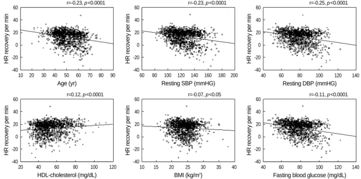
HR recovery was lower in men (12.2±9.8 vs. 14.6±9.4 per minute for women, p<0.0001) and in smokers (10.7± 10.6 vs. 13.5±10.2 per minute for nonsmoker, p<0.0001) (Fig. 1). HR recovery inversely correlated to age, (r=-0.25,
BMI, body mass index; SBP/DBP, systolic/diastolic blood pressure; HDL, high-density lipoprotein; FBS, fasting blood sugar; HR, heart rate; NS, nonspecific.
Metabolic syndrome (-)
(n=1,184) (+) (n=248) Total
(n=1,434)
p- value
Age (yr) 51±8.7 50±8.3 52±8.6 <0.01
Male 68% 65% 82% <0.01
Smoker 19% 19% 22% NS
BMI (kg/m2) 24.1±2.8 23.5±2.6 27.1±2.5 <0.01 SBP (mmHg) 122±17 120±16 133±15 <0.01 DBP (mmHg) 79±12 77±11 88±12 <0.01 Total cholesterol 201±34 200±34 209±31 <0.01
(mg/dL)
Triglyceride (mg/dL) 139±81 122±65 218±99 <0.01 HDL-cholesterol 53±13 55±13 44±10 <0.01
(mg/dL)
FBS (mg/dL) 95±17 93±14 104±21 <0.01 Vo2 peak (mL/kg/min) 20.3±5.3 20.5±5.3 19.3±5.0 <0.01 Resting HR (min) 62±9 62±9 64±10 <0.01 Peak HR (min) 151±16 152±15 148±17 <0.01 HR recovery (min) 13.1±10.1 13.6±9.7 10.3±11.6 <0.01 Table 1.Demographic and clinical characteristics
p<0.0001) resting SBP, (r=-0.23, p<0.0001) resting DBP, (r=-0.25, p<0.0001) and BMI, (r=-0.07, p<0.05). HR recov- ery correlated positively to HDL-cholesterol (r=0.12, p<
0.0001) (Fig. 2). The resting HR and maximal oxygen uptake showed no correlation with HR recovery.
Metabolic syndrome and HR recovery
Subjects with metabolic syndrome had significantly im- paired HR recovery as compared to those without metabolic syndrome (10.3±11.6 vs. 13.6±9.7 per min, p<0.0001, Fig. 1). The resting HR, a parameter reflecting the balance between sympathetic and parasympathetic tone in the basal state, was higher in the presence of metabolic syndrome (64.3
±10.3 vs. 61.6±9.1 per min, p<0.0001). There was a sta- tistically significant difference in HR recovery with increasing number of criteria of the metabolic syndrome present (p<
0.05). The post-hoc multiple comparison showed that HR recovery was significantly different between those who had 0 and 1 factors, who had 0 and 3 factors, and who had 2 and 3 factors of metabolic syndrome (Fig. 3). By multivariate anal- ysis, the presence of metabolic syndrome was independently associated with delayed HR recovery after an adjustment for age, gender, smoking and resting HR, which is a marker of sympathetic activity (p<0.01, Table 2). This result was not changed by analysis excluding 12% of subjects who were on any antihypertensive medication (p<0.01, data not shown).
DISCUSSION
This study demonstrates for the first time that subjects with metabolic syndrome show a delayed HR recovery as a suggested measure of vagal activity, and that HR recovery is
HR recovery per min
30
20
10
0
Male Female
Fig. 1.Relationship between HR recovery, and gender, current smoking status, and the presence of metabolic syndrome. p<0.001 between each group.
*
HR recovery per min
30
20
10
0
(+) (-)
Smoking
*
HR recovery per min
30
20
10
0
(+) (-)
Metabolic syndrome
*
HR recovery per min
60 40 20 0 -20 -40
10 20 30 40 50 60 70 80 90
Age (yr)
60 80 100 120 140 160 180 200 Resting SBP (mmHG)
40 60 80 100 120 140
Resting DBP (mmHG) r=-0.23, p<0.0001
HR recovery per min
60 40 20 0 -20 -40
r=-0.23, p<0.0001
HR recovery per min
60 40 20 0 -20 -40
r=-0.25, p<0.0001
HR recovery per min
60 40 20 0 -20 -40
20 40 60 80 100 120
HDL-cholesterol (mg/dL)
10 15 20 25 30 35 40
BMI (kg/m2)
40 60 80 100 120 140
Fasting blood glucose (mg/dL) r=0.12, p<0.0001
HR recovery per min
60 40 20 0 -20 -40
r=-0.07, p<0.05
HR recovery per min
60 40 20 0 -20 -40
r=-0.11, p<0.0001
Fig. 2.Correlation between HR recovery and individual parameters associated with metabolic syndrome.
delayed further in subjects having an increasing number of metabolic syndrome criteria met. This relationship was clear and persistent after we adjusted the results for several vari- ables that can influence HR recovery, including the resting HR, thereby suggesting that there is a link between metabolic syndrome and impaired vagal activity, regardless of the pres- ence of sympathetic overactivity.
Metabolic syndrome is a clinical concept that facilitates the identification of patients who have a metabolic derange- ment, this making them prone to atherosclerosis and thus, at risk for adverse cardiovascular events (18). Previous stud- ies have suggested the relationship existing between auto- nomic dysfunction and hyperinsulinemia or insulin resis- tance. Obesity and associated hyperinsulinemia correlated with sympathetic overactivity, and this is reflected in the parameters of heart rate variability (10). Moreover, HR recov- ery after exercise was related to insulin sensitivity using the hyperinsulinemic eugleucemic clamp (13). These studies suggest that insulin resistance, which is thought to be an underlying abnormality for metabolic syndrome, is related to HR recovery. Recently, the association of metabolic syn- drome with poor exercise capacity and poor heart rate recov- ery was demonstrated in patients who have established coro- nary heart disease (21). However, the relationship between metabolic syndrome and HR recovery after exercise has not been shown yet. We investigated a free-living population without cardiovascular disease or overt diabetes who agreed to take a health screening check up. A significant proportion of the subjects (17%) had metabolic syndrome, although less than 5% of them had obesity (BMI of over 30) and none had severe morbid obesity (BMI of over 40), and slightly less than half were just ‘overweight’ by Western standard. The aver- age maximal oxygen uptake was 20.3 mg/kg/min, which seems to be quite poor for the average 51-yr-old person (19).
A majority of subjects did not reach their 100% exercise per-
formance level and it is highly likely that they were mostly sedentary, as judging from their frequency of sports activity, which might contribute to the development of metabolic syndrome.
The mechanism by which HR recovery is related to meta- bolic syndrome is largely a matter of speculation. One expla- nation involves the poorer aerobic fitness of the metabolic syndrome group. But in our data, the correlation between aerobic fitness and HR recovery is low. This result is proba- bly because of the narrow range of maximal oxygen uptake, and the relationship between HR recovery and metabolic syndrome was persistent even after maximal oxygen uptake was forced into a multiple regression model (data not shown).
Obesity, an important component of metabolic syndrome, is characterized by autonomic dysfunction in the parasympathetic system (12, 22), which can be lessened by weight loss (23).
There is also good evidence that the parasympathetic nervous system participates in the release of free fatty acids, thereby influencing insulin sensitivity and fat synthesis (24).
We did not make any sophisticated estimation of autonomic function, such as heart rate variability, but resting HR can be used as a crude estimate of sympathetic tone. Higher rest- ing heart rate was shown to be associated with sympathetic overactivity, various cardiovascular risk factors including hyper- tension and higher fasting blood glucose (25, 26) and mor- tality even after an adjustment for other risk factors (27, 28).
Our data showed, as was expected, that a higher resting HR is associated with metabolic syndrome, but it is not related to HR recovery. Delayed HR recovery was independently associated with metabolic syndrome after an adjustment for the resting HR. These findings suggest that metabolic syn- drome is associated with impaired vagal reactivation in addi- tion to the previously known relationship with sympathetic overactivity. When considering the abundance of robust data regarding the prognostic value of delayed HR recovery in various populations (1-6), and also our current findings of its association with metabolic syndrome, it is likely that im- paired vagal tone as well as sympathetic overactivity contri- butes to the cardiovascular risk of metabolic syndrome. Aer- obic fitness correlated to HR recovery (29) and it can be im- proved with exercise training, even in patients with existing
HR recovery per min
40
20
0
-20
-40
0 1 2 ≥3
Number of risk factors of metabolic syndrome Fig. 3.Delayed HR recovery and the number of factors indicating metabolic syndrome (mean-SD). *p<0.05 for post-hoc multiple comparison test, p value for overall difference between the group was <0.0001.
*
*
*
*Subjects with metabolic syndrome have lower HR recovery than those without it.
Independent variables
Regression
coefficient SE F-value p value
Intercept 29.60 2.67
Age 2.05 0.54 81.25 <0.001
Gender (men) -1.36 0.63 4.79 <0.05
Smoking -3.30 0.71 17.87 <0.001
Resting HR -0.04 0.03 2.76 0.14
Metabolic syndrome* -2.16 0.73 13.32 <0.01 Table 2.Multivariate regression analysis using delayed HR reco- very as dependent variable
cardiovascular disease (30). Exercise training is beneficial in overcoming various aspects of metabolic syndrome and imp- roving the vagal tone may also be an important mechanism for benefiting from exercise training. The exercise protocols for measuring HR recovery were differently applied in pre- vious studies. For example, Cole et al. (1) employed at least 2 min of cool-down period immediately after exercise which was considered as recovery phase, whereas Morshedi-Meibodi et al. (3) had patients to get off the treadmill immediately after peak exercise. Whether this difference influenced final results is not known. We used the protocol that allowed 30 sec of cool-down on treadmill after peak exercise to minimize the possible risk of blood pressure drop after sudden stopping of exercise (31).
Our study has a few limitations. About 12% of our sub- jects were on treatment with antihypertensive agents, and some of them may be on a drug that can interfere with the interpretation of HR response during stress tests such as beta- blockers. Because we can speculate that only a small propor- tion of our subjects were on beta-blockers, its influence on the final results of analysis was probably minimal. And the use of beta-blockers may not have a significant influence on the predictive value of HR recovery (32). Analysis excluding those on antihypertensive medication shows the same results in our study. Second, we did not have data on abdominal cir- cumference, a better indicator of visceral fat than BMI alone (33). Substitution of BMI for abdominal circumference in defining metabolic syndrome might have had some influence on our results, but we consider BMI as a reasonable alterna- tive having some predictive value for visceral adiposity (33).
Finally, measurement of HR recovery can be done in various ways, but the arbitrary definition of HR decrease during the first minute after exercise has been frequently used because this parameter showed the predictive value for outcomes (1-3).
Because we did not have the HR data at 1 min post-exercise, the slope of HR decrease during 3 min was used. In other studies, HR recovery at 2 min showed the maximal predic- tive value for mortality, but HR recovery at 3 min also had a significant predictive value (31, 33, 34). Therefore, it is not necessary to only look at average 1 min HR recovery as it assumes that the fall in heart rate after exercise is a simple linear function, which it is not. Some subjects had negative heart rate recovery, i.e., paradoxical heart rate increase dur- ing recovery period. While simple errors during data input is not likely because all the data were electronically transferred from exercise testing work station to research database, it is highly probable that extreme values such as -30/min is due to miscalculation of heart rate from RR interval by artifacts or ectopic beats. We were not able to reconfirm this error but such cases were very rarely seen in our data, and consid- ering the sample size, this does not seem to influence the overall results. On the other hand, ‘mild’ negative value itself can be seen as in a previous study also reporting negative val- ues in a minority of patients. For example, Morshedi-Meibodi
et al. (3) reported that HR recovery ranges in lowest quin- tile are -10 to 8 and -2 to 20 per minute for men and women respectively. Other studies usually reported binary result of normal and abnormal HR recovery but did not show the actual range of HR recovery values. This unusual finding has not been discussed in detail before. As a speculation, those with very poor vagal reactivation may have actual increase of heart rate after exercise because plasma catecholamine dur- ing early recovery (about 90 sec) is actually higher than at peak exercise (35).
In conclusion, metabolic syndrome is significantly associ- ated with impaired vagal reactivation. Therefore, cardiovas- cular risks associated with metabolic syndrome may also be mediated by the failure of vagal reactivation, in addition to sympathetic overactivity.
ACKNOWLEDGEMENT
We appreciate Dr Michael S. Lauer (Cleveland Clinic, U.S.A.) for criticism and kind comments on the paper.
REFERENCES
1. Cole CR, Blackstone EH, Pashkow FJ, Snader CE, Lauer MS. Heart- rate recovery immediately after exercise as a predictor of mortality.
N Engl J Med 1999; 341: 1351-7.
2. Cole CR, Foody JM, Blackstone EH, Lauer MS. Heart rate recovery after submaximal exercise testing as a predictor of mortality in a car- diovascularly healthy cohort. Ann Intern Med 2000; 132: 552-5.
3. Morshedi-Meibodi A, Larson MG, Levy D, O’Donnell CJ, Vasan RS. Heart rate recovery after treadmill exercise testing and risk of cardiovascular disease events (The Framingham Heart Study). Am J Cardiol 2002; 90: 848-52.
4. Kim HS, Lee JH, Kwon YS, Lee HS, Yang DH, Park HS, Jo YK, Chae SC, Jun JE, Park WH. Changes in heart rate during and after exercise treadmill test as prognostic factor in cardiovascular disease.
Korean Circ J 2004; 34: 170-7.
5. Ju DU, Kang HJ, Kim SW, No TM, Son HS, Kang BJ, Kim SR, Lee BR, Jung BC, Lee JJ. The difference of heart rate recovery in ischemic heart disease comparing to normal. Korean J Med 2004; 66: 586-92.
6. Vivekananthan DP, Blackstone EH, Pothier CE, Lauer MS. Heart rate recovery after exercise is a predictor of mortality, independent of the angiographic severity of coronary disease. J Am Coll Cardiol 2003; 42: 831-8.
7. Cheng YJ, Lauer MS, Earnest CP, Church TS, Kampert JB, Gibbons LW, Blair SN. Heart rate recovery following maximal exercise test- ing as a predictor of cardiovascular disease and all-cause mortality in men with diabetes. Diabetes Care 2003; 26: 2052-7.
8. Rosenwinkel ET, Bloomfield DM, Arwady MA, Goldsmith RL. Exer- cise and autonomic function in health and cardiovascular disease.
Cardiol Clin 2001; 19: 369-87.
9. Arai Y, Saul JP, Albrecht P, Hartley LH, Lilly LS, Cohen RJ, Colucci
WS. Modulation of cardiac autonomic activity during and immedi- ately after exercise. Am J Physiol 1989; 256: 132-41.
10. Emdin M, Gastaldelli A, Muscelli E, Macerata A, Natali A, Camastra S, Ferrannini E. Hyperinsulinemia and autonomic nervous system dysfunction in obesity: effects of weight loss. Circulation 2001; 103:
513-9.
11. Esler M, Rumantir M, Wiesner G, Kaye D, Hastings J, Lambert G.
Sympathetic nervous system and insulin resistance: from obesity to diabetes. Am J Hypertens 2001; 14: 304-9S.
12. Peterson HR, Rothschild M, Weinberg CR, Fell RD, McLeish KR, Pfeifer MA. Body fat and the activity of the autonomic nervous sys- tem. N Engl J Med 1988; 318: 1077-83.
13. Lind L, Andren B. Heart rate recovery after exercise is related to the insulin resistance syndrome and heart rate variability in elderly men. Am Heart J 2002; 144: 666-72.
14. Gottsater A, Ahmed M, Fernlund P, Sundkvist G. Autonomic neu- ropathy in type 2 diabetic patients is associated with hyperinsuli- naemia and hypertriglyceridaemia. Diabet Med 1999; 16: 49-54.
15. Kawahara E, Ikeda S, Miyahara Y, Kohno S. Role of autonomic ner- vous dysfunction in electrocardiographic abnormalities and cardiac injury in patients with acute subarachnoid hemorrhage. Circ J 2003;
67: 753-6.
16. Lindmark S, Wiklund U, Bjerle P, Eriksson JW. Does the autonomic nervous system play a role in the development of insulin resistance?
A study on heart rate variability in first-degree relatives of type 2 diabetes patients and control subjects. Diabet Med 2003; 20: 399- 405.
17. Laitinen T, Vauhkonen IK, Niskanen LK, Hartikainen JE, Lansimies EA, Uusitupa MI, Laakso M. Power spectral analysis of heart rate variability during hyperinsulinemia in nondiabetic offspring of type 2 diabetic patients: evidence for possible early autonomic dysfunc- tion in insulin-resistant subjects. Diabetes 1999; 48: 1295-9.
18. National Cholesterol Education Program (NCEP) Expert Panel on Detection, Evaluation, and Treatment of High Blood Cholesterol in Adults (Adult Treatment Panel III). Third Report of the National Cho- lesterol Education Program (NCEP). Expert Panel on Detection, Eval- uation, and Treatment of High Blood Cholesterol in Adults (Adult Treatment Panel III) final report. Circulation 2002; 106: 3143-421.
19. Ferguson C, Myers J, Foroelicher V. Overview of exercise testing. In:
PD T, ed. Exercise and sports cardiology. 1st ed. New York: McGraw- Hill 2001: 92.
20. Borg G. Perceived exertion as an indicator of somatic stress. Scand J Rehab Med 1970; 2: 92-8.
21. Spies C, Otte C, Kanaya A, Pipkin SS, Schiller NB, Whooley MA.
Association of metabolic syndrome with exercise capacity and heart
rate recovery in patients with coronary heart disease in the heart and soul study. Am J Cardiol 2005; 95: 1175-9.
22. Scheurink AJ, Balkan B, Nyakas C, van Dijk G, Steffens AB, Bohus B. Energy homeostasis, autonomic activity and obesity. Obes Res 1995; 3 (Suppl 5): 721-7.
23. Rissanen P, Franssila-Kallunki A, Rissanen A. Cardiac parasympa- thetic activity is increased by weight loss in healthy obese women.
Obes Res 2001; 9: 637-43.
24. Boden G, Hoeldtke RD. Nerves, fat, and insulin resistance. N Engl J Med 2003; 349: 1966-7.
25. Palatini P. Elevated heart rate as a predictor of increased cardiovas- cular morbidity. J Hypertens 1999; 17 (Suppl 3): 3-10.
26. Palatini P, Casiglia E, Pauletto P, Staessen J, Kaciroti N, Julius S.
Relationship of tachycardia with high blood pressure and metabolic abnormalities: a study with mixture analysis in three populations.
Hypertension 1997; 30: 1267-73.
27. Greenland P, Daviglus ML, Dyer AR, Liu K, Huang CF, Goldberger JJ, Stamler J. Resting heart rate is a risk factor for cardiovascular and noncardiovascular mortality: the Chicago Heart Association Detection Project in Industry. Am J Epidemiol 1999; 149: 853-62.
28. Mensink GB, Hoffmeister H. The relationship between resting heart rate and all-cause, cardiovascular and cancer mortality. Eur Heart J 1997; 18: 1404-10.
29. Darr KC, Bassett DR, Morgan BJ, Thomas DP. Effects of age and training status on heart rate recovery after peak exercise. Am J Physi- ol 1988; 254: 340-3.
30. Tiukinhoy S, Beohar N, Hsie M. Improvement in heart rate recovery after cardiac rehabilitation. J Cardiopulm Rehabil 2003; 23: 84-7.
31. Holtzhausen LM, Noakes TD. The prevalence and significance of post-exercise (postural) hypotension in ultramarathon runners. Med Sci Sports Exerc 1995; 27: 1595-601.
32. Shetler K, Marcus R, Froelicher VF, Vora S, Kalisetti D, Prakash M, Do D, Myers J. Heart rate recovery: validation and methodologic issues. J Am Coll Cardiol 2001; 38: 1980-7.
33. Janssen I, Heymsfield SB, Allison DB, Kotler DP, Ross R. Body mass index and waist circumference independently contribute to the prediction of nonabdominal, abdominal subcutaneous, and visceral fat. Am J Clin Nutr 2002; 75: 683-8.
34. Shishehbor MH, Hoogwerf BJ, Lauer MS. Association of triglyceride- to-HDL cholesterol ratio with heart rate recovery. Diabetes Care 2004; 27: 936-41.
35. Krock LP, Hartung GH. Influence of post-exercise activity on plasma catecholamines, blood pressure and heart rate in normal subjects.
Clin Auton Res 1992; 2: 89-97.