INTRODUCTION
Autosomal recessive limb-girdle muscular dystrophy (ARL- GMD) is a clinically and genetically heterogeneous disorder affecting mainly the pelvic and shoulder girdle muscles. Accor- ding to the genetic classification of ARLGMD, it has been divided into ten subtypes, from LGMD type 2A to LGMD type 2J (1). LGMD type2B (LGMD2B) is caused by muta- tions in the dysferline gene (DYSF, MIM*603009) located on chromosome 2p13.3, which induces a dysfunction of dysfer- lin at the protein level (2). Dysferlin is a member of the FER- 1 protein family and contains six putative C2 domains, which can bind to phospholipids, inositol polyphosphates, Ca2+and intracellular proteins (3). Dysferlin is expressed predominant- ly in the skeletal muscle and localizes to the plasma mem- brane of the muscle fibers. In addition, it has been suggest- ed to be involved in membrane fusion (4, 5). A recent ani- mal study has suggested that dysferlin plays a role in the sar- colemma repair process (6). The DYSF has been also shown to cause Miyoshi myopathy (MM), which is a rare form of distal myopathy with the calf muscles weakness. Identical mutations in patients with LGMD2B or Miyoshi myopathy have been noted in several reports, suggesting a role for the modifier gene(s) (7-11). Both LGMD2B and Miyoshi myopa-
thy are referred to as “dysferlinopathy” because of the deficien- cy of the same protein.
Because of the clinical and genetic heterogeneity of the LGMD, the LGMD subtypes are classified based on genetic or protein analysis. The development of antibodies directed against dysferlin has recently been shown to be useful for making a diagnosis of LGMD2B by an examination of a mus- cle biopsy with immunostaining or immunoblotting tech- niques (4, 5, 10). In the current study, immunocytochemi- cal examination for dysferlin was tested on a muscle biopsies from 17 Korean ARLGMD patients, and the clinical and pathological characteristics of four Korean LGMD2B patients, who showed the complete loss of the dysferlin protein on a muscle biopsy, were investigated.
MATERIALS AND METHODS Patients Selection and Muscle biopsy
From March 2001 to November 2002, 17 unrelated, non- consanguineous LGMD patients were enrolled in this study.
The diagnosis of LGMD was established by the clinical his- tory and physical examination, the family history, a normal
Seung-Hun Oh*,�, Seong-Woong Kang�, Jin-Goo Lee*, Sang-Jun Na*, Tai-Seung Kim�, Young-Chul Choi*
Department of Neurology*and Rehabilitation Medicine�, Yongdong Severance Hospital, Seoul; Department of Medicine, Euijong-bu Collectional Institution�, Euijong-bu; Department of Pathology�, Severance Hospital, Brain Korea 21 Project for Medicine, Yonsei University College of Medicine, Seoul, Korea
Address for correspondence Young-Chul Choi, M.D.
Department of Neurology, Yongdong Severance Hospital, 146-92 Dogok-dong, Kangnam-gu, Seoul 135-270, Korea
Tel : +82.2-3497-3323, Fax : +82.2-3462-5904 E-mail : [email protected]
447
Clinical and Pathological Characteristics of Four Korean Patients with Limb-Girdle Muscular Dystrophy type 2B
Limb-girdle muscular dystrophy type 2B (LGMD2B), a subtype of autosomal reces- sive limb-girdle muscular dystrophy (ARLGMD), is characterized by a relatively late onset and slow progressive course. LGMD2B is known to be caused by the loss of the dysferlin protein at sarcolemma in muscle fibers. In this study, the clinical and pathological characteristics of Korean LGMD2B patients were investigated. Seven- teen patients with ARLGMD underwent muscle biopsy and the histochemical exami- nation was performed. For the immunocytochemistry, a set of antibodies against dystrophin, , , , -sarcoglycans, dysferlin, caveolin-3, and -dystroglycan was used. Four patients (24%) showed selective loss of immunoreactivity against dysfer- lin at the sarcolemma on the muscle specimens. Therefore, they were classified into the LGMD2B category. The age at the onset of disease ranged from 9 yr to 33 yr, and none of the patients was wheelchair bound at the neurological examination. The serum creatine kinase (CK) was high in all the patients (4010-5310 IU/L). The patho- logic examination showed mild to moderate dystrophic features. These are the first Korean LGMD2B cases with a dysferlin deficiency confirmed by immunocytochem- istry. The clinical, pathological, and immunocytochemical findings of the patients with LGMD2B in this study were in accordance with those of other previous reports.
Key Words : Muscular Dystrophies; Dysferlin; Immunocytochemistry
Received : 18 December 2003 Accepted : 2 March 2004
nerve conduction study and the myopathic pattern on elec- tromyography (12). All the patients underwent an open biop- sy from the quadriceps femoris or biceps brachii muscle under local anesthesia. In all the cases, informed consents were ob- tained from the patients or their parents. Transverse serial fro- zen muscle sections (7 m thickness) were stained with hema- toxylin and eosin (H&E), modified Gomori trichrome, and a battery of histochemical techniques (NADH-TR, ATPase pH 9.4/4.6/4.3) were applied. All of them showed normal immunostaining for three domains of dystrophin (N-termi- nus, C-terminus and Rod-domain) on the muscle specimens, and the deletion was not found on multiplex PCR for select- ed exons of dystrophin gene.
Immunocytochemical staining of muscle specimens
The tissues were processed for immunocytochemistry as follows: the 7 m serial sections were fixed in acetone at 4℃ for 10 min, rinsed in 0.05 mol/L Tris-buffered saline (pH 7.5) for 15 min, and incubated for 30 min with a blocking solution containing 2% bovine serum albumin and 5% nor- mal goat serum as described (13). The sections were then
incubated overnight at 4℃with one of the following anti- bodies: -sarcoglycan (NCL- -SARC, Novocastra), -sarco- glycan (NCL- -SARC, Novocastra), -sarcoglycan (NCL- - SARC, Novocastra), -sarcoglycan (NCL- -SARC, Novo- castra), dysferlin (NCL-DYSF, Novocastra), -dystroglycan (NCL- -DG, Novocastra, Newcastle Upon Tyne, U.K.), and caveolin-3 (Transduction laboratory, Lexington, KY, U.S.A.).
These affinity-purified antibodies were well characterized and did not cross-react with each other. After washing for 30 min in Tris-buffered saline, the sections were examined using sec- ondary goat anti-mouse IgG antibodies conjugated to perox- idase and visualized by a DAB (diaminobenzidine)-peroxidase reaction (Vector Laboratories, CA., U.S.A.). The sections were also incubated with biotinylated goat anti-mouse IgG, which was followed by fluorescent isothiocyanate (FITC) avidin D (Vector Laboratory, CA., U.S.A.) for dysferlin.
RESULTS
Four (Patient No. 1-4) of the 17 ARLGMD patients group (24%) showed a selective loss of dysferlin by both DAB and
DAB, diaminobenzidine; FITC, fluorescent isothiocyanate; DPH, dystrophin; -SG, -sarcoglycan; -SG, -sarcoglycan; -SG, -sarcoglycan; - SG, -darcoglycan; Dysf, dysferlin; Cav3, caveolin-3; -DG, -dystroglycan; +/-, decreased or weakly positive.
Patient No. Sex/Age (yr) DPH (DAB) -SG (DAB) -SG (DAB) -SG (DAB) -SG (DAB) Dysf (DAB) Dysf (FITC) Cav3 (DAB) -DG (DAB)
1 F/35 + + + + + - - + +
2 F/41 + + + + + - - + +
3 F/43 + + + + + - - + +
4 M/15 + + + + + - - + +
5 F/21 + + + + +/- + + + +
6 M/40 + + + + + + + + +
7 M/13 + + + + +/- + + + +
8 F/25 + + + + - +/- + + +
9 M/5 + + +/- + - + + + +
10 M/7 + + +/- + - + + + +
11 M/17 + + + + + + +/- + +
12 M/10 + + + + + + + + +
13 M/5 + + + + + + + + +
14 F/10 + + + + + + +/- + +
15 F/9 + + - +/- - + +/- + +
16 M/9 + + + + +/- + + + +
17 M/23 + + + + + +/- + + +
Table 1.The Immunocytochemical findings of the 17 ARLGMD patients
U/Ex, upper extremity; L/Ex, lower extremity; prox, proximal; dist, distal; CK, creatine kinase.
prox dist prox dist
U/Ex L/Ex
Face Neck
Serum CK level (IU/L) Calf muscle
hypertrophy Bony
abnormality Cardiac/pulmonary
insufficiency Distribution of muscle weakness
Age at onset (yr) Sex/
Age (yr) Patient No.
1 F/35 27 - - 4 4+ 4 4+ - - + (symmetric) 5310
2 F/41 26 - - 4 4+ 4- 4 - - - 4010
3 F/43 33 - - 4+ 5 4 4+ - - - 4460
4 M/15 9 - - 4- 4+ 4- 4 - - + (symmetric) 4689
Table 2.The clinical and laboratory characteristics of the four LGMD2B patients
FITC methods on the muscle specimen (Table 1). Therefore, they were classified as having LGMD type 2B (LGMD2B).
Although three patients (patient No. 8-10) showed isolated
deficiency of -sarcoglycan, it was regarded as a technical error since the -sarcoglycanopathy had always showed com- bined deficiency of other sarcoglycans ( , , and ) at exam-
Fig. 1. Muscle specimens of the quadriceps femoris muscle (from patient No. 1) stained by hematoxylin-eosin (A) and modified Gomori Trichrome (B). There were great variation in the size of fibers, fiber splitting with internal nuclei, and clumps of small fibers (×200).
A B
Fig. 2.Immunostaining against -sarcoglycan ( -SG), -sarcoglycan ( -SG), caveolin-3 (Cav-3), and -dystroglycan ( -DG) on the muscle specimen in the four LGMD2B patients by the diaminobenzidine (DAB)-peroxidase method. All four LGMD2B patients showed a normal immunoreactivity against -sarcoglycan, -sarcoglycan, caveolin-3, and -dystroglycan (×200).
Case 1
-SG -SG Cav-3 -DG
Case 2
Case 3
Case 4
ination by immunocytochemistry (14). One patient (patient No. 15) showed loss of and -sarcoglycan with partial defi- ciency of -sarcoglycan. This patient might be tentavely clas- sified as or -sarcoglycanopathy although molecular genet- ic diagnosis was not performed.
Clinical characteristics and course of disease in four patients with LGMD2B
The age of patients at the onset of the disease varied from 9 yr to 33 yr, and the disease duration ranged from 6 yr to 15 yr (Table 2). All four patients showed symmetrical proxi- mal weakness. Although all the patients showed a function- al disability in walking or running, none was wheelchair- bound at the examination. While symmetrical hypertrophy of the calf muscle was noted in two patients (patient No. 1 &
4), muscle atrophy was not found in any of the four patients.
None of the four patients had a cardiac or respiratory insuf- ficiency nor did they have any bony abnormalities. The serum creatine kinase (CK) level was elevated to approximately 20 times of the normal level in all the patients. (4010-5310 IU/L). One patient (patient No. 1) had a positive family his- tory. One of her elder brother (41 yr old) had similar symp- toms, which presented as a slowly progressive proximal weak- ness over 15 yr.
Pathological and immunocytochemical findings of muscle biopsy in four patients with LGMD2B
All the patients showed the following non-specific myo- pathic changes in the muscle: increased internal nuclei, a few
or many atrophic fibers, degenerating fibers with regenerating fibers, and an increased fiber size variation (Fig. 1). Type 1 fiber predominance (>60%) was noted in one patient (patient No. 1). There was no endomysial or perivascular inflamma- tory cell infiltrations in all the patients.
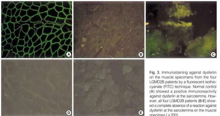
In all the patients with LGMD2B, immunocytochemical reaction against all the subtypes of sarcoglycans, -dystro- glycan, and caveolin-3 was normal (Fig. 2, Table 1), while the reaction against dysferlin showed complete loss of reac- tivity at the sarcolemma of the muscle fibers (Fig. 3).
DISCUSSION
The clinical features of our patients with LGMD2B was similar to those of previous reports (12, 15-17). Symptoms of the disease generally are manifested during the early adult- hood (average onset is noted between the age of 17-30 yr), and the disease generally has a benign course, and confinement to a wheelchair may occur in approximately 10% of patients (15, 18). Despite of the slow clinical evolution, extremely high CK level (10-20 times of normal value) was characteris- tic of LGMD2B, being indicative of protein’s role in normal muscle homeostasis (“leaky” membrane) rather than being essential for structural stabilization of the skeletal muscle (6, 19). A few LGMD2B patients showed symmetrical or asymmetrical calf muscle hypertrophy, as was observed in two of the patients in this study. Up to date, only several esti- mates of the frequency of dysferlinopathy exist. The propor- tion of LGMD2B is thought to be relatively high in Brazil- ian and Japanese population (estimated to be 19-25% of all
Fig. 3.Immunostaining against dysferlin on the muscle specimens from the four LGMD2B patients by a fluorescent isothio- cyanate (FITC) technique. Normal control (A) showed a positive immunoreactivity against dysferlin at the sarcolemma. How- ever, all four LGMD2B patients (B-E) show- ed a complete absence of a reaction against dysferlin at the sarcolemma on the muscle specimen (×200).
A
D
B
E
C
ARLGMD cases) (15, 20) and there is founder mutation of DYSF appears to be present in the Libyan Jews (17). How- ever, another recent study reported a relatively lower frequen- cy of the dysferlin deficiency in Caucasian patients with the LGMD phenotype (approx. 1%), indicating a frequency varia- tion according to the ethnicity (21). Although it is not enough to estimate the frequency of LGMD2B in the Korean ARL- GMD patients due to a small number of subjects, these results suggest that the frequency of LGMD2B is relatively high among ARLGMD in Korea. A detailed population-based study will be needed to determine the exact frequency of the LGMD2B in Korea.
In all the cases with dysferlin deficiency, the immunocyto- chemical study showed normal reactivity against dystrophin, -, -, -, -sarcoglycans, -dystroglycan on muscle speci- men, which suggests that there are no interactions between dysferlin and the dystrophin-glycoprotein complex (DGC).
This finding is in accordance with previous studies (22, 23).
The DYSF is large, comprising 55 exons that span the geno- mic region of >150 kb (24). Although direct gene analysis provides the most reliable diagnosis, it is costly, time-con- suming, and labor intensive because of the large size of the DYSF. Moreover, defects in the dysferlin gene involve most- ly single nucleotide changes with no common mutations, gross rearrangements, or mutational hotspots that could aid detection (2). For this reason, it would be better to initiate screening for the dysferlin deficiency using antibodies against this protein in muscle biopsies. Complete loss of dysferlin without deficiency of other proteins appears to be specific for primary dysferlinopathy on immunocytochemistry (24).
However, it should be noted that some cases with sarcogly- canopathy, caveolinopathy can show partial deficiency of dys- ferlin (secondary deficiency) (25, 26). Therefore, it is recom- mendable to perform immunocytochemical analysis using antibodies against other sarcolemmal proteins in addition to the antibodies against dysferlin. When partial deficiency of dysferlin is present on immunocytochemistry, direct sequenc- ing of DYSF will be needed.
In conclusion, the clinical, pathological, and immunocy- tochemical findings of patients with LGMD2B in this study were in accordance with those of previous reports.
ACKNOWLEDGMENTS
This work was supported by the Brain Korea 21 Project for Medical Science, Yonsei University.
REFERENCES
1. Zatz M, de Paula F, Starling A, Vainzof M. The 10 autosomal reces- sive limb-girdle muscular dystrophies. Neuromuscul Disord 2003;
13: 532-44.
2. Liu J, Aoki M, Illa I, Wu C, Fardeau M, Angelini C, Serrano C, Urtiz- berea JA, Hentati F, Hamida MB, Bohlega S, Culper EJ, Amato AA, Bossie K, Oeltjen J, Bejaoui K, McKenna-Yasek D, Hosler BA, Schurr E, Arahata K, de Jong PJ, Brown RH Jr. Dysferlin, a novel skeletal muscle genes, is mutated in Miyoshi myopathy and limb girdle muscular dystrophy. Nat Genet 1998; 20: 31-6.
3. Britton S, Freeman T, Vafiadaki E, Keers S, Harrison R, Bushby K, Bashir R. The third human FER-1-like protein is highly similar to dysferlin. Genomics 2000; 68: 313-21.
4. Matsuda C, Aoki M, Hayashi YK, Ho MF, Arahata K, Brown RH Jr. Dysferlin is a surface membrane-associated protein that is absent in Miyoshi myopathy. Neurology 1999; 53: 1119-22.
5. Anderson LV, Davison K, Moss JA, Young C, Cullen MJ, Walsh J, Johnson MA, Bashir R, Britton S, Keers S, Argov Z, Mahjneh I, Fougerousse F, Beckmann JS, Bushby KM. Dysferlin is a plasma membrane protein and is expressed early in human development.
Hum Mol Genet 1999; 8: 855-61.
6. Bansal D, Miyake K, Vogel SS, Groh S, Chen CC, Williamson R, McNeil PL, Campbell KP. Defective membrane repair in dysferlin- deficient muscular dystrophy. Nature 2003; 423: 168-72.
7. Weiler T, Greenberg CR, Nylen E, Halliday W, Morgan K, Eggert- son D, Wrogemann K. Limb-girdle muscular dystrophy and Miyoshi myopathy in an aboriginal Canadian kindred map to LGMD2B and segregate with the same haplotype. Am J Hum Genet 1996; 59: 872-8.
8. Illarioshkin SN, Ivanova-Smolenskaya IA, Tanaka H, Vereshchagin NV, Markova ED, Poleshchuk VV, Lozhnikova SM, Sukhorukov VS, Limborska SA, Slominsky PA, Bulayeva KB, Tsuji S. Clinical and molecular analysis of a large family with three distinct pheno- types of progressive muscular dystrophy. Brain 1996; 119: 1895- 909.
9. Illarioshkin SN, Ivanova-Smolenskaya IA, Greenberg CR, Nylen E, Sukhorukov VS, Poleshchuk VV, Markova ED, Wrogemann K. Iden- tical dysferlin mutation in limb-girdle muscular dystrophy type 2B and distal myopathy. Neurology 2000; 55: 1931-3.
10. Weiler T, Bashir R, Anderson LV, Davison K, Moss JA, Britton S, Nylen E, Keers S, Vafiadaki E, Greenberg CR, Bushby CR, Wroge- mann K. Identical mutation in patients with limb girdle muscular dystrophy type 2B or Miyoshi myopathy suggests a role for modifier gene(s). Hum Mol Genet 1999; 8: 871-7.
11. Linssen WH, Notermans NC, Van der Graaf Y, Wokke JH, Van Doorn PA, Howeler CJ, Busch HF, De Jager AE, De Visser M. Miyo- shi-type distal muscular dystrophy. Clinical spectrum in 24 Dutch patients. Brain 1997; 120: 1989-96.
12. Bushby KM. Making sense of the limb-girdle muscular dystrophies.
Brain 1999; 122: 1403-20.
13. Choi YC, Park GT, Kim TS, Sunwoo IN, Steinert PM, Kim SY. Spo- radic inclusion body myositis correlates with increased expression and cross-linking by transglutaminases 1 and 2. J Biol Chem 2000;
275: 8703-10.
14. Bushby KM. The limb-girdle muscular dystrophies-multiple genes, multiple mechanisms, Hum Mol Genet 1999; 8: 1875-82.
15. Passos-Bueno MR, Vainzof M, Moreira ES, Zatz M. Seven autosomal recessive limb-girdle muscular dystrophies in the Brazilian popula- tion: from LGMD2A to LGMD2G. Am J Med Genet 1999; 82: 392-8.
16. Ueyama H, Kumamoto T, Horinouchi H, Fujimoto S, Aono H, Tsuda T. Clinical heterogeneity in dysferlinopathy. Intern Med 2002; 41:
532-6.
17. Argov Z, Sadeh M, Mazor K, Soffer D, Kahana E, Eisenberg I, Mit- rani-Rosenbaum S, Richard I, Beckmann J, Keers S, Bashir R, Bush- by K, Rosenmann H. Muscular dystrophy due to dysferlin deficien- cy in Libyan Jews. Clinical and genetic features. Brain 2000; 123:
1229-37.
18. Zatz M, Vainzof M, Passos-Bueno MR. Limb-girdle muscular dys- trophy: one gene with different phenotypes, one phenotype with dif- ferent genes. Curr Opin Neurol 2000; 13: 511-7.
19. Lennon NJ, Kho A, Bacskai BJ, Perlmutter SL, Hyman BT, Brown RH Jr. Dysferlin interacts with annexins A1 and A2 and mediates sarcolemmal wound-healing. J Biol Chem 2003; 278: 50466-73.
20. Tagawa K, Ogawa M, Kawabe K, Yamanaka G, Matsumura T, Goto K, Nonaka I, Nishino I, Hayashi YK. Protein and gene analyses of dysferlinopathy in a large group of Japanese muscular dystrophy patients. J Neurol Sci 2003; 211: 23-8.
21. Fanin M, Pegoraro E, Matsuda-Asada C, Brown RH Jr, Angelini C.
Calpain-3 and dysferlin protein screening in patients with limb-gir- dle dystrophy and myopathy. Neurology 2001; 56: 660-5.
22. Vainzof M, Anderson LV, McNally EM, Davis DB, Faulkner G, Valle G, Moreira ES, Pavanello RC, Passos-Bueno MR, Zatz M.
Dysferlin protein analysis in limb-girdle muscular dystrophies. J Mol Neurosci 2001; 17: 71-80.
23. Anderson LV, Harrison RM, Pogue R, Vafiadaki E, Pollitt C, Davi- son K, Moss JA, Keers S, Pyle A, Shaw PJ, Mahjneh I, Argov Z, Greenberg CR, Wrogemann K, Bertorini T, Goebe HH, Beckmann JS, Bashir R, Bushby KM. Secondary reduction in calpain 3 expres- sion in patients with limb girdle muscular dystrophy type 2B and Miyoshi myopathy (primary dysferlinopathies). Neuromuscul Dis- ord 2000; 10: 553-9.
24. Aoki M, Liu J, Richard I, Bashir R, Britton S, Keers SM, Oeltjen J, Brown HE, Marchand S, Bourg N, Beley C, McKenna-Yasek D, Arahata K, Bohlega S, Cupler E, Illa I, Majneh I, Barohn RJ, Urtiz- berea JA, Fardeau M, Amato A, Angelini C, Bushby K, Beckmann JS, Brown RH Jr. Genomic organization of the dysferlin gene and novel mutations in Miyoshi myopathy. Neurology 2001; 57: 271-8.
25. Piccolo F, Moore SA, Ford GC, Campbell KP. Intracellular accu- mulation and reduced sarcolemmal expression of dysferlin in limb- girdle muscular dystrophies. Ann Neurol 2000; 48: 902-12.
26. Matsuda C, Hayashi YK, Ogawa M, Aoki M, Murayama K, Nishi- no I, Nonaka I, Arahata K, Brown RH Jr. The sarcolemmal proteins dysferlin and caveolin-3 interact in skeletal muscle. Hum Mol Genet 2001; 10: 1761-6.