INTRODUCTION
Adult onset Still’s disease (AOSD) is a chronic systemic inflammatory disorder of unknown etiology, and its major clinical manifestations include high spiking fever, polyarthral- gia, salmon colored evanescent rash, and neutrophilic leuko- cytosis. Life threatening conditions such as hepatic involve- ment, cardiac tamponade, disseminated intravascular coagu- lation (DIC), respiratory distress syndrome or pancytopenia were occasionally developed in the course of AOSD (1), and in some cases were often associated with hemophagocytic syn- drome (HS) (2-4). Most patients with HS have rapid and fatal outcomes unless the diagnosis is made early and followed by prompt therapeutic intervention. There have been some fatal- ities due to a delayed recognition in reported AOSD patients with HS, because many of the symptoms of HS are overlapped with those of AOSD and mimic sepsis. We report a success- fully treated case of a patient with AOSD in which DIC and multiple organ dysfunctions were presumed.
CASE REPORT
A 49-yr-old woman with a 7-yr history of AOSD was ad- mitted to emergency room due to deterioration of conscious- ness with a few hours of duration. Four years before the admis- sion, she had been admitted for high spiking fever, evanescent morbilliform rash, polyarthritis, neutrophilic leukocytosis and hyperferritinemia (21,239 ng/mL). At that time she had been diagnosed as a flare-up of AOSD by Yamaguchi’s criteria (5), and the course of the disease had been improved with mod- erate dose of prednisolone (PSL).
Since the first admission, she had had recurrent episodes of fever, rash and polyarthritis mimicked rheumatoid arthri- tis. Her symptoms were dependent on PSL, and the course of the disease did not change in spite of concurrent treatment with sulfasalazine, methotrexate, bucillamine, azathioprine, cyclosporine and cyclophosphamide. One month prior to the admission, she had been treated with famciclovir (750 mg/day) for 1 week due to acute herpes zoster rash on left forearm.
Until 4 days before the admission, she had received PSL (5 mg/day), hydroxychloroquine (400 mg/day) and sulindac (200
Jae-Hong Park, Joong Ho Bae, Yeon-Soo Choi, Hye-Soon Lee, Jae-Bum Jun, Sungsoo Jung, Dae-Hyun Yoo, Sang-Cheol Bae, Tae-Hwan Kim
Division of Rheumatology, The Hospital for Rheumatic Diseases, Hanyang University Medical Center, Seoul, Korea
Address for correspondence Tae-Hwan Kim, M.D.
The Hospital for Rheumatic Diseases, Hanyang University Medical Center, 17 Haengdang-dong, Seongdong-gu, Seoul 133-792, Korea Tel : +82.2-2290-9246, Fax : +82.2-2298-8231 E-mail : [email protected]
137
Adult-onset Still's Disease with Disseminated Intravascular
Coagulation and Multiple Organ Dysfunctions Dramatically Treated with Cyclosporine A
Severe systemic manifestations of adult onset Still’s disease (AOSD) are often fatal and occasionally related to hemophagocytic syndrome (HS). We describe the case of a 49-yr-old woman with AOSD presenting with non-remitting high fever, confusion, jaundice, hepatosplenomegaly, serositis, azotemia, pancytopenia, coagulopathy with disseminated intravascular coagulation (DIC), hyperferritinemia, acute acalcu- lous cholecystitis and ileocolitis noted in computed tomographic images. The patient had a history of herpes zoster developed prior to the admission, but there is no his- tory of diarrhea or abdominal pain. Although bone marrow examination was not per- formed due to hemorrhagic diathesis, we suspected AOSD-associated HS on the basis of clinical course without detectable infectious agents in cultures or serologic studies. Intravenous immunoglobulin, pulse methylprednisolone, oral cyclosporine A (CsA) and ceftriaxone brought about transient improvement of fever and confusion, but the disease progressed. After increasing CsA dose, all previously mentioned abnormalities disappeared rapidly. Accordingly, we believe that DIC and multiple organ dysfunctions might have been the complications of HS but not that of sepsis, and that CsA can be used as a first-line therapy in case of life-threatening situations.
Key Words : Still’s Disease, Adult-onset; Hemophagocytic Syndrome; Cyclosporins
Received : 5 December 2002 Accepted : 27 February 2003
mg/day) for a month, as well as elemental iron (80 mg/day) for 8 months because of iron deficiency anemia.
On admission (4th day after the onset), she appeared acute- ly ill with a confused mental status. Her vital signs were the blood pressure of 90/60 mmHg, the heart rate of 100/min, the temperature of 39.0℃, and the respiratory rate of 30/min.
Physical examination revealed facial rash without coalescence, icteric sclera, dehydrated tongue, equivocal neck stiffness, sp- lenomegaly, purpuras over the limbs, scabs of zoster on left forearm and severe tenderness in the right upper and lower
quadrant of the abdomen with positive Murphy’s sign. There was no definite abdominal rigidity or palpable lymphadenopa- thy. Initial laboratory results were as follows: WBC 7,100/ L (neutrophil 62%, bands 10%, lymphocyte 15%, monocyte 12%), hemoglobin 9.5g/dL, mean corpuscular volume 74.2 fL (normal 79-95), platelet 17,000/ L, reticulocytes 0.2%, iron 164 g/dL (normal 50-150), TIBC 218 g/dL (normal 250- 400), ferritin>1,831 ng/mL (normal 10-291), ESR 10 mm/hr, C-reactive protein 16.5 mg/dL (normal 0.1-0.8), total biliru- bin 3.7 mg/dL, direct bilirubin 2.8 mg/dL, AST 453 U/L, ALT 154 U/L, ALP 356 U/L, LDH 2,350 U/L, CK 1,547 U/L, BUN 41 mg/dL, creatinine 2.3 mg/dL, total cholesterol 79 mg/dL (normal 130-250), triglyceride 335 mg/dL (normal 50-150), HDL-cholesterol 13 mg/dL (normal 30-70), C3 88.8 mg/dL (normal 79-152) and C4 13.8 mg/dL (normal 16-38).
Coagulation tests revealed the following results: PT 18 s (con- trol 12), aPTT 98 s (normal 23-39), fibrinogen 52 mg/dL (normal 190-430), FDP 40 g/mL (normal<10), antithrom- bin III 33.9 % (normal 80-120) and D-dimer 4 mg/L (normal
<0.3). Direct and indirect Coomb’s tests were negative. Blood smear revealed polychromasia, combined with normocytic and microcytic anemia and severe thrombocytopenia. Anti- bodies to nuclear antigens, dsDNA, cardiolipin, VDRL and rheumatoid factor were negative. Urine examination revealed protein 1.41 g/day with granular casts, fractional excretion of sodium measuring 1% and urine sodium concentration 21 mmol/L. Antistreptolysin O and Widal test were normal. Hep- atitis B surface antigen, hepatitis C virus, and HIV antibod- ies were negative. Serological tests for varicella-zoster virus (VZV), Epstein-Barr virus, cytomegalovirus and herpes sim- plex viruses revealed no signs of recent infections. Chest radio-
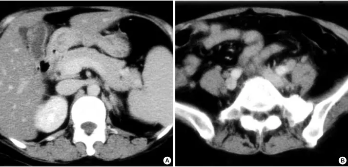
Fig. 1.Computed tomographic findings of abdomen appear acute acalculous cholecystitis and ileocolitis. (A) A thickened wall of gall blad- der with pericholecytic fluid collection, (B) An unenhanced wall thickenings of cecum and terminal ileum.
A B
Fig. 2.Clinical course and treatment of this patient. FFP, fresh frozen plasma; PC, platelet concentrate; PRC, packed red cell; PSL, pred- nisolone; IVIG, intravenous immunoglobulin; ND, not done.
Zoster rash
Famciclovir Sulindac Hydroxychloroquine PSL (5 mg/day) Iron (80 mg/day)
FFP PC
PRC Ceftriaxone IVIG (0.4 g/kg/day)
Methyl-PSL (1g/day)(62.5 mg/day) PSL (50 mg/day) Oral cyclosporine A (2-3 mg/kg/day) Fever
Confusion
Abdominal tenderness Confusion
Fever
WBC (×103/ L) 4.0 1.5 2.4 2.7 4.6 9.8
Hb (g/dL) 7.6 5.7 10.9 11.1 11.3 11.6
Platelet (×103/ L) 11 17 49 70 106 180
GOT (U/L) 412 203 93 62 24 27
GPT (U/L) 179 147 88 93 47 24
Total bilirubin (mg/dL) 4.2 2.8 2.8 2.3 2.0 1.2
Cr (mg/dL) 2.3 1.5 0.8 0.6 0.6 0.8
LDH (U/L) 2,495 1,570 697 409 194 200
Fibrinogen (mg/dL) 52 ND 170 142 143 198
D-dimer (mg/L) 4 ND 7.4 1.2 0.4 0.1
Ferritin (ng/mL) >1,831 ND 1,330 287 ND 112
-30 -3 0 1 2 3 4 5 6 7 8 9 10 16 42
Days (after admission)
graph and electrocardiogram appeared normal. Abdominal computed tomography (CT) images showed acute acalculous cholecystitis, ileocolitis (Fig. 1), hepatosplenomegaly, a small amount of right pleural effusion and ascites. The patient had no history of recurrent episode of abdominal pain or diarrhea.
Bone marrow (BM) biopsy and cerebrospinal fluid examina- tion were not performed due to hemorrhagic diathesis.
The combination of anemia, thrombocytopenia, coagulopa- thy, DIC, hepatic and renal dysfunctions, neurological symp- toms, and capillary leakage signs was compatible with severe systemic inflammatory response syndrome. A differential diagnosis of hemophagocytic syndrome and septic DIC was considered. Fluid and electrolytes replacement, fresh frozen plasma and platelet concentrates transfusions were immedi- ately initiated. Empirical antibiotics (ceftriaxone, 3 g/day) and intravenous immunoglobulin (IVIG, 400 mg/kg/day) therapy were also initiated. On the next day after the admission, con- fused mentality diminished, and serum creatinine level was normalized. Negative results for blood, throat, urine and stool cultures were noted. On the basis of patient’s clinical presen- tations, marked elevated ferritin, hypertriglyceridemia, and no evidence of infectious etiology, a presumptive diagnosis of reactive hemophagocytic syndrome (RHS) associated with AOSD was made. Intravenous pulsed methylprednisolone (Methyl-PSL, 1 g/day) and oral cyclosporine A (CsA, 2 mg/
kg/day) were added. Two day after the admission, fever was disappeared rapidly, and abdominal tenderness was also de- creased. On fourth day of the admission, high fever and neu- rological dysfunctions such as confusion, irritability, disorien- tation abruptly recurred, and abdominal tenderness re-aggra- vated despite treatment with IVIG (for 3 days), pulsed methyl- PSL (for 3 days) followed by methyl-PSL 62.5 mg/day, con- tinuous oral CsA and blood component replacements. Alth- ough levels of transaminase and LDH were gradually decreas- ed, pancytopenia and DIC progressed. On the sixth day of the admission, we increased the dosage of oral CsA from 2 mg/kg/day to 3 mg/kg/day. Fever and confusion were disap- peared dramatically within a day. She no longer needed any transfusion, with pancytopenia and DIC rapidly improving.
Subsequently, abdominal tenderness also disappeared within 3 days. On day 42 of the hospitalization, all laboratory abnor- malities except lipid profile returned to normal (Fig. 2). Previ- ous radiological abnormalities were also normalized on follow- up study. CsA administration was stopped, and PSL was ta- pered. She has remained clinically well with low-dose PSL alone for over one year.
DISCUSSION
HS is an uncommon disorder characterized by inappropriate systemic proliferation of benign histiocytes throughout the reticuloendothelial system and hemophagocytosis. It has been suggested to be caused by extensively activated T cells (Th1
cells) and macrophages, and subsequent overproduction of cytokines such as IL-1, IL-6 and IFN- (6). It can occur as a primary hemophagocytic lymphohistiocytosis, but more com- monly is secondary (also called RHS) to a variety of infections, neoplasms, drugs, autoimmune diseases or various immuno- deficiencies. Among the rheumatic disease, systemic onset juvenile rheumatoid arthritis (SOJRA) is most often associat- ed with HS, and it has been named macrophage activation syndrome (MAS) (7). As AOSD is similar to Still’s disease, it seems to be prone to developing reactive HS in adults (8).
Although we did not identify tissue demonstration of he- mophagocytosis in our case, typical clinical and laboratory find- ings of defining HS, such as the sudden onset of non-remit- ting high fever, change in mental state, hepatosplenomegaly, serositis, cytopenia, elevated serum transaminase, hyperbiliru- binemia, high concentrations of LDH and triglyceride, coag- ulopathy with prolonged PT and aPTT, hypofibrinogenemia, the presence of fibrin degradation products, hyperferritinemia, complete resolutions of all symptoms and abnormal findings by immunosuppressive therapy, can reasonably establish the diagnosis of HS (6, 8-10). In fact, the absence of histological confirmation of hemophagocytsis must not be allowed to delay treatment, which is urgently required. Our chronic AOSD patient with HS became acutely ill, and the clinical picture mimicked septic DIC or a flare-up of AOSD with complicat- ing DIC. However, patterns of non-remitting fever and pur- puritic or petechial rash were different from the remitting high- spiking fever and evanescent maculopapular rash of AOSD.
The most dramatic clue was the fall in the ESR inspite of both the face of a worsening clinical situation and the very high levels of ferritin. Low ESR is a strikingly unusual feature in active phase of inflammatory disease, and thought be related to hypofibrinogenemia. Hyperferritinemia is an important laboratory hallmark for diagnosis of HS and disease activity of AOSD. Patients with HS showed markedly elevated serum ferritin levels ranging from 1,000 to 250,000 ng/mL (2, 3).
In the appropriate clinical setting, a serum ferritin level exceed- ing 1,000 ng/mL may assist in establishing the diagnosis of HS (6).
The pathognomic feature of the HS is seen on BM exami- nation, which reveals numerous well-differentiated macrpha- ges actively phagocytosing hematopoietic cells. Association between massive hyperferritinemia and hemophagocytosis is strong, but BM specimen may be inconclusive in case of early sampling time or hemophagocytosis sparing bone mar- row (8, 11, 12). However, BM analysis is recommended in all patients with hyperferritinemias, not only for diagnostic accuracy but also given the high percentage of underlying malignancies such as malignant histiocytosis or lymphoma (13, 14). In our patient, the dramatic restoration of general condition by immunosuppressive treatment without a specific chemotherapy would support the unlikeness of malignancy associated HS inspite of omitting BM examination.
HS commonly occurs in patients with pre-existing immuno-
logical abnormalities due to various infections (15). In this case, herpes zoster preceded HS a month before the admission. Pre- sumed acute acalculous cholecystitis and ileocolitis were found in CT images on admission. Therefore, we suspected infection associated hemophagocytic syndrome at first. Viral infection may be a delayed trigger of HS whether antiviral agent was used or not (10). In fact, viral-associated HS frequently result- ed in severe pancytopenia, DIC and fulminant clinical course due to multiple organ failure. Enterocolitis and acute non-cal- culous cholecystitis can be concurrently caused by same pa- thogen (16). However, no active infection of viruses includ- ing VZV, bacteria, fungi, and parasites were detected in our case. Actually, it is difficult to confirm the absence of any infec- tious agents thoroughly. Although we treated this patient with a moderate dose of single broad-spectrum antibiotic as well as immunosuppressive agents, the lack of aggravation or relapse of infection under aggressive immunosuppression would also favor the unlikeness of underlying non-viral infection.
On the one hand, diffuse wall thickenings in colon, cecum and terminal ileum observed on CT suggests that inflamma- tory bowel disease such as Crohn’s disease cannot be ruled out.
HS associated with inflammatory bowel disease (IBD) is very rare and we are aware of only one previous case report of ful- minant ulcerative colitis associated HS (17). However, our patient had no previous history of recurrent episodes of abdom- inal pain or diarrhea at presentation of HS. After recovery, the patient has not shown a sign or symptom of IBD yet. Therefore, it is possible to consider the bowel lesion as a result of inflam- matory response by hypercytokinemia or tissue infiltrations by histiocytes associated with HS rather than a feature of IBD itself. The classical feature of acute acalculous cholecytitis revealed in our case is atypical, but patients with severe AOSD or HS often present with an acute abdomen. Abdominal pain is supposed to be due to peritoneal inflammation, acute enlar- gement of mesenteric lymph node, or functional obstruction of the intestine (1). In our case, there is a possibility that acute serositis mimics acute cholecystitis. We think that it is impor- tant to recognize this as a clinical feature of RHS with AOSD in order to avoid unnecessary surgical exploration. A number of triggers for MAS in SOJRA have been proposed, including aspirin or other non-steroidal anti-inflammatory drug toxicity, a second injection of gold salts, sulfasalazine and methotrex- ate therapy, as well as a viral infection (7). In our case, the patient had received treatment with sulindac, hydroxychloro- quine and oral ferrous sulfate at the onset of RHS, but there have been no reports of RHS related to these drugs. However, a case of acute hepatitis in AOSD apparently resulting from oral iron substitution was recently reported, and there was a suggestion that the iron exacerbated the macrophage hyper- activity in active phase of AOSD (18). In our case, RHS did not develop during the long-term iron supplement period for more than 8 months despite several flare-ups of AOSD. There- fore, RHS triggered by iron would be less likely.
Because RHS may have a fatal outcome, prompt recognition
and treatment are of the uppermost importance. The treatment strategy for RHS is usually based on the parenteral adminis- tration of high doses of corticosteroids. However, corticosteroid is effective in most but not all cases, and the use of high-dose IVIG, cyclophosphamide, plasma exchange, and etoposide has provided conflicting results. TNF- blocker may also be an effective therapeutic agent in conjunction with corticosteroid, and a steroid-sparing agent (19). CsA proved effective in treat- ing severe or corticosteroid-resistant HS (20, 21). In some pa- tients with MAS, the introduction of this drug leads a dramat- ic effect on the process of the disease, leading to resolution of the fever and improvement of the laboratory abnormalities within 12 to 24 hr (20). Although the exact mechanism by which CsA achieves immunosuppression is unknown, it is believed to exert its major effects by the suppression of the early steps in T-cell activation, leading to failure to activate the transcription of early genes such as those encoding for cytokines (22). CsA has also been shown to affect macrophage production of IL-6, IL-1 and TNF- and to inhibit the expres- sion of inducible nitric oxide synthetase and cyclooxygenase-2 in macrophages (23-25). CsA also inhibits the expression of key cell surface co-stimulatory molecules, thus altering the antigen-presenting function of dendritic cells for T cell acti- vation (26).
To our knowledge, this is the first case of AOSD associated HS masquerading as acute acalculous cholecystitis and ileo- colitis. We suggest that CsA can be considered as first-line treatment with steroids in life threatening HS such as DIC and multiple organ dysfunctions.
REFERENCES
1. Ohta A, Yamaguchi M, Kaneoka H, Nagayoshi T, Hiida M. Adult Still’s disease: review of 228 cases from the literature. J Rheumatol 1987; 14: 1139-46.
2. Coffernils M, Soupart A, Pradier O, Feremans W, Neve P, Decaux G. Hyperferritinemia in adult onset Still’s disease and the hemophago- cystic syndrome. J Rheumatol 1992; 19: 1425-7.
3. Kumakura S, Ishikura H, Munemasa S, Adachi T, Murakawa Y, Kobayashi S. Adult onset Still’s disease associated hemophagocyto- sis. J Rheumatol 1997; 24: 1645-8.
4. Takeshita A, Takeuchi T, Nakagawa A, Tsuda Y, Fukuda A, Nariya- ma K, Shibayama Y. Adult onset Still’s disease with hemophagocytic syndrome and severe liver dysfunction. Hepatol Res 2000; 17: 139- 44.
5. Yamaguchi M, Ohta A, Tsunematsu T, Kasukawa R, Mizushima Y, Kashiwagi H, Kashiwazaki S, Tanimoto K, Matsumoto Y, Ota T, Akizuki M. Preliminary criteria for classification of adult Still’s dis- ease. J Rheumatol 1992; 19: 424-30.
6. Imashuku S. Differential diagnosis of hemophagocytic syndrome:
underlying disorders and selection of the most effective treatment. Int J Hematol 1997; 66: 135-51.
7. Ravelli A. Macrophage activation syndrome. Curr Opin Rhematol
2002; 14: 548-52.
8. Emmenegger U, Frey U, Reimers A, Fux C, Semela D, Cottagnoud P, Spaeth PJ, Neftel KA. Hyperferritinemia as indicator for intravenous immunoglobulin treatment in reactive macrophage activation syn- dromes. Am J Hematol 2001; 68: 4-10.
9. Emmenegger U, Reimers A, Frey U, Fux C, Bihl F, Semela D, Cot- tagnoud P, Cerny A, Spaeth PJ, Neftel KA. Reactive macrophage activation syndrome: a simple screening strategy and its potential in early treatment initiation. Swiss Med Wkly 2002; 132: 230-6.
10. Stephan JL, Kone-Paut I, Galambrun C, Mouy R, Bader-Meunier B, Prieur AM. Reactive hemophagocytic syndrome in children with inflammatory disorders: a retrospective study of 24 patients. Rheuma- tology 2001; 40: 1285-92.
11. Henter JI, Arico M, Elinder G, Imashuku S, Janka G. Familial hemo- phagocytic lymphohistiocytosis: primary hemophagocytic lymphohis- tiocytosis. Hematol Oncol Clin North Am 1998; 12: 417-33.
12. Ost A, Nilsson-Ardnor S, Henter JI. Autopsy findings in 27 children with haemophagocytic lymphohistiocytosis. Histopathology 1998;
32: 310-6.
13. Janka G, Imashuku S, Elinder G, Schneider M, Henter JI. Infection and malignancy associated hemophagocytic syndromes: secondary hemophagocytic lymphohistiocytosis. Hematol Oncol Clin North Am 1998; 12: 435-44.
14. Miyahara M, Sano M, Shibata K, Matsuzaki M, Ibaraki K, Shimamoto Y, Tokunaga O. B-cell lymphoma-associated hemophagocytic syn- drome: clinicopathological characteristics. Ann Hematol 2000; 79:
378-88.
15. Fisman DN. Hemophagocytic syndromes and infection. Emerg Infect Dis 2000; 6: 601-8.
16. Otero Anton E, Lado Abeal J, Losada Arias E, Barrio Gomez E. Ente- rocolitis and acute non-calculous cholecystitis caused by Salmonella enteritidis. Med Clin (Barc) 1996; 106: 155-6.
17. Kanaji S, Okuma K, Tokumitsu Y, Yoshizawa S, Nakamura M, Niho Y. Hemophagocytic syndrome associated with fulminant ulcerative colitis and presumed acute pancreatitis. Am J Gastroenterol 1998;
93: 1956-9.
18. Maclachlan D, Tyndall A. Acute hepatitis in adult Still’s disease apparently resulting from oral iron substitution: a case report. Clin Rheumatol 2000; 19: 222-5.
19. Prahalad S, Bove KE, Dickens D, Lovell DJ, Grom AA. Etanercept in the treatment of macrophage activation syndrome. J Rheumatol 2001; 28: 2120-4.
20. Ravelli A, Viola S, De Benedetti F, Magni-Manzoni S, Tzialla C, Martini A. Dramatic efficacy of cyclosporine A in macrophage acti- vation syndrome. Clin Exp Rheumatol 2001; 19: 108.
21. Mouy R, Stephan J-L, Pillet P, Haddad E, Hubert P, Prieur A-M. Effi- cacy of cyclosporine A in the treatment of macrophage activation syndrome in juvenile arthritis: report of five cases. J Pediatr 1996;
129: 750-4.
22. Schreiber SL, Crabtree GR. The mechanism of action of cyclosporine A and FK506. Immunol Today 1992; 13: 136-42.
23. Garcia JE, Lopez AM, de Cabo MR, Rodriguez FM, Losada JP, Sa- rmiento RG, Lopez AJ, Arellano JL. Cyclosporin A decreases human macrophage interleukin-6 synthesis at post-transcriptional level. Medi- ators Inflamm 1999; 8: 253-9.
24. Garcia JE, de Cabo MR, Rodriguez FM, Losada JP, Lopez AJ, Arel- lano JL. Effect of cyclosporin A on inflammatory cytokine production by U937 monocyte-like cells. Mediators Inflamm 2000; 9: 169-74.
25. Attur MG, Patel R, Thakker G, Vyas P, Levartovsky D, Patel P, Naqvi S, Raza R, Patel K, Abramson D, Bruno G, Abramson SB, Amin AR.
Differential anti-inflammatory effects of immunosuppressive drugs:
cyclosporin, rapamycin and FK-506 on inducible nitric oxide synthase, nitric oxide, cyclooxygenase-2 and PGE2 production. Inflamm Res 2000; 49: 20-6.
26. Lee JI, Ganster RW, Geller DA, Burckart GJ, Thomson AW, Lu L.
Cyclosporine A inhibits the expression of costimulatory molecules on in vitro-generated dendritic cells: association with reduced nuclear translocation of nuclear factor kappa B. Transplantation 1999; 68:
1255-63.