349
©The Korean Society of Food Science and Technology
혈관내피세포에서 산양삼 추출물과 진세노사이드 Rg5의 혈관신생 효과
김나은
1·이미옥
1·장미희
1·정병희
1,*
1(주)해송KNS 기업부설연구소
Angiogenic effects of wood-cultivated ginseng extract and
ginsenoside Rg5 in human umbilical vein endothelial cells
Na-Eun Kim1, Mi-Ok Lee1, Mi-Hee Jang1, and Byung-Hee Chung1,*
1Research and Development Institute, Haesong KNS
Abstract Ginsenoside Rg5, one of the protopanaxadiol ginsenosides of wood-cultivated ginseng, has been implicated in various diseases, such as diabetes, cancer, and hypertension; however, its angiogenic activity and molecular mechanisms have not yet been elucidated. Here, we found that wood-cultivated ginseng extract and ginsenoside Rg5 increase in vitro proliferation, migration, and tube-like structure formation, which are typical phenomena associated with angiogenesis, in cultured human umbilical vein endothelial cells (HUVECs). Moreover, Ginsenoside Rg5 stimulated the phosphorylation of Akt, endothelial nitric oxide (NO) synthase (eNOS), and extracellular-regulated kinase (ERK)1/2, which are well-known signal mediators of the angiogenic pathway. Furthermore, Ginsenoside Rg5 did not accelerate the activation of ICAM-1 and VCAM-1 which are inflammatory response mediators. These results suggest that wood-cultivated ginseng extract and ginsenoside Rg5 stimulated in vitro angiogenesis by activating the Akt/eNOS- and ERK1/2-dependent signal pathways without inducing vascular inflammation.
Keywords: angiogenesis, wood-cultivated ginseng, ginsenoside Rg5, vascular endothelial growth factor, signal cascade
서
론
혈관신생(Angiogenesis)은 기존에 존재하고 있던 내피조직에서 새롭게 혈관이 형성되는 과정으로 상처치유나 배아발달과 같은 다양한 생리학적 혹은 만성 염증이나 종양 발생과 같은 병리학 적 상태의 기초단계라고 볼 수 있다(Folkman, 1971; Folkman, 1995; Folkman 등, 1997; Hanahan과 Folkman, 1996; Risau, 1997). 이 중에서 상처 치유와 같은 생리학적 상태는 새롭게 모 세혈관이 성장하는 과정에서 혈관신생에 의해 조절을 받게 되는 데, 새로운 모세혈관은 상처발생 3-5일 후에 형성되기 시작하며, 혈관의 증식과 섬유아세포의 이동, 새로운 콜라겐 침착이 나타나 게 된다(Folkman, 1971; Folkman, 1995). 내피세포 내부에서는 혈 관신생과 관련된 다양한 성장 인자들이 수용체에 결합하여 다양 한 신호들을 체내로 전달하며 세포의 증식(proliferaition)과 이동 (migration), 관 형성(tube formation)을 유도하게 된다(Bussolino 등, 1997; Folkman, 1971; Folkman, 1995). 혈액순환장애로 인한 허혈성 조직은 심근경색이나 뇌 허혈성 손상, 사지 허혈과 같은 여러 질환들을 야기하므로 이에 대한 최 소한의 치료방법으로서의 접근법이 필요하며, 이러한 과정을 통 해 혈관신생 현상을 유도하면 허혈성 조직 손상과 같은 질환들 을 치료할 수 있으므로 이를 위한 다양한 소재개발 연구가 진행 되고 있다(Chung 등, 2008). 그러므로 혈관내피세포생장인자 (vascular endothelial growth factor, VEGF)와 같은 혈관신생을 유 도하는 소재를 찾아 이를 활용하면 허혈성 질환이나 상처 치료, 조직 재생에 있어서 치료제로서의 개발전망이 높은 상황이다 (Chung 등, 2008).
장뇌삼이라고도 불리는 산양삼(Korean Panax ginseng, wood-cultivated ginseng)은 야생상태에서 자연적으로 발아한 산삼의 종 자나 묘삼을 깊은 산속의 그늘지고 습기가 많은 곳에 뿌려 생육 환경에 가깝게 방치해 두었다가 일정 기간이 경과한 후에 캐낸 삼을 말한다(Lim 등, 2007; Lui와 Staba, 1980). 재배 인삼에 비 해 향기가 강한 것이 특징이며, 약리활성이 자연 산삼 다음으로 뛰어나다고 알려져 있고, 진세노사이트 함량이 높아 항독성과 항 암, 혈압과 혈당강하, 항산화, 항비만, 간독성 등에 효능이 있다 고 알려져 있다(Choi 등, 2006; Jang 등, 2008; Jang 등, 2010; Kim 등, 2009; Kim과 Kwon, 2007; Kim과 Son, 2012; Kwon 등, 2003; Kwon, 2010; Min 등, 2007; Park 등, 2005; Rhim 등, 2009). 따라서 산에서 자연 발아하여 생산량이 적고 고가인 산삼 에 비해 산에서 인위적으로 재배한 산양삼이 산삼을 대체할 수 있는 약물로 주목받고 있으며, 산양삼에 대한 관심이 높아지면서 이에 대한 연구의 필요성이 대두됨에 따라 효능에 관한 다양한 연구들이 최근에 이루어지고 있는 실정이다(Kim 등, 2012; Lee 등, 2008). 이러한 산양삼의 주요 약리활성 물질인 사포닌은 글리코사이 드(glycoside)의 하나로 인삼의 사포닌인 진세노사이드는 다른 식 물계에는 거의 존재하지 않는 다마레인(dammarane) 계열의 트라 이터페노이드(triterpoenoid)인 프로토파낙사다이올(protopanaxadiol, *Corresponding author: Byung-Hee Chung, Research and
Develop-ment Institute, Haesong KNS, Chuncheon, Gangwon, 24232, Korea Tel: +82-33-258-6350
Fax: +82-33-258-6351
E-mail: [email protected]
Received February 1, 2018; revised April 2, 2018; accepted April 4, 2018
PPD)과 프로토파낙사트라이올(protopanaxatriol, PPT)의 글리코사 이드이다(Baek 등, 2015). 현재까지 연구된 진세노사이드에 대한 연구를 살펴보면, 진세노사이드 Rb1은 위염과 위궤양을 예방하 고 산화방지 작용이 있으며, 진세노사이드 Rb2는 암 독소 호르 몬에 대한 길한 작용, Rc는 항암효과, Rd는 신 기능부전 치료효 과와 신경계 보호 작용이 보고되었다(Jung 등, 2002; Choi, 1991; Hideo 등, 1998; Choi 등, 2003; Yokozawa와 Wu Liu, 2000). 또 한 희귀 진세노사이드인 Rg3와 Rh2는 항암작용과 항전이작용 등 이 연구되었으며(Popovich와 Kitts, 2004; Wang 등, 2007; Xin 등, 2006), 단일 진세노사이드의 효과보다는 복합체인 사포닌의 생리 활성이 더 높다는 보고도 나오고 있다(Attele 등, 1999; Morita, 1986). 하지만 산양삼과 Rb1이 람노사이드 가수분해효소(rham-nosidase)와 글루코사이드 가수분해효소(glucosidase)에 의해 가수 분해되어 생성되는 희귀 진세노사이드인 Rg5에 대한 연구는 많 이 않으며, 특히 혈관신생에 관한 연구는 전무한 실정이다. 따라서 본 연구에서는 산양삼 추출물과 희귀 진세노사이드인 Rg5가 혈관신생 과정에서 미치는 효과와 그 메커니즘에 대해 연 구하고자 하였다.
재료 및 방법
실험재료 산양삼은 조재범 홍천산양산삼에서 5-6년산의 국내산 원물을 구입하였으며, 진세노사이드 Rg5는 앰보연구소(Ambo Institute, Seoul, Korea)에서 구입하여 사용하였다. 인간 제대정맥 내피세포 와 배양에 필요한 배지는 Lonza (Walkersville, MD, USA)에서 구 입하여 사용하였으며, 혈관내피세포생장인자(vascular endothelial growth factor, VEGF)는 코마(KOMA Biotech, Seoul, Korea), 단 백질 항체는 Cell Signaling Technology (Danvers, MA, USA)와 BD Biociences (San Diego, CA, USA)에서 각각 구입하여 사용하 였으며, 기타 시약들은 Sigma-Aldrich (St. Louis, MO, USA)에서 구입하여 사용하였다. 산양삼 추출물 제조와 세포 배양 산양삼은 세척 후 자연건조하고 95% 주정(에탄올)을 용매로 하여 70oC에서 12시간 동안 추출한 후 고형물을 제거하고 추출 물을 회수하였다. 이후 60oBx가 될 때까지 농축한 후 동결건조 하여 산양삼 추출물 시료를 얻었다. 인간 제대정맥 내피세포인 HUVEC은 EGM Plus Bulletkit (Lonza, Walkersville, MD, USA) 를 이용하여 젤라틴이 코팅된 판에 옮겨 37oC, 5% 이산화탄소배양기에서 배양하였으며, 세포가 증식하면 계대배양을 실시하고 passage 3-8을 사용하였다. 실험 직전에는 1% M199 (Lonza, Walkersville, MD, USA) 배지에서 6시간 동안 starvation을 진행하 여 세포의 주기를 일정하게 조절하였다. 혈관내피세포의 증식도 조사 산양삼 추출물과 진세노사이드 Rg5를 농도별로 혈관내피 세포 에 처리하고 혈관 신생에 주요 인자로 알려진 VEGF를 양성대조 군으로 처리하여 세포의 성장 촉진을 얼마나 유도하는지를 살아 있는 세포의 미토콘드리아에서 반응하는 MTT법으로 확인하였다 (Carmichael 등, 1987). 2×10⁴ cells/well로 동일하게 배양한 세포에 시료를 농도별로 처리하고 20시간 동안 배양한 후 MTT solution (tetrazolium salt, Sigma-Aldrich, St. Louis, MO, USA)을 처리하
고 4시간 동안 다시 배양하였다. 이후 배지를 제거하고 DMSO 를 넣어 용해시킨 후 570 nm에서 분광광도계를 이용하여 흡광 도를 측정하였다.
혈관내피세포의 이동성 분석
세포의 이동성 분석은 Boyden’s chamber (Transwell, 6.5-mm-diameter polycarbonate filter, 8-µm pore size, Corning, NY, USA)를 이용하여 화학적인 자극에 반응하여 세포가 움직이는 성 질인 주화성을 이용한 chemotaxis assay법을 이용하여 측정하였 다(Chung 등, 2008; Lee 등, 2006). 24 well plates에 각각 600 µL의 1% M199 배지를 넣고, Transwell의 아래쪽 면에 젤라틴(1 mg/mL)을 도말한 후 위쪽 면에 1×10⁵ cells/well의 세포를 부착시 켰으며, 양성대조군인 VEGF와 산양삼 추출물, 그리고 진세노사 이드 Rg5를 농도별로 처리하였다. 4시간 후 transwell의 아래쪽으 로 이동한 세포를 H&E 염색법으로 염색하고 메탄올로 고정시켰 다. 이후 이동하지 않은 위쪽면의 세포를 면봉으로 제거하고 이 동한 세포를 현미경으로 확인하여 그 이미지를 eXcope (Dixi Science, Daejeon, Korea)으로 캡처하였으며 세포의 수는 Image J 프로그램(Wayne Rasband, National Institutes of Health, USA)을 이용하여 측정하였다.
혈관내피세포의 관 형성도 분석
Growth factor-reduced Matrigel (BD Biociences)에서 산양삼 추 출물과 진세노사이드 Rg5가 혈관내피세포의 관 형성에 어떠한 영향을 미치는지 측정하였다(Chung 등, 2008; Lee 등, 2006). 24 well plates에 matrigel 250 µL를 도말한 다음 2×10⁵ cells/well의 세포를 matrigel 위에 부착시키고 양성대조군인 VEGF와 시료인 산양삼 추출물 및 진세노사이드 Rg5를 농도별로 처리하여 배양 하였다. 18시간이 경과한 후 관 형성이 일어난 정도를 현미경으 로 확인하고 이미지를 캡처한 후 eXcope (Dixi Science)를 사용 하여 관의 길이를 측정하였다.
혈관신생 관련 단백질 발현도 분석
혈관신생에 관여하는 것으로 알려진 단백질인 Akt, endothelial nitric oxide synthase (eNOS)와 ERK (Extracellular Signal-regu-lated Kinase) 1/2의 인산화 정도와 발현량을 웨스턴 블랏으로 확 인하였다(Chung 등, 2008; Lee 등, 2005). 혈관내피세포를 배양한 후 RIPA buffer를 이용하여 lysis시키고 BCA protein assay (Ther-moscientific, IL, USA)로 정량한 후 동일한 양의 단백질을 SDS-PAGE gel에 전기영동하였다. 이후 폴리바이닐리덴다이플루오라 이드(polyvinylidene difluoride, PVDF, Millipore, Bed-ford, MA, USA) 막에 이동시키고 타깃 단백질 각각의 항체를 이용하여 배 양한 후 enhanced chemiluminescence (ECL) solution (Intron Biotechnology, Seongnam, Korea)을 이용하여 단백질의 발현 정도 와 인산화 정도를 확인하고 Image J 프로그램을 이용하여 단백 질의 인산화 정도를 정량하여 판단하였다.
통계처리
모든 측정은 독립적으로 3회 반복실험을 진행하고 측정값을 mean±SD으로 표시하였다. 측정값의 통계처리는 Student’s t-test를 이용하여 독립적인 두 집단간의 통계학적 비교를 수행하였으며, p<0.05인 것을 통계학적 유의성이 있는 것으로 판단하였다.
결과 및 고찰
산양삼 추출물과 진세노사이드 Rg5의 혈관내피세포 증식 효과 혈관신생에 있어서 가장 기본적인 단계는 세포의 증식이 활발 해지는 현상이므로(Chung 등, 2008) 산양삼 추출물(wood-cultivated ginseng extract, WGE)과 진세노사이드 Rg5를 혈관의 기저막 (basement membrane) 상에 존재하며 혈액과 직접 접하고 있는 혈 관내피세포에 처리하고 혈관신생의 주요 인자로 잘 알려져 있는 혈관내피세포생장인자를 양성대조군으로 처리하여 세포의 성장 촉진을 얼마나 유도하는지를 MTT 방법으로 확인하였다(Carmichael 등, 1987). 산양삼 추출물의 경우, 대조군과 비교하여 10 ng/mL에 서는 유의적으로 증가하지 않았으나, 20-100 ng/mL에서는 대조군 대비 유의적인 세포증식을 확인하였으며 양성대조군으로 사용한 VEGF와 유사한 수준으로 증가하는 것을 확인하였으며, 그 이상 의 농도에서는 세포의 증식효과는 나타나지 않았다(Fig. 1A). Rg5 의 경우, 10 nM에서는 거의 세포의 증식이 나타나지 않았으나 20-100 nM 수준에서 모두 양성대조군인 VEGF와 유사한 수준으 로 유의적인 세포 증식을 확인하였으며, 산양삼 추출물과 마찬가 지로 200 ng/mL 농도에서는 더 이상 세포의 증식은 확인되지 않 았다(Fig. 1B). 산양삼 추출물과 진세노사이드 Rg5 처리에 따른 혈관내피세 포의 이동성 증대 혈관신생에서 세포의 증식이 발생하면 그 이후 세포가 주화성 (chemotaxis) 현상에 의해 이동하게 되므로(Lee 등, 2006) 산양삼 추출물과 Rg5가 혈관내피세포의 이동과정에 영향을 미치는지 확 인하기 위하여 transwell plate를 이용하여 주화성 분석을 수행한 후 주화성에 의해 이동한 세포를 염색하고 그 수를 측정하였다 (Chung 등, 2008). 산양삼 추출물의 경우, 대조군에 비하여 10, 20 ng/mL에서 유의적으로 증가햐였으며 20 ng/mL세포의 이동성이 낮은 농도인 10, 20 ng/mL에서 유의적으로 높았으며 특히 20 ng/ mL의 경우 양성대조군인 VEGF보다 세포의 이동량이 높았다. 반 면에 50, 100 ng/mL에서는 산양삼 추출물을 처리하는 않은 세포 에 비해 이동성은 약간 증가하였으나 유의적인 차이는 없었다 (Fig. 2A-B). Rg5의 경우, 10, 20 nM에서 양성대조군인 VEGF와 유사한 수준의 유의적 증가를 나타내었으며, 그 이상의 농도에서 는 산양삼 추출물과 마찬가지로 대조군에 비해 세포의 이동이 약 간 증가하는 것을 확인하였으나 유의적인 아닌 것으로 나타났다 (Fig. 2C-D). 산양삼 추출물과 진세노사이드 Rg5의 혈관내피세포 관 형성 도 증식효과 혈관신생을 조절하는 것을 측정할 수 있는 방법 중에 하나로 증식 후 이동한 혈관내피세포가 관(tube) 형성을 유도하는지 확 인하기 위하여(Folkman, 1971; Folkman, 1995) matrigel을 이용하 여 산양삼 추출물과 Rg5가 관 형성에 영향을 미치는지 확인하였 다. 그 결과, 산양삼 추출물의 경우 추출물을 처리하지 않은 대 조군에 비해 관 형성이 10-100 ng/mL까지 모든 농도에서 증가하 였고, 양성대조군인 VEGF와 유사한 수준의 유의적 증가했다. 특 히 세포의 증식과 이동에서도 산양삼 추출물에서 증가하는 효과 를 보였지만 특히 관 형성도의 증가는 뚜렷하게 확인할 수 있었 다(Fig. 3A-B). Rg5는 처리하지 않은 세포에 비해 관 형성도가 10-100 nM에서 농도 의존적으로 유의적인 수준으로 증가하였으 며, 산양삼 추출물과 마찬가지로 세포의 관 형성도에 큰 영향을 미치는 것을 알 수 있었다(Fig. 3C-D). 이상의 결과로 볼 때 산 양삼 추출물과 Rg5는 세포증식과 이동, 관 형성에서 혈관신생 효 과가 유사한 수준으로 증가하였으며, 저농도에서 그 효과가 통계 학적 유의성을 가졌으므로, 산양삼 중에 존재하는 희귀 진세노사 이드인 Rg5가 혈관내피세포의 증식에 주요 영향을 미치는 인자 라는 인식을 하게 되었다. 또한 일정농도 이상에서는 산양삼 추 출물과 Rg5 모두 세포의 증식이 일어나는 않는 결과로 미루어 볼 때 정상세포내에서의 혈관신생 효과는 저농도에서 효과적임 을 유추해볼 수 있었으며, 추후 암세포들을 이용한 비정상적인 혈관의 생성에 있어서 세포의 증식이 어떻게 변화하는지 확인하 고 진세노사이드 중 항암효과로 가장 잘 알려진 Rg3와의 비교연 구를 통한 원인 규명의 필요성을 인식하게 되었다. 진세노사이드 Rg5 처리에 따른 혈관신생 신호전달 메커니즘 확인
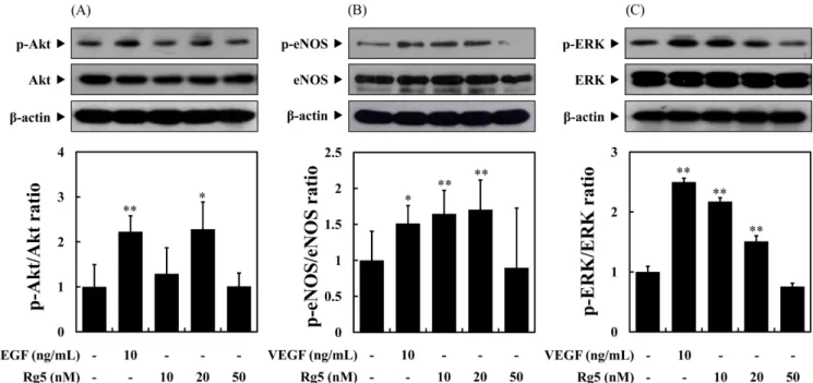
생존 신호전달 메커니즘인 phosphoinositide 3-kinase (PI3K)/Akt 활성화와 그 하부 신호전달 체계이며 내피세포에 특이적인 eNOS/ nitric oxide (NO) pathway는 혈관의 재생과 혈관신생에 밀접한 관 련이 있다고 보고되어 있다(Papapetropoulos 등, 1999). 특히 eNOS 는 성인의 신체기관에서 혈관신생에 중요한 역할을 담당한다고 알려져 있으며(Dulak과 Józkowicz, 2002; Namkoong 등, 2005), Fig. 1. WGE and Rg5 stimulate cell proliferation in human umbilical vein endothelial cells (HUVECs). HUVECs were induced with the indicated concentrations of wood-cultivated ginsen (WGE, A) and Ginsenoside Rg5 (B) or vascular endothelial growth factor (VEGF, positive control, 10 ng/mL) for 24 h, and cell proliferation was determined by MTT assay. All graphical data shown are the mean±SD (n=3). *p<0.05 and **p<0.01 versus control.
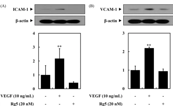
신체 내에서 eNOS가 감소하면 동물모델에서 신생 혈관의 형성 이 감소한다는 연구결과가 보고된 바 있다(Dulak과 Józkowicz, 2002; Gratton 등, 2003; Murohara 등, 1998). 또한 ERK 1/2의 경우 혈관신생 유도인자로 자극된 내피세포에서 세포의 증식과 이동, 그리고 관 형성에 중요한 인자로 보고되어 있으며, ERK 인산화가 억제될 경우 내피세포와 동물모델 모두에서 혈관신생 효과가 감소된다고 많은 연구자료에서 밝히고 있다(Dong 등, 2001; Huang 등, 2008; Lee 등, 2006). 따라서 혈관신생 메커니즘에서 중요한 위치를 차지하는 단백질인 Akt/eNOS와 ERK의 인산화를 Rg5가 유도하는지 확인하고자 하였으며, in vitro 혈관신생(혈관 내피세포의 증식, 이동, 관 형성) 실험에서 유의적인 증가를 보 였던 농도에서 단백질 발현량을 확인하였다(Fig. 4). 그 결과, Rg5 를 혈관내피세포에 10분간 처리한 경우 Akt의 인산화는 대조군 에 비하여 10, 20 nM에서 증가하였으며, 특히 20 nM에서는 양 성대조군인 VEGF와 유사한 수준의 유의적인 증가를 확인하였다. eNOS의 인산화는 아무것도 처리하지 않은 대조군에 비해 10, 20 nM에서 유의적으로 증가하였으며, 이는 양성대조군인 VEGF보다 높은 통계학적 유의성을 나타냈다. ERK의 인산화는 Rg5를 혈관 내피세포에 10분간 처리하였을 때 아무것도 처리하지 않은 세포 에 비해 인산화가 유도되었으며, 그 수준은 VEGF와 유사한 수 준으로 통계학적 증가량을 보였다. 반면에 Rg5를 50 nM에서 처 리하였을 때에는 대조군과 비교하여 단백질의 인산화에는 크게 영향을 미치지 못했으며, eNOS 인산화의 경우 표준편차가 커서 유의적인 증가는 확인할 수 없었다. 진세노사이드 Rg5 처리가 혈관염증인자에 미치는 영향 확인 혈관신생의 대표적인 유도인자로 알려져 있는 VEGF는 강력한 혈관신생 효과를 나타내지만 혈관 염증이나 혈관세포의 투과성 을 증가시키는 부작용에 대한 연구가 보고되어 있어 허혈성 심 질환이나 상처지연에 대한 치료제로서의 한계에 부딪친 상황이 다(Gavard와 Gutkind, 2006; Kim 등, 2001). 따라서 부작용을 나 타내지 않고 혈관신생 효능을 보이는 잠재적 인자들을 찾는 연 구가 지속되고 있으며, 본 연구에서는 Rg5가 혈관내피세포에서 혈관염증의 전형적인 지표로 알려져 있는 세포사이 부착분자-1 (intercellular adhesion molecule-1, ICAM-1)과 혈관세포 부착분자-1 (vascular cell adhesion molecule-부착분자-1, VCAM-부착분자-1)의 발현에 있어서 연관성이 있는지 확인하고자 하였다(Chung 등, 2008; Kim 등, 2001). 그 결과, 양성대조군인 VEGF가 ICAM-1, VCAM-1의 발 현을 유의적으로 증가시킨 것과 대조적으로 Rg5는 혈관신생 신 호전달 메커니즘이 가장 강한 농도인 20 nM을 처리하였을 경우, ICAM-1과 VCAM-1의 발현이 모두 증가하지 않았으며, 대조군과 유사한 수준으로 낮게 나타났다(Fig. 5). 따라서 Rg5는 혈관신생 의 강력한 유도인자이며 양성대조군으로 사용한 VEGF와 유사한 수준의 통계학적 유의성이 있는 혈관신생 효과를 갖고 있으며, Fig. 2. WGE and Rg5 induce cell migration in HUVECs. HUVEC suspensions were pre-treated for 20 min with the indicated concentrations of Wood-cultivated ginsen (WGE, A) and ginsenoside Rg5 (B) or vascular endothelial growth factor (VEGF, positive control, 10 ng/mL), and loaded into the upper wells of transwell plates for 4 h. Cell migration was quantified by counting the cells that migrated and stained (hematoxylin and eosin) to the lower side of the filter with optical microscopy at 200× magnification. All graphical data shown are the mean±SD (n=3). *p<0.05 and **p<0.01 versus control.
Fig. 3. WGE and Rg5 increase cell tube formation in HUVECs. HUVECs were cultured on a layer of Matrigel in the presence or absence of ood-cultivated ginsen (WGE, A) and ginsenoside Rg5 (B) or vascular endothelial growth factor (VEGF, positive control, 10 ng/mL) for 18 h. Tube formation was determined using an inverted phase contrast microscope with a video graphic system. The area covered by the tube network was quantitated using eXcope software. All graphical data shown are the mean±SD (n=3). *p<0.05 and **p<0.01 versus control.
Fig. 4. Rg5 stimulates the phosphorylation of Akt/eNOS and ERK1/2 in HUVECs. HUVECs were treated with the indicated concentrations of ginsenoside Rg5 or vascular endothelial growth factor (VEGF, positive control, 10 ng/mL) for 10 min. The phosphorylated levels of Akt (A), eNOS (B), and ERK (C) were determined by Western blot analysis using their specific antibodies. The area covered by the protein intensity was quantitated using Image J software. All graphical data shown are the mean±SD (n=3). *p<0.05 and **p<0.01 versus control.
VEGF의 부작용을 확인할 수 있는 지표인 혈관 염증관련 인자인 ICAM-1과 VCAM-1가 발현되지 않아 혈관신생 조절자로서의 역 할이 있는 것으로 확인되었다.
요
약
본 연구에서는 상처치유(wound healing)와 같은 허혈성 심뇌혈 관 질환의 잠재적 치료제로서 산양삼 추출물과 진세노사이드 Rg5 의 가능성을 인간 제대정맥 내피세포인 HUVEC에서 확인하고자 하였다. 그 결과, 산양삼 추출물과 Rg5는 10-100 nM의 저농도에 서 혈관신생 과정에서 발생하는 세포의 증식이나 이동, 관 형성 과정을 유의적으로 증진시켰으며, 그 증가현상은 산양삼 추출물 과 Rg5가 유사한 수준으로 발생하였다. 따라서 Rg5를 이용하여 혈관신생 과정에 관여하는 신호전달 메커니즘을 확인한 결과, Akt/eNOS와 ERK1/2의 인산화는 양성대조군으로 사용한 VEGF 와 유사한 수준으로 증가되는 것을 확인하였다. 마지막으로 혈관 신생 유도인자이며 양성대조군인 VEGF의 혈관염증 관련 부작용 이 Rg5의 혈관신생 효과에도 작용하는지 확인하기 위하여 혈관 염증 관련 단백질인 ICAM-1과 VCAM-1의 발현량을 확인한 결 과, ICAM-1과VCAM-1의 발현이 양성대조군인 VEGF에서는 유 의적으로 증가하였으나 Rg5를 처리한 경우에는 일반 대조군과 유사한 수준으로 낮게 나타났다. 따라서 본 연구는 산양삼 추출 물과 Rg5가 혈관신생 유도효과가 있으며, 이러한 현상은 Akt/ eNOS와 ERK 관련 신호전달 메커니즘을 통해 진행되고 이러한 효과가 혈관염증은 유도하지 않는다는 것을 입증하였으며, 잠재 적 치료제로서의 가능성을 확인하는 계기가 되었다.감사의 글
본 연구는 2017년도 중소벤처기업부의 지역주력산업육성 창의 융합 R&D 사업(R0005390)의 지원을 받아 수행된 것으로 이에 감사를 드립니다.References
Attele AS, Wu JA, Yuan CS. Ginseng pharmacology: multiple con-stituents and multiple actions. Biochem. Pharmacol. 58: 1685-1693 (1999)
Baek SH, Lee IH, Kim MJ, Kim EJ, Ha IH, Lee JH. Component analysis and toxicity study of combined cultivated wild ginseng pharmacopuncture. J. Int. Korean Med. 36: 189-199 (2015) Bussolino F, Mantovani A, Persico G. Molecular mechanisms of
blood vessel formation. Trends Biochem. Sci. 22: 251-256 (1997) Carmichael J, DeGraff WG, Gazdar AF, Minna JD, Mitchell JB.
Evaluation of a tetrazolium-based semiautomated colorimetric assay: Assessment of radiosensitivity. Cancer Res. 47: 943-946 (1987)
Choi KJ. The constituent of material ginseng and management of quality. Korean J. Ginseng Sci. 15: 247-256 (1991)
Choi SS, Lee JK, Han EJ, Han KJ, Lee HK, Lee J, Suh HW. Effect of ginsenoside Rd on nitric oxide system induced by lipopolysac-charide plus TNF-α in C6 rat glioma cells. Arch. Pharm. Res. 26: 375-382 (2003)
Choi EJ, Lee JM, Won SH, Kwon KR. Effects of cultivated wild ginseng pharmacopuncture on lowering lipid and oxidative capac-ity in biochemical and molecular biological study in obese rats. J. Pharmacopuncture. 9: 5-20 (2006)
Chung BH, Kim JD, Kim CK, Kim JH, Won MH, Lee HS, Dong M S, Ha KS, Kwon YG, Kim YM. Icariin stimulates angiogenesis by activating the MEK/ERK-and PI3K/Akt/eNOS-dependent sig-nal pathways in human endothelial cells. Biochem. Bioph. Res. Co. 376: 404-408 (2008)
Chung BH, Lee JJ, Kim JD, Jeoung D, Lee H, Choe J, Ha KS, Kwon YG, Kim YM. Angioginic activity of sesamin through the activation of multiple signal pathways. Biochem. Bioph. Res. Co. 391: 254-260 (2010)
Dong G, Chen Z, Li ZY, Yeh NT, Bancroft CC, Van Waes C. Hepa-tocyte growth factor/scatter factor-induced activation of MEK and PI3K signal pathways contributes to expression of proangiogenic cytokines interleukin-8 and vascular endothelial growth factor in head and neck squamous cell carcinoma. Cancer Res. 61: 5911-5918 (2001)
Dulak J, Józkowicz A. Nitric oxide and angiogenic activity of endot-helial cells: direct or VEGF-dependent effect? Cardiovasc. Res.
Fig. 5. Rg5 does not affect the expression of adhesion molecules in HUVECs. HUVECs were incubated with ginsenoside Rg5 (20 nM) or vascular endothelial growth factor (VEGF, positive control, 10 ng/mL) for 6 h. The protein lovels of ICAM-1 and VCAM-1 were determined by Western blot analysis. All graphical data shown are the mean±SD (n3). *p<0.05 and **p<0.01 versus control.
56: 487-488 (2002)
Folkman J. Tumor angiogenesis: therapeutic implications. New Engl. J. Med. 285: 1182-1186 (1971)
Folkman J. Angiogenesis in cancer, vascular, rheumatoid and other diseas. Nat. Med. 1: 27-31 (1995)
Folkman S, Chesney M, Grief. Vancouver Conference Review. Aids Care. 9: 39-43 (1997)
Gratton JP, Lin MI, Yu J, Weiss ED, Jiang ZL, Fairchild TA, Iwakiri Y, Groszmann R, Claffey KP, Cheng YC, Sessa WC. Selective inhibition of tumor microvascular permeability by cavtratin blocks tumor progression in mice. Cancer cell. 4: 31-39 (2003) Gavard J, Gutkind JS. VEGF controls endothelial-cell permeability
by promoting the β-arrestin-dependent endocytosis of VE-cad-herin. Nat. Cell Biol. 8: 1223-1234 (2006)
Hanahan D, Folkman J. Patterns and emerging mechanisms of the angiogenic switch during tumorigenesis. Cell. 86: 353-364 (1996) Hideo H, Seong JH, Yasatosi M, Masamori W, Hur JD. Metabolites
of ginseng saponin by enterobacteria and anticancer substance include it’s useful constituent. Korea Plant. 10-164266: 0000 (1998)
Huang D, Ding Y, Luo WM, Bender S, Qian CN, Kort E, Zhang ZF, VandenBeldt K, Duesbery NS, Resau JH, Teh BT. Inhibition of MAPK kinase signaling pathways suppressed renal cell carci-noma growth and angiogenesis in vivo. Cancer Res. 68: 81-88 (2008)
Jang HY, Park HS, Kwon KR, Rhim TJ. A study on the comparison of antioxidant effects among wild ginseng, cultivated wild gin-seng, and cultivated ginseng extracts. J. Pharmacopuncture. 11: 67-78 (2008)
Jang SB, Lim CS, Jang JH, Kwon KR. Anti-metastatic mechanism of mountain cultivated wild ginseng in human cancer cell line. J. Pharmacopuncture. 13: 37-43 (2010)
Jung CS, Hyun JE, Kim YS. Anti-oxidative Effect of Ginsenoside Rb1 on the HCL·Ethanol-Induced Gastric Tissue in Rats. Kor. J. Pharmacogn. 33: 252-256 (2002)
Kim YJ, Son DY. Antioxidant and inhibitory effects of Korean Panax ginseng extract on pro-inflammatory mediators in LPS-stimulated RAW264. 7 macrophages. J. Korean Soc. Food Sci. Nutr. 41: 1371-1377 (2012)
Kim BW, Kwon KR. The effect of cultivated wild ginseng extract on preadipocyte proliferation. J. Pharmacopuncture. 10: 29-35 (2007) Kim EL, Kim CS, Lee HY, Lee HR, Kim EY, Yoon MC, Shin SS.
Mountain cultivated ginseng water boiled extract decreases blood glucose level and improves lipid metabolism in male db/db mice. Korea J. Herb. 27: 69-75 (2012)
Kim I, Moon SO, Kim SH, Kim HJ, Hoh YS, Koh GY. Vascular endothelial growth factor expression of intercellular adhesion molecule 1(ICAM-1), vascular cell adhesion molecule 1(VCAM-1), and E-selectin through nuclear factor-κB activation in endot-helial cells. J. Biol. Chem. 276: 7614-7620 (2001)
Kim YJ, Park DI, Kwon KR. Case report on the improvement of liver functions by mountain cultivated wild ginseng pharma-copuncture. J. Pharmapharma-copuncture. 12: 107-112 (2009)
Kwon KR. Anticancer effect of mountain ginseng Pharmacopuncture to the nude mouse of lung carcinoma induced by NCI-H460 human non-small cell lung cancer cells. J. Pharmacopuncture. 13: 5-14 (2010)
Kwon KR, Cho AL, Lee SG. The study on acute and subacute toxic-ity and anti-cancer effects of cultivated wild ginseng herbal acu-puncture. J. Pharmacoacu-puncture. 6: 7-27 (2003)
Lee SJ, Kim KM, Namkoong S, Kim CK, Kang YC, Lee H, Ha KS, Han JA, Chung HT, Kwon YG, Kim YM. Nitric oxide inhibition of homocysteine-induced human endothelial cell apoptosis by down-regulation of p53-dependent Noxa expression through the formation of S-nitrosohomocysteine. J. Biol. Chem. 280: 5781-5788 (2005)
Lee JH, Kwon KR, Cha BC. Component analysis of cultivated gin-seng, red gingin-seng, cultivated wild gingin-seng, and red wild ginseng using HPLC method J. Pharmacopuncture. 11: 87-95 (2008) Lee SJ, Namkoong S, Kim YM, Kim CK, Lee H, Ha KS, Chung
HT, Kwon YG, Kim YM. Fractalkine stimulates angiogenesis by activating the Raf-1/MEK/ERK-and PI3K/Akt/eNOS-dependent signal pathways. Am. J. Physiol.-Heart C. 291: H2836-H2846 (2006).
Lim W, Mudge KW, Weston LA. Utilization of RAPD markers to assess genetic diversity of wild populations of North American ginseng (Panax quinquefolium). Planta Med. 73: 71-76 (2007) Lui JHC, Staba EJ. The ginsenosides of various ginseng plants and
selected products. J Nat Prod. 43: 340-346 (1980)
Min BI, Kim HH, Seo IB, Kwon KR. Antitumor effects and protec-tive effects against doxorubicin-induced testicular toxicity of cul-tivated wild ginseng extract in the B16/F10 melanoma-bearing C57BL/6 mice. J. Pharmacopuncture. 10: 85-100 (2007)
Morita T. Chemical studies on Panax genus plants grown in Asia. Hiroshima Univ. 6-7 (1986)
Murohara T, Asahara T, Silver M, Bauters C, Masuda H, Kalka C, Kearney M, Chen D, Symes JF, Fishman MC, Huang PL, Isner JM. Nitric oxide synthase modulates angiogenesis in response to tissue ischemia. J. Clin. Invest. 101: 2567 (1998)
Namkoong S, Lee SJ, Kim CK, Kim YM, Chung HT, Lee H, Han JA, Ha KS, Kwon YG. Prostaglandin E2 stimulates angiogenesis by activating the nitric oxide/cGMP pathway in human umbilical vein endothelial cells. Exp. Mol. Med. 37: 588-600 (2005)
Papapetropoulos A, Rudic RD, Sessa WC. Molecular control of nitric oxide synthases in the cardiovascular system. Cardiovasc. Res. 43: 509-520 (1999)
Park WP, Lee E, Kwon KR. Effects of distilled Cultivated Wild Gin-seng Herbal Acupuncture in Rats with Diabetes Induced by High Fat Diet. J. Pharmacopuncture. 8: 97-108 (2005)
Popovich DG, Kitts DD. Ginsenosides 20 (S)-protopanaxadiol and Rh2 reduce cell proliferation and increase sub-G1 cells in two cultured intestinal cell lines, Int-407 and Caco-2. Can. J. Physiol. Pharm. 82: 183-190 (2004)
Rhim TJ, Jeong HS, Kim YJ, Kim DY, Han YJ, Kwon HY, Kwon KR. A study on the comparison of antioxidant effects among cul-tivated ginseng and culcul-tivated wild ginseng extracts-Using the measurement of superoxide and hydroxy radical scavenging activ-ities. J. Pharmacopuncture. 12: 7-12 (2009)
Risau W. Mechanisms of angiogenesis. Nature 386: 671-674 (1997) Tonnesen MG, Feng X, Clark RA. Angiogenesis in wound healing. J.
Invest. Derm. Symp. P. 5: 40-46 (2000)
Wang W, Zhao Y, Rayburn ER, Hill DL, Wang H, Zhang R. In vitro anti-cancer activity and structure–activity relationships of natural products isolated from fruits of Panax ginseng. Cancer chemoth. Pharm. 59: 589-601 (2007)
Xin Y, Ni JS, Jiang X, Wang XR, Shi B, Wu JX. Inhibitory effect of 20 (S)-ginsenoside Rg3 on tumor growth. J. Jilin Univ.(Medicine Edition). 1: 024 (2006)
Yokozawa T, Wu Liu Z. The role of ginsenoside-Rd in cisplatin-induced acute renal failure. Renal failure. 22: 115-127 (2000)