INTRODUCTION
Epidemiological studies have analyzed the impact of dietary components on the incidence of breast cancer (1-7). While there has been no clear consensus on the specific link between breast cancer development and diet, one study reported that soya products rich in phytoestrogens may protect against breast cancer in premenopausal women (1). Foods containing soybean and ginseng contain large quantities of the iso- flavones genistein and daidzein, which are phytoestrogens with weak estrogen agonist activity that may interfere with the breast cancer promoting effects of physiologic estrogen (3). Increased soy protein consumption is significantly cor- related with a reduction in the risk of sex steroid implicated cancers such as breast cancer and prostate cancer (1, 4). While a meta-analysis supported a reduced risk of breast cancer development with intake of vegetables and to a lesser extent fruit (6), a pooled analysis of cohort studies could verify no significant reduction in breast cancer risk with either fruit or vegetable consumption in the adult years (7).
The molecular basis of the effects of ginseng on cell prolif- eration has not been identified. Previous studies have shown that American ginseng induces the expression of pS2, an
estrogen regulated gene (8). AG has also been shown to exert inhibitory growth effects on the hormone dependent MCF- 7 breast cancer cell line (8, 9).
This present investigation focuses on determining a pos- sible mechanism of action that may account for the breast cancer cell growth inhibitory properties of AG. The shift from quiescence (G0) to an actively growing state is a pre- requisite for entry into the cell cycle in most cells, and it is crucial step for cancer cells (10). Cell cycle progression is modulated by certain types of regulators known as cyclin kinase inhibitors. The first of these proteins to be identified and cloned was p21 (11-13). The p21 protein, a universal cell cycle inhibitor, binds cyclin-CDK complexes and pro- liferating cell nuclear antigen, thereby serving as a potent growth inhibitor and effector of cell-cycle checkpoint (14).
Induction of p21 expression has been linked to growth inhibition by p53 (11), and also has been found to signal growth arrest, independent of p53 in cells undergoing dif- ferentiation (15-17).
The purpose of this investigation was to determine if Amer- ican ginseng induces the cell cycle inhibitor p21 in hormone sensitive MCF-7 and insensitive MDA-MB-231 breast can- cer cell lines.
Rosemary B. Duda, Sung-Soo Kang, Sonia Y. Archer, Shufen Meng, Richard A. Hodin
Department of Surgery, Beth Israel Deaconess Medical Center, Harvard Medical School, Boston, U.S.A.
Address for correspondence Rosemary B. Duda, M.D.
Associate Professor of Surgery, Beth Israel Deaconess Medical Center Surgical Oncology, RW 871, 330 Brookline Avenue, Boston, MA. 02215, U.S.A.
Tel : 617-667-2169, Fax : 617-667-2978 E-mail : [email protected]
* This investigation was funded by the Surgical Oncology Research Fund, BIDMC. The American ginseng extract was donated by Chai Na Ta, Corp., Langley, British Columbia.
S54
American Ginseng Transcriptionally Activates p21 mRNA in Breast Cancer Cell Lines
American ginseng (AG) has been demonstrated to inhibit breast cancer cell growth in vitro. p21 protein, a universal cell cycle inhibitor, binds cyclin-CDK complexes, an important mechanism in cell cycle regulation. The purpose of this investigation was to determine if AG induces p21 gene expression in hormone sensitive (MCF-7) and insensitive (MDA-MB-231) breast cancer cell lines. Cells grown in steroid stripped medium (SSM) were treated with AG, 17- -estradiol (E2), genistein or cycloheximide (CHX). Northern blot analyses were performed using human p21Cip1 and 36B4 cDNA probes. Cell lines were transiently trans- fected with select mouse p21 CAT reporter constructs, including those lacking a p53 binding site. Cell cycle analyses was performed by FACScan. The results revealed that AG induced p21 mRNA expression in MCF-7 and MDA-MB-231 cells (p=0.0004; p≤0.0001, respectively). Neither E2nor genistein alter p21 mRNA expression. CHX, a protein synthesis inhibitor, did not block p21 mRNA expression induced by AG, indicating that p21 is induced as an immediate early gene. AG activated p21 reporter constructs in transfected cells, independent of p53 binding sites. The cell cycle proliferative phase was significantly decreased by AG and increased by E2(p≤0.0001). AG may inhibit breast cancer cell growth by transcriptional activation of the p21 gene, independent of p53.
Key Words : p21; American Ginseng; Breast Neoplasms
METHODS Cells and cell culture
MCF-7 (estrogen sensitive) and MDA-MB-231 (estrogen insensitive) breast cancer cell lines and the HT-29 colon cancer cell line were purchased from American Type Culture Collection (ATCC, Rockville, MD). The cells were grown in 75 cm2flasks for northern blot analyses, 100 mm culture plates for transient transfection and 25 cm2 flasks for cell cycle analyses, respectively, at 37℃and 5% CO2in Eagle’s Minimum Essential Medium (EMEM, Biowhittaker, MD) and supplemented with 10% fetal bovine serum, 584 mg/L L-glutamine and 5×10⁻²mg/mL gentamicin until the cells were grown to 70% confluence. The medium was changed every 2 to 3 days and the cells were split by trypsinization when they reached confluence.
The breast cancer cell lines were stripped of endogenous steroid hormone by successive passages in phenol red free Dulbeco’s Modified Eagle’s Medium (DMEM, Biowhittak- er, MD) containing 5% charcoal and dextran treated fetal bovine serum, 584 mg/mL L-glutamine, 0.2 IU/mL insulin and 5×10⁻²mg/mL gentamicin (18).
After 7 days of steroid withdrawal, the breast cancer cell lines were treated with select concentrations (50 to 2,000 g/mL) of a standardized American ginseng (AG) extract (Panax quinquefolium grown in Canada), (gift, CNT2000, Chi Na Ta Corp, Langley, British Columbia), 17- -estradi- ol (E2), (10-9 M, Sigma Chemical Company, St. Louis, MO), genistein (Gn), (10-6 M, Sigma) or cycloheximide (CHX), (10 g/mL, Sigma) for 1 to 48 hr and the medium and test compounds were replenished at 24 hr (19).
HT-29 colon cancer cells were treated with 5 mM of sodi- um butyrate for four hours as the positive control group for p21 mRNA expression (20).
Northern blot analyses
Total RNA was extracted by the single step method using Ultraspec RNA Isolation system (BIOTECX Laboratories, Houston, TX). Twenty micrograms of total RNA was elec- trophoresed onto a 1% agarose gel containing formaldehyde and transferred to a nylon membrane. The transferred RNA was crosslinked to a nylon membrane using UV stratalinker (Stratagene, La Jolla, CA). p21, a 1.009 kb Xho1/Ecor1 frag- ment derived from the human Cip1 cDNA (ATCC) and 36B4 cDNA were labeled with 32P-dCTP by the random primer method (21) to a specific activity of 5×10⁸cpm/ g DNA using Oligolabelling kit (Pharmacia Biotech, Piscat- away, NJ). The membranes were prehybridized for 16 hr, and then hybridized in 50% formamide/6×standard saline citrate (SSC)/0.5% sodium dodecyl sulfate (SDS)/5% Den- hart’s solution/100 g/mL salmon sperm DNA/0.001 M EDTA at 42℃for 16 hr. The cells were washed twice in 2
×SSC/0.2% SDS, twice in 1×SSC/0.2% SDS at room temperature and twice in 0.5×SSC/0.2% SDS at 65℃. The washed filters were exposed to Kodak BioMax MR film at - 70℃. The level of p21 mRNA expression was determined by using laser densitometry and Image Quant software (Molecular Dynamics 400-B 2D, Sunnyvaly, CA) and nor- malized for RNA content using the 36B4 cDNA control.
Transient transfection and Chloramphenicol Acetyl- transferase (CAT) Assays
The cells were grown at 50-60% confluence in 100 mm tissue culture plates and were transiently transfected with 10-20 g of p21 CAT reporter plasmids containing selected portions of the p21 promoter constructs (gift, Tayler Jacks, Howard Hughes Medical Institute, Center for Cancer Re- search, MIT, Cambridge, MA.) from 0 to 4.7 kb upstream of the transcriptional start site or empty vector, pJFCAT by using the CaPO4/DNA coprecipitation technique (Strata- gene) (22). After 24 hr of transfection, the transfectants were treated with or without AG 2,000 g/mL for 24 hr, then harvested by direct scraping followed by washing with phos- phate buffered saline (PBS). Total protein was isolated in 150-200 L of 0.25 M Tris-HCl. The concentration of the isolated protein was measured by CobasBio system (ROCHE Diagnostic, Nutley, NJ). Reaction products were applied onto a thin layer chromatography plate (23) and results of thin layer chromatography were recorded by standard instant photography using 55 positive negative film (Polaroid, Cam- bridge, MA) and were analyzed by densitometry scanning.
Flow cytometry
When the confluence of the cells reached 70% in 25 cm2 flasks in standard or steroid stripped medium, the cells were treated with 10-9M E2or 60 to 2,000 g/mL AG. Culture media and test compounds were replenished at 24 hr. Forty- eight hours after either E2or AG treatment, cells were har- vested by trypsinization, collected in 15 mL polyethylene tubes, and washed twice with phosphate buffered saline (PBS), and fixed in 70% ethanol. For DNA analyses, cells were centrifuged, and the pellet was stained with propidi- um iodide (PI, 50 g/mL) dissolved in 50 g/mL DNAse free RNAse and 0.1% Tritan X-100 (24-25). The cell pel- lets were dispersed by repeated pipetting and were placed at room temperature in the dark for 30 min. The DNA con- tent fluorescence due to PI was measured by flow cytometry (FACScan, Becton Dickinson, San Jose, CA) on a minimum of 10,000 cells in the band above 620 nm. Data were recorded on Cell Quest software (Becton Dickinson) after an electronic gating of nuclear aggregates by the doublet discriminating module. The percentage of each cell cycle component was calculated from the histogram with ModFit (Becton Dick- inson).
Statistical analyses
Fisher’s PLSD of the ANOVA test and mean±s.d. were used for statistical analyses and a p-value less than 0.05 was considered statistically significant.
RESULTS American ginseng induces p21 mRNA
All experiments for northern blot analyses were performed in steroid stripped medium (SSM). Positive and negative controls, including cells grown in standard medium (SM)
were utilized for all experiments. The doses of American ginseng (AG) tested ranged from 50 to 2,000 g/mL and the time of treatment ranged from 1 to 48 hr.
AG induced a dose-dependent increase in p21 mRNA expression in MCF-7 and MDA-MB-231 breast cancer cell lines. The greatest increase in p21 mRNA expression was identified with the AG 2,000 g/mL treatment dose (p=
0.0004; p≤0.0001, respectively). Neither E2nor Gn induced p21 mRNA expression in the MCF-7 or MDA-MB-231 breast cancer cell lines.
p21 mRNA induction by AG 2,000 g/mL was identified as early as 1 hr after initiation of treatment in both MCF-7 and MDA-MB-231 cell lines and continued to be detected at an increased level at 48 hr of treatment. At 4 hr of treat- ment, AG induced a peak p21 mRNA expression by approx- imately 4-fold in the MCF-7 cells and 3-fold in the MDA- MB-231 cells (p≤0.0001, both cell lines) in this time course experiment in comparison to the untreated SSM control group.
This increase in p21 mRNA expression induced by the treatment of breast cancer cells by AG was equivalent to the increase in p21 mRNA induced in the HT-29 colon cancer cell line positive control group treated with sodium butyrate.
At 48 hr of treatment, there was no detection of p21 mRNA induced by either E2or Gn in the MCF-7 or MDA-MB-231
p21
Fig. 1. Cycloheximide (CHX), the protein synthesis inhibitor, does not block p21 mRNA expression by American ginseng.
Both MCF-7 (A) and MDA-MB-231 (B) breast cancer cell lines were treated with AG 2,000 g/mL in the presence or absence of 10 g/mL CHX for 4 hr and 24 hr. p21 mRNA expression induced by AG was not blocked by concurrent treatment with CHX. There was no induction of p21 mRNA by E2alone or in combination with CHX. There was a 3.8-fold increase in p21 expression in MCF-7 cells treated with AG alone for 4 hr and a 4.2-fold increase in p21 expression when cells were treated with AG and CHX. At 24 hr of treatment, there was a 2.0-fold increase in p21 mRNA expression induced when MCF-7 cells were treated with AG alone and a 14.8- fold increase when treated with AG and CHX compared to the untreated control group. A 2-fold increase in p21 expression is noted for HT-29 cells treated with both sodium butyrate and CHX in comparison to sodium butyrate alone. In (A), the lanes corre- spond to the following treatments: a=SSM controls; b=E2(10-9 M) 4 hr; c=E2(10-9 M) 24 hr; d=AG 4 hr; e=AG 24 hr; f=E2and CHX 4 hr; g=E2and CHX 24 hr; h=AG and CHX 4 hr; i=AG and CHX 24 hr; j=HT-29 cells untreated control; k=HT-29 cells treated 4 hr with sodium butyrate; l=HT-29 cells treated 24 hr with sodium butyrate;
m=HT-29 cells and sodium butyrate and CHX 4 hr; n=HT-29 cells and sodium butyrate and CHX 24 hr.
A 3.3, 1.2 and 1.5 fold increase in p21 mRNA expression was detected for MDA-MB-231 cells treated with AG 2,000 g/mL for 4, 24 and 48 hr, respectively, in comparison to untreated controls.
There was a 4.7, 5.8, and 4.7 fold increase in p21 expression when CHX was added to AG for the same time periods, respectively.
In (B), the lanes correspond to the following treatments: a=SSM controls; b=AG 4 hr; c=AG and CHX 4 hr; d=AG 24 hr; e=AG and CXH 24 hr; f=AG 48 hr; g=AG and CHX 48 hr; h=CHX alone.
p36B4
a b c d e f g h i j k
a b c d e f g h i j
p21 p36B4
Increase of p21 CAT activity (%)
250
200
150
100
50
0
p21 (1.4 kb) CAT p21 (4.7 kb) CAT p21 (1.4 kb) CAT p21 (4.7 kb) CAT
control
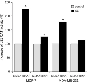
Fig. 2. American ginseng induces p21 promoter activity.
MCF-7 and MDA-MB-231 breast cancer cells were transiently transfected with 10 to 20 g of murine p21 CAT reporter plas- mid containing selected portions of p21 promoter constructs (from 0 to 4.7 kb upstream of the transcriptional start site). Trans- fectants were treated with AG 2,000 g/mL for 24 hr. This his- togram depicts the percent CAT induction of AG treated cells compared with the untreated negative control of p21 CAT activi- ty in MCF-7 and MDA-MB-231 cell line, arbitrarily taken as 100%. *p≤0.05.
AG
*
*
*
MCF-7 MDA-MB-231
A
B
breast cancer cell lines.
Fig. 1A and B illustrates the results of the concomitant treatment of the MCF-7 and MDA-MB-231 breast cancer cell lines with AG and the protein synthesis inhibitor, CHX, respectively. This experiment demonstrated that p21 mRNA expression induced by AG treatment is not blocked by CHX.
Both AG and CHX increased p21 levels greater than either AG or CHX alone, indicating that p21 is induced as an im- mediate early gene and is superinducible by cycloheximide in wild type cells.
American ginseng induces p21 gene expression in breast cancer cells, independent of p53 binding sites
The CAT reporter plasmids under the control of various portions of the mouse p21 gene 5′flanking region (from 0 to 4.7 kb upstream of the transcriptional initiation site) were transiently transfected into MCF-7 and MDA-MB-231 cells.
The transfectants were treated with AG 2,000 g/mL. AG induced p21 promoter activity in both MCF-7 and MDA-
MB-231 cells, from 1.4 to 4.7 kb, even in those plasmids lacking the two p53 binding sites, located at -2.85 and -1.95 upstream from the transcriptional start site. AG activated the mouse p21 CAT plasmids in both cell lines, in compari- son to that of the pJFCAT empty vector. The experiments were repeated four times to confirm the results. The results are shown in the histogram Fig. 2.
American ginseng induces growth arrest in breast cancer cells
Cell cycle analysis was performed in MCF-7 and MDA- MB-231 cell lines in steroid stripped medium (SSM) and standard medium (SM). It has been previously demonstrat- ed that culture medium contains various growth factors and steroid hormones that may influence the outcome of hor-
%-S-Phase
30,000
20,000
10,000
0.000 Control
Fig. 3. Effect of American Ginseng and Estradiol on the Prolifera- tive Phase (% S-Phase) of the Cell Cycle in Breast Cancer Cell Lines.
MCF-7 and MDA-MB-231 breast cancer cell lines were grown in either standard medium (SM) or steroid stripped medium (SSM) and treated for 48 hr with either AG (2,000 g/mL), E2(10-9 M) or SM/SSM as a control group. DNA content was determined by FACScan flow cytometry (n=4). The graph represents the mean
±s.d. For both cell lines grown in SM, AG significantly decreased the % S-phase (p≤0.001 for both) while AG had no significant effect on cells grown in SSM in comparison to the control groups.
E2significantly increased the %-S-phase in MCF-7 cells grown in SM (p=0.022) and SSM (p≤0.001). E2had no significant effect on MDA-MB-231 breast cancer cells grown in either SM or SSM.
AG E2 Control AG E2 Control AG E2 Control AG E2
Treatment
SM SSM SM SSM
MCF-7 MDA-MB-231
G0/G1 Phase
75.00
50.00
25.00
0.00 Control
Fig. 4. Effect of American Ginseng and Estradiol on the Quies- cent Phase (G0/G1) of the Cell Cycle in Breast Cancer Cell Lines.
MCF-7 and MDA-MB-231 breast cancer cell lines were grown in standard medium (SM) and steroid stripped medium (SSM) and treated for 48 hr with either AG (2,000 g/mL), E2(10-9 M) or SM/SSM as a control group. DNA content was determined by FACScan flow cytometry (n=4). The graph represents the mean
±standard deviation. For MCF-7 and MDA-MB-231 breast can- cer cell lines grown in SM, AG significantly increased the G0/G1 phase (p≤0.0001 for both cell lines) in comparison to the respective control groups. AG also increased the G0/G1 phase in MCF-7 cells grown in SM (p≤0.01) but had no significant effect on MDA-MB-231 cells grown in SSM. E2had no significant effect on the G0/G1 phase of MCF-7 cells grown in SM or in MDA-MB-231 cells grown in SM or SSM. E2did significantly decrease the G0/G1 phase (p≤0.0001) in MCF-7 cells grown in SSM in comparison to the control group.
AG E2 Control AG E2 Control AG E2 Control AG E2
Treatment
SM SSM SM SSM
MCF-7 MDA-MB-231
monally related experiments (26), hence the effect on the proliferative (% S-phase) and resting phases (G0/G1) of the cell cycle was evaluated and compared in both mediums.
Both cells lines were treated with either E2(10-9 M) or AG (2,000 g/mL) and a negative control was used for all exper- iments.
In the MCF-7 cells grown in SM, E2significantly increased the % S-phase (p=0.022) and AG significantly decreased the % S-phase (p≤0.001) compared to the untreated con- trol groups, as demonstrated in Fig. 3. E2had no effect on the G0/G1phase while AG significantly increased this phase (p≤0.0001). For MCF-7 cells grown in SSM, E2significant- ly increased the % S-phase (p≤0.001) while AG exhibited a non-significant decrease of the proliferative phase in this medium (p=0.10). AG but not E2 increased the G0/G1phase (p≤0.01) in SSM cells.
The effect of AG on the % S-phase in MDA-MB-231 cells grown in SM was similar to that of the MCF-7 cells grown in SM. AG decreased (p≤0.001) the %-S-phase and increased the resting phase (p≤0.0001) while E2(p=NS) had no effect on either cell cycle phase in this estrogen insensitive cell line.
Neither AG nor E2had any effect on the % S-phase or G0/G1 phase of MDA-MB-231 cells grown in SSM. The differences in apoptotic cell counts were minimal between controls and AG treated breast cancer cells (data not shown). Of note is the observation that the % S-phase for the control groups are significantly higher when the cell lines are grown in SM in comparison to SSM. Fig. 3 and 4 are histogram depictions of the results of the studies of the % S-phase and the G0/G1 cell cycle phases in MCF-7 and MDA-MB-231 cell lines.
DISCUSSION
The results from this study suggest that one possible mech- anism of action of inhibition of breast cancer cell growth by AG may be through a molecular pathway involving the in- duction of the p21 gene. Induction of p21 by sodium butyrate in the HT-29 colon cancer cell line served as a model for this investigation. Butyrate induction of p21 may represent a molecular link between a high fiber diet and the preven- tion of colon cancer (20).
One epidemiologic study reported that ginseng may con- tribute to a reduced risk of cancer development (27), but there is a paucity of data regarding the molecular mechanism involving the effects of ginseng on cell proliferation. In this present investigation, northern blot analyses were performed to examine the effects of American ginseng on the p21 expres- sion. These studies established that AG induced p21 mRNA expression in a time and dose dependent manner in both hormone sensitive (MCF-7) and insensitive (MDA-MB-231) breast cancer cell lines. Induction of p21 mRNA expression was identified as early as 1 hr and peaked at 4 hr after AG treatment in MCF-7 and MDA-MB-231 cells. The protein
synthesis inhibitor, cycloheximide (CHX) did not block American ginseng induced p21 expression, indicating that p21 is an immediate early gene.
This rapid p21 induction is similar to that seen with many early genes such as Egr-1 (28), c-fos (29), or c-jun (30) that are induced by mitogenic or other stimuli in the absence of de novo protein synthesis and thus constitute the first step in such a cascade. Macleod et al. showed that expression of the p21 mRNA following serum stimulation is superinducible by cycloheximide in wild type primary mouse embryonic fibroblasts (22), similar to the results of these present studies.
Estrogenic steroids perform several major roles in mam- malian physiology which include control of the development of the reproductive tract and secondary sex organs. They are also intimately linked with the development and progres- sion of a number of human cancers, particularly breast can- cer, by stimulating progression through G1phase of the cell cycle (31). Planas-Silva et al. (32) have shown that cyclin D1 has an important role in steroid-dependent cell proliferation and that estrogen, by stimulating the activities of G1cyclin- dependent kinases, can control the proliferation of breast cancer cells.
17- -estradiol (E2) was shown to exhibit no effect on p21 mRNA expression in either estrogen sensitive or insensitive cell lines. E2increased the % S-phase fraction in MCF-7 cells but not in MDA-MB-231 cells. These cell cycle studies sup- port previous studies that indicate that the MCF-7 breast cancer cell line is not only dependent on estrogen but that estradiol also has mitogenic effects in this estrogen depen- dent breast cancer cell line (19). This mitogenic property appears to be independent of p21 activation.
Several in vivo studies have revealed that estrogen and estro- gen agonists, such as genistein and daidzein, can act as chemo- preventive agents to inhibit the development of carcinogen induced mammary tumors (33-35). Hsieh et al. reported a biphasic proliferative effect of genistein on MCF-7 cells (36).
This study suggested that low dose genistein (as low as 10-8 M) enhanced the proliferation of MCF-7 cells in vitro, with a concentration of 10-7 M achieving proliferative effects sim- ilar to those of 10-9M estradiol. At concentrations above 2
×10-5 M, however, genistein significantly inhibited MCF-7 cell growth. Since genistein had no effect on p21 expression, the mechanism of action for cell growth regulation must in- volve a different molecular event.
It has been shown that induction of p21 following expo- sure to ionizing radiation or other DNA damaging agents requires p53 function (37). p53 mediates p21 gene induc- tion by transactivation through cis-elements located 1.95 and 2.85 kb upstream from the transcriptional initiation site in the mouse and 2.4 kb upstream in human p21 gene (11, 22). p53 appears to be independent of p21 expression during normal mammalian development and cellular differ- entiation. This data from this present investigation suggests that AG may possibly achieve its growth inhibition in
breast cancer cells by transcriptional upregulation of cyclin dependent kinase inhibitor, p21, through one or more cis- element, in a p53 independent fashion.
Since AG induced p21 mRNA expression in a time and dose dependent manner, flow cytometry studies were per- formed to determine whether AG would induce growth arrest in breast cancer cells. This data demonstrate that the
% S-phase fraction of the cell cycle is significantly decreased by AG in the cells grown in standard medium and to a less- er degree in steroid stripped medium.
In the northern blot analyses, induction of p21 mRNA expression by AG begins at a concentration of 50 g/mL in MCF-7 cells and 500 g/mL in MDA-MB-231 cells. The peak increase is found at 2,000 g/mL in both MCF-7 and MDA-MB-231 cells. This is the same dose of this standard- ized AG extract that has been previously shown to induce mRNA expression of the estrogen regulated gene pS2 to equal levels as E210-9 M induced pS2 expression (38). In pre- vious experiments, it had been shown that higher doses of E2did not further increase the levels of pS2 mRNA expres- sion in MCF-7 cells, hence this was the dose used in these current experiments. This increasing dose response of AG on p21 mRNA expression is inversely related to the decrease of S-phase fraction of the cell cycle in standard medium, with the higher doses of AG resulting in a larger decrease in % S-phase. For both cell lines, the basal % S-phase fraction was higher in standard medium than steroid stripped medium.
This is most likely secondary to the lack of growth factors and steroid hormones in SSM. Passage of cells with phenol red free medium containing 5% dextran charcoal treated fetal bovine serum followed by washing with phosphate buffered saline (PBS) removes not only endogenous steroids but also various growth factors (26).
Some investigators have shown that p21 binds tightly to the G1and S-phase kinases, cyclin E/Cdk2, cyclin D/Cdk 4, and cyclin A/Cdk2 and inhibits their activity, whereas p21 is a relatively poor inhibitor of the G2/M phase kinase, Cyclin B/Cdc2 (39). G0/G1phase was analyzed to investigate cyclin dependent kinase (CDK) inhibitor of cell cycle progression as a potential mechanism by which American ginseng neg- atively regulate cell proliferation. As shown in Fig. 4, AG induces G0/G1arrest in both MCF-7 and MDA-MB-231 cells grown in standard medium, suggesting that the induction of the CDK inhibitor may be at least partly responsible for growth arrest induced by AG. Conversely, estradiol signifi- cantly decreases G0/G1phase and increases % S-phase of the cell cycle in MCF-7 cells grown in standard medium, indi- cating a G1/S transition and a mitogenic effect of E2on hor- mone dependent breast cancer cells.
The role of p21 in apoptosis is still not clear. Some inves- tigators have shown that induction of apoptosis is associated with upregulation of endogenous p21 (40). However, others have reported that the p21 gene is not essential for apoptosis (41) or protects the cells from apoptosis (42, 43). As deter-
mined by FACScan, this study found minimal differences in apoptotic cell counts between control and American ginseng treated cells. This result is consistent with the report of Hague et al., which showed minimal apoptosis in butyrate treated adherent colon cancer cells, but extensive apoptosis in those that were floating (44). Since adherent cells and not floating cells were harvested and subjected to FACScan in order to identify apoptosis, additional studies will be needed to deter- mine if American ginseng induces apoptosis.
In conclusion, this investigation demonstrates that Amer- ican ginseng exhibits breast cancer cell growth inhibitory properties by a mechanism that may involve transcriptional upregulation of cyclin dependent kinase inhibitor, p21, independent of p53.
REFERENCES
1. Lee HP, Gourley L, Duffy SW, Esteve J, Lee J, Day NE. Dietary effects on breast cancer risk in Singapore. Lancet 1991; 337: 1197- 200.
2. Willett WC, Stampfer MJ, Colditz GA, Rosner BA, Hennekens CH, Speizer FE. Dietary fat and the risk of breast cancer. N Engl J Med 1987; 316: 22-8.
3. Ingram D, Sanders K, Kolybaba M, Lopez D. Case control study of phyto-estrogen and breast cancer. Lancet 1997; 350: 990-4.
4. Adler-Creutz H, Honjo H, Higashi A, Fotsis T, Hamalainen E, Hasegawa T, Okada H. Urinary excretion of lignans and isoflavonoid phytoestrogens in Japanese men and women consuming a traditional Japanese diet. Am J Clin Nutr 1991; 54: 1093-100.
5. Mills PK, Beeson WL, Phillips RL, Fraser GE. Dietary habits and breast cancer among Seventh-day Adventists. Cancer 1989; 64:
582-90.
6. Gandini S, Merzenich H, Robertson C, Boyle P. Meta-analysis of stud- ies on breast cancer risk and diet. Eur J Cancer 2000; 36: 636-46.
7. Smith-Warner SA, Spiegelman D, Yaun SS, Adami HO, Beeson WL, Van den Brundt PA, Folsom AR, Fraser GE, Freudenhein JL, Goldbohm RA, Graham S, Miller AB, Potter JD, Rohan TE, Speiter FE, Toniolo P, Willett WC, Wolk A, Zeleniuch-Jacquotte A, Hunter DJ. Intake of fruits and vegetables and risk of breast cancer-A pooled analysis of cohort studies. JAMA 2001; 285: 769-76.
8. Duda RB, Tabak B, Kessel B, Dooley DD, Yang H, Marchiori J, Slomovic BM, Alvarez JG. pS2 expression induced by American ginseng in MCF-7 breast cancer cells. Ann Surg Oncol 1996; 3:
515-20.
9. Duda RB, Dooley DD, Tabak B, Slomovic BM, Alvarez JG. Estro- genic effects of American ginseng in MCF-7 breast cancer cells. Proc Annu Meet Am Assoc Cancer Res 1996; 37: A1947.
10. Nurse P, Masui Y, Hartwell L. Understanding the cell cycle. Nature Med 1998; 4: 1103-6.
11. el-Deiry WS, Tokino T, Velculescu VE, Levy DB, Parsons R, Trent JM, Lin D, Mercer WE, Kinzler KW, Vogelstein B. WAF1, a poten- tial mediator of p53 tumor suppression. Cell 1993; 75: 817-25.
12. Harper JW, Adami GR, Wei N, Keyomarsi K, Elledge SJ. The p21
cdk-interacting protein Cip1 is a potent inhibitor of G1 cyclin-depen- dent kinase. Cell 1993; 75: 805-16.
13. Xiong Y, Hannon GJ, Zhang H, Casso D, Kobayashi R, Beach D.
p21 is a universal inhibitor of cyclin kinases. Nature 1993; 366: 701-4.
14. Sherr CJ, Roberts JM. Inhibitors of mammalian G1 cycle dependent kinases. Genes & Dev 1995; 9: 1149-63.
15. Zhang W, Grasso L, McClain CD, Gambel AM, Cha Y, Travali S, Deisseroth AB, Mercer WE. p53-independent induction of WAF1/
CIP1 in human leukemia cell is correlated with growth arrest accom- panying monocyte/macrophage differentiation. Cancer Res 1995;
55: 668-74.
16. Parker SB, Eichele G, Zhang P, Rawls A, Sands AT, Bradley A, Olson EN, Harper JW, Elledge SJ. p53-independent expression of p21Cip1in muscle and other terminally differentiating cells. Science 1995; 267: 1024-7.
17. Somasundaram K, Zhang H, Zeng YX, Houvras Y, Peng Y, Zhang H, Wu GS, Licht JD, Weber B, EL-Deiry WS. Arrest of the cell cycle by the tumor-suppressor BRCA1 requires the CDK inhibitor p21WAF1/ Cip1. Nature 1997; 389: 187-90.
18. Berthois Y, Katzenellenbogen JA, Katzenellenbogen BS. Phenol red in Tissue culture is a weak estrogen: implications concerning the study. Proc Natl Acad Sci USA 1986; 83: 2496-500.
19. Saceda M, Lippman ME, Lindsey RK, Puente M, Martin MB. Role of an estrogen receptor-dependent mechanism in the regulation of estrogen receptor mRNA in MCF-7 cells. Mol Endocrinol 1989; 3:
1782-7.
20. Archer SY, Meng S, Shei A, Hodin RA. P21WAF1is required for butyrate-mediated growth inhibition of human colon cancer cells.
Proc Natl Acad Sci USA 1998; 95: 6791-6.
21. Fineberg AP, Vogelstein B. A technique for radiolabelling DNA restriction endonuclease fragment to high specific activity. Anal Biochem 1984; 137: 266.
22. Macleod KF, Sherry N, Hannon G, Beach D, Tokino T, Kinzler K, Vogelstein B, Jacks T. p53-dependent and independent expression of p21 during cell growth, differentiation, and DNA damage. Genes
& Dev 1995; 9: 935-44.
23. Gorman CM, Moffat LF, Howard BH. Recombinant genomes which express CAT in mammalian cells. Mol Cell Biol 1982; 2: 1044-51.
24. Chinery R, Brockman JA, Peeler MO, Shyr Y, Beauchamp RD, Coffey RJ. Antioxidants enhance the cytotoxicity of chemotherapeu- tic agents in colorectal cancer: A p53-independent induction of p21WAF/CIP1via C/EBPbeta. Nature Med 1997; 3: 1233-41.
25. Brunner N, Bronzert D, Vindelov LL, Rygaard K, Spang-Thomsen M, Lippman ME. Effect on growth and cell cycle kinetics of estradi- ol and tamoxifen on MCF-7 human breast cancer cells grown vitro and in nude mice. Cancer Res 1989; 49: 1515-20.
26. Darbre PD, Curtis S, King RJ. Effect of estradiol and tamoxifen on human breast cancer cells in serum-free culture. Cancer Res 1984;
44: 2790-3.
27. Yun TK, Choi SY. Non-organ specific cancer prevention of ginseng:
a prospective study in Korea. Int J Epidemiol 1998; 27: 359-64.
28. Gashler AL, Swaminatan S, Sukhatme VP. A novel repression mod- ule, an extensive activation domain, and a bipartite nuclear local- ization signal defined in the immediate early transcription factor
Egr-1. Mol Cell Biol 1993; 13: 4556-71.
29. Kruijer W, Cooper JA, Hunter T, Verma IM. Platelet-derived growth factor induced rapid but transient expression of the c-fos gene and protein. Nature 1984; 312: 711-6.
30. Rysec RP, Harai SI, Yaniv M, Bravo R. Transcriptional activation of c-jun during the G0G1 transition in mouse fibroblasts. Nature 1988; 334: 535-7.
31. Prall OWJ, Sarcevic B, Musgrove EA, Watts CKW, Sutherland RL.
Estrogen-induced activation of cdk4 and cdk2 during G1-S phase progression is accompanied by increased cyclin D1 expression and decreased cyclin dependent kinase inhibitor association with cyclin E-cdk2. J Biol Chem 1997; 272: 10882-94.
32. Planas-Silva MD, Weinberg RA. Estrogen-dependent cyclin E cdk-2 activation through p21 redistribution. Mol Cell Biol 1997; 17: 4059-69.
33. Setchell KDR, Borriello SP, Hulme P, Kirk DN, Axelson M. Nons- teroidal estrogens of dietary origin: possible roles on hormone-depen- dent disease. Am J Clin Nutr 1984; 40: 569-78.
34. Adlercreutz H. Western diet and Western disease: some hormonal and biochemical mechanisms and associations. Scand J Lab Inves- tig 1990; 201: 3-23.
35. Barnes S, Grubbs C, Setchell KDR, Carlson J. Soybeans inhibit mam- mary tumors in model of breast cancer. Prog Clin Biol Res 1990; 347:
239-53.
36. Hsieh CY, Santell RC, Haslam SZ, Helferich WG. Estrogenic effect of genistein on the growth of estrogen receptor-positive human breast cancer (MCF-7) cells in vitro and in vivo. Cancer Res 1998; 58:
3833-8.
37. Di Leonardo A, Linke SP, Clarkin K, Wahl GM. DNA damage trig- gers a prolonged p-53 dependent G1 arrest and long term induction of Cip 1 in normal human fibroblasts. Genes & Dev 1994; 8: 2540-51.
38. Duda RB, Zhong Y, Navas V, Li MZL, Toy BR, Alavarez JG. Amer- ican ginseng and breast cancer therapeutic agents synergistically inhibit MCF-7 breast cancer cell growth. J Surg Oncol 1999; 72:
230-9.
39. Harper JW, Elledge SJ, Keyomarsi K, Dynlacht B, Tsai LH, Zhang P, Dobrowolski S, Bai C, Connell-Crowley L, Swindell E, Fox MP, Wei N. Inhibition of cyclin dependent kinases by p21. Mol Biol Cell 1995; 6: 387-400.
40. Duttaroy A, Qian JF, Smith JS, Wang E. Up-regulated p21 Cip1 expression is part of the regulation quantitatively controlling serum deprivation-induced apoptosis. J Cell Biochem 1997; 64: 434-46.
41. Attardi LD, Lowe SW, Brugarolas J, Jacks T. Transcriptional acti- vation by p53, but not induction of the p21 gene, is essential for oncogenic-mediated apoptosis. EMBO J 1996; 15: 3693-701.
42. Gorospe M, Cirielli C, Wang X, Seth P, Capogrossi MC, Holbrook NJ. P21WAF1/Cip1protects against p53-mediated apoptosis of human melanoma cells. Oncogene 1997; 14: 929-35.
43. Bissonette N, Hunting DJ. p21 induced cycle arrest in G1 protects cells from apoptosis induced by UV-radiation or RNA polymerase II blockage. Oncogene 1998; 16: 3461-9.
44. Hague A, Elder DJE, Hicks DJ, Paraskeva C. Apoptosis in colorec- tal tumor cells: induction by the short chain fatty acids butyrate, propionate and acetate and the bile salt deoxycholate. Int J Cancer 1995; 60: 400-6.