ARTICLE
J Korean Thyroid Assoc 2013 November 6(2): 96-100 http://dx.doi.org/10.11106/jkta.2013.6.2.96Received October 10, 2012 / Accepted March 6, 2013
Correspondence: Sun Wook Cho, MD, PhD, Department of Internal Medicine, Seoul National University College of Medicine, 101 Daehak-ro, Jongno-gu, Seoul 110-744, Korea
Tel: 82-2-2072-4761, Fax: 82-2-762-2292, E-mail: [email protected]
Copyright ⓒ 2013, the Korean Thyroid Association. All rights reserved.
This is an open-access article distributed under the terms of the Creative Commons Attribution Non-Commercial License (http:// creative- commons.org/licenses/by-nc/3.0/), which permits unrestricted non-commercial use, distribution, and reproduction in any medium, provided the original work is properly cited.
면역세포와 암세포의 상호작용
서울대학교 의과대학 내과학교실, 서울대학교병원 내과
조선욱
Interactions between Immune Cells and Tumor Cells
Sun Wook Cho
Department of Internal Medicine, Seoul National University College of Medicine, Department of Internal Medicine, Seoul National University Hospital, Seoul, Korea
Tumor microenvironment is defined as a heterogeneous complex composed of cancer cells, vascular endothelial cells, fibroblasts, and diverse immune cells. Cancer immunology is the study of interactions between the immune system and cancer cells which is applied to develop therapeutic strategies for human cancers.
This review focused on tumor promoting myeloid derived cells such as tumor associated macrophages (TAM) and myeloid derived suppressor cells (MDSC) and their therapeutic applications.
Key Words: Tumor microenvironment, Tumor associated macrophages (TAM), Myeloid derived suppressor cells (MDSC), Cancer immunology
면역학이란
암 면역학(Cancer immunology)은 1957년 Burnet이 주 장한 “종양 면역감시체계(tumor immunosurveillance)”
라는 개념에서 처음 시작되었다.1) 종양 면역감시체계 란, 종양세포가 그 발생 및 성장 단계에서 숙주의 면역 세포 중 T-세포에 의해 처음 인식이 되고, 이 T-세포에 서 인터페론 감마 등이 분비되어 면역세포들을 동원 하여 종양세포를 죽이게 된다는 내용이다. 그리고 종 양세포는 적극적으로 T-세포의 관용(T-cell tolerance) 을 유도하여 재분포되고 성장을 하는 “암 면역편집 (immunoediting)” 과정을 겪게 된다.2) 이후, 암 면역학 분야에서는 이러한 암세포와 면역세포의 상호작용을 응용하여 종양에 대한 면역요법(immune therapy)을 개 발하고자 하는 노력이 지속되어 왔다. 최근에는 다양 한 면역세포 중에서 고전적인 숙주 방어기전과는 반대
로, 적극적으로 종양의 성장을 보조 및 촉진하는 성격 을 가진 세포들이 존재함을 밝혔고, 이들의 조절을 통 하여 암세포의 성장을 억제하는 치료법 개발 분야가 활발히 연구되고 있다.
종양 미세환경에서의 골수성 세포
골수성 세포(Myeloid cells)는 조혈모줄기세포(hema- topoietic stem cell)에서 기원한다. 이는 우리 몸에 가장 많이 존재하는 조혈모세포로, 골수 및 림프 조직에 주 로 존재한다. 최종적으로는 대식세포(macrophage), 수 지상 세포(dendritic cell) 그리고 과립구(granulocyte)로 분화하나, 이들은 특정 계층 구조를 띄지 않고 다양한 단계의 분화도를 가진 골수성 세포가 조직과 환경에 특이적으로 다양하게 분포되는 특징을 가지고 있다.
지난 100여 년 동안 이들은 외부에서 침입한 병원균에 대하여 우리 몸을 보호, 즉 죽어가는 세포를 제거하고,
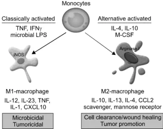
Fig. 1. Macrophage activation and polarization. “Classically activated” M1 macrophages are activated by TNF, IFNγ or bacterial products such as microbial lipopolysaccharide (LPS), express high levels of IL12, IL-23, TNF, IL-1 or CXCL10 and low levels of IL10. By contrast, “alternatively activated” M2 macrophages are activated by IL4, IL10, IL13, M-CSF or glucocorticoid hormones, express high levels of IL10, IL-13, IL-4, CCL2, scavenger receptor-A or mannose receptor and low levels of IL12. Functionally, M1 macrophages are microbicidal or tumoricidal and M2 macrophages play a role in cell clearance/wound healing or tumor promotions.
조직의 재생을 촉진시키는 고전적인 면역기능을 하는 것으로 알려졌다. 또한, 이들이 종양의 미세환경 (microenvironment)에 존재하며, 종양의 형성 및 성장 에 어떤 역할을 할 것임은 오래 전부터 알려져 있었으 나, 구체적으로 종양 혈관의 신생을 촉진시키고, 종양 세포의 침입과 전이를 보조하는 역할이 있음이 알려진 것은 최근의 일이다. 이들 중, 종양 관련 대식세포 (tumor associated macrophage, TAM) 및 골수유래억제 세포(myeloid derived suppressor cell, MDSC)에 대해 고 찰하고자 한다.
종양 관련 대식세포(TAM)
대식세포는 골수성 세포가 최종적으로 분화된 표현 형의 하나로 주로 골수 내 존재하는 골수성 세포가 혈 액을 타고 종양 미세환경으로 이동하여 분화하는 것으 로 이해되고 있다. 최근에는 이들을 대식세포가 생체 내 다양한 생리적 병적 조건에서 역동적으로 M1-대식 세포와 M2-대식세포로 분극화(polarization)됨이 제시 되었다.3) M1-대식세포는 대식세포가 고전적으로 활성 화(classically activated)된 상태로 외부 병원균과 인터페 론 감마 등의 자극에 의해 활성화되고, IL-12를 분비하
여 염증 반응을 일으키며 종양 미세 환경에서는 종양 억제 작용이 있다. 이에 반해 M2-대식세포는 대체 활 성화(alternatively activated)된 대식세포로 IL-4, IL-10, IL-13, 그리고 부신피질호르몬에 의해 활성화되어 항 염증 작용을 하는 것으로 알려져 있으며, IL-4, IL-10, IL-13을 분비하여 종양의 성장을 촉진시킨다(Fig. 1).
최근 이들 M2-대식세포의 표현형을 가지면서 종양 미 세환경에 존재하는 대식세포를 종양 관련 대식세포 (TAM)라 일컬으며 많은 연구가 진행되고 있다.4) 종양 미세환경에서 종양 관련 대식세포는 면역작용으로 TH1 세포 대신 TH2 세포의 증식을 촉진하고 regulatory T 세포를 활성화시켜서 면역관용을 일으키며, 비면역 작용으로 혈관신생을 직접 촉진시키고 종양세포의 침 습력과 전이능을 보조하며, 항암제로 인한 세포 사멸 을 억제하여 종양의 성장을 유도한다. 또한, 종양 관련 대식세포의 분극화는 종양 미세환경 내의 여러 면역세 포들뿐만 아니라 종양세포 자체에서 분비되는 IL-4, IL-10, IL-13, TGFβ-1, PGE2 등의 각종 사이토카인에 의해서도 촉진되는 것으로 알려져, 이들 면역세포와 종양세포는 서로 유기적인 관계를 가지는 것으로 이해 되고 있다. 최근, 실제 임상에서 종양 관련 대식세포의 존재가 임상적으로 나쁜 예후와 연관됨이 여러 암에서 보고된 이후,5,6) 종양 관련 대식세포의 분극화를 차단 하여 종양의 성장을 억제하는 치료법 개발이 활발히 연구되고 있다(Table 1).
골수유래억제세포(MDSC)
골수유래억제세포(MDSC)는 골수성 세포 중에서 면역억제 작용을 가진 세포들로서, 매우 광범위한 미 분화 골수성 세포를 포함하는 세포군으로, 종양이나 염증의 발생 상태에서 증가하게 된다. 일반적으로 마 우스에서는 CD11b와 Gr1을 동시에 발현하는 것을 표지자로 사용하며, 전체 골수세포의 20-30%를 차지 하는 것으로 알려져 있다.7) 종양 관련 대식세포는 분 화도가 높아 GR1−F4/80+임에 비해 골수유래억제세 포는 미분화세포로 구성되어 F4/80−의 표현형을 보 인다. 최근에는 더욱 특이적으로 GR1의 두 개 항원결 정기인 LY6G와 LY6C를 구분하여 과립구 형태를 가 진 CD11b+LY6G+LY6Clow와 단핵구(monocyte) 형태의 CD11b+LY6G−LY6Chigh로 나누어 구분하기도 한다.7,8) 사람에서는 CD14−CD11b+ 표지자를 일반적으로 사용 하며, 더 좁게는 CD14−CD11b+CD33+를 사용하기도
한다.9,10) 혈액에서는 CD15+ 세포로 확인할 수 있으며,
Table 1. Therapeutic strategies to target TAMs
Therapeutic agents Study subjects References
Blocking the differentiation and recruitment of macrophages c-fms inhibitor
Yondelis
Anti-CCL2 blocking antibody CNTO 888
MLN1202 COX inhibitor, DFU
Killing of macrophages in the tumor microenvironment Clonodrate-loaded liposomes (CLIPs)
Zoledronic acid Repolarization of TAMs
Proton pump inhibitor pantoprazole IL-12
NF-κb signaling inhibitor Histidine-rich glycoprotein (HRG) Inhibition of M2 macrophage functions Prednisolone liposomes
Silibinin
Rituximab (anti-CD20 Ab)
Human
Mice Human
Human
Mice Mice
Human Mice Mice Mice
Mice Human Human
NCT01004861 NCT01316822 NCT01346358
19,20)
NCT00992186 NCT01204996 NCT01015560
21-23)
24-26) 27)
NCT01163903
28,29) 30) 31)
32)
NCT01129570
33)
NCT: clinical trial registry numbers in ClinicalTrials.gov, DFU: 5,5-dimethyl-3-(3-fluorophenyl)-4-(4-methylsulphonyl)phenyl- 2(5H)-furanone, TAMs: tumor-associated macrophages
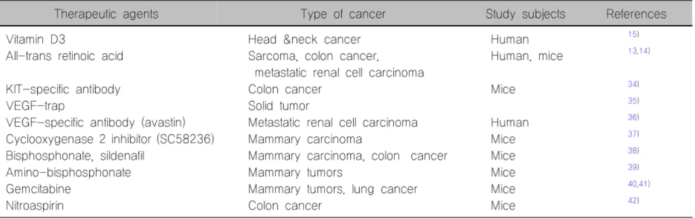
Table 2. Therapeutic strategies to target MDSCs
Therapeutic agents Type of cancer Study subjects References
Vitamin D3
All-trans retinoic acid
KIT-specific antibody VEGF-trap
VEGF-specific antibody (avastin) Cyclooxygenase 2 inhibitor (SC58236) Bisphosphonate, sildenafil
Amino-bisphosphonate Gemcitabine
Nitroaspirin
Head &neck cancer Sarcoma, colon cancer, metastatic renal cell carcinoma Colon cancer
Solid tumor
Metastatic renal cell carcinoma Mammary carcinoma
Mammary carcinoma, colon cancer Mammary tumors
Mammary tumors, lung cancer Colon cancer
Human Human, mice
Mice Human Mice Mice Mice Mice Mice
15) 13,14)
34) 35) 36) 37) 38) 39) 40,41) 42)
MDSCs: myeloid derived suppressor cells
건강한 상태에서는 말초 혈액세포의 ~0.5%를 차지하 는 것으로 보고되었다.11)
이들 골수유래억제세포는 대부분 세포-세포 간의 직 접 접촉을 통해 면역억제 작용을 하는 것으로 알려져 있어, 반감기가 짧은 사이토카인 등의 물질을 분비하여 면역억제 기능을 수행하는 것으로 이해되고 있다. 현재 까지 알려진 작용물질로는 Arginase I과 inducible nitric oxide synthesis (iNOS), 활성산소물질(reactive oxygen species, ROS), 그리고 peroxynitrite 등이 있다. Arginase I과 iNOS는 대표적인 T-세포 억제 물질로, 직접적으로
T-세포의 증식을 억제하며, ROS와 peroxynitrite는 T-세포 수용체의 번역 후 수정(post-translational modification) 과정을 통하여 항원 인식능을 억제한다.12) 이러한 골수 유래억제세포의 기능과 작용기전에 대한 연구를 바탕 으로 최근에는 이들의 조절을 통해 새로운 암 치료법 을 개발하고자 하는 노력이 가속화되고 있다. 첫째로 는 골수유래억제세포를 M1-대식세포로의 분화를 촉진 하여 오히려 종양 억제 효과를 기대하는 전략으로 all-trans retinoic acid (ATRA)13,14) 또는 vitamin D315)를 이용한 치료가 시도되고 있으며, 또 다른 전략으로는
골수유래억제세포의 증식을 억제하거나 세포독성 항 암제를 이용하여 제거하는 방법, 그리고 기능을 억제 하는 방법 등이 응용되고 있다(Table 2).
갑상선암에서의 면역억제 골수성 세포
갑상선암에서 이들 면역억제 골수성 세포에 대한 연 구는 많지 않은 실정이다. 그러나 최근 몇몇 보고에 의 하면 갑상선암에서도 종양 관련 대식세포가 존재하며, 이들이 임상적인 예후 인자들과 관련이 되어 있어, 이 들 세포가 갑상선암의 형성 및 진행에 역할이 있음을 추론할 수 있겠다. 최근 보고에 의하면, 종양 관련 대식 세포는 갑상선 역형성암16)과 분화도가 나쁜 갑상선암17) 에서 양성 조직(갑상선 선종, adenomatous goiter)에 비 하여 상대적으로 많이 존재하고, extrathyroidal extension 의 증가와 암 관련 생존율의 감소와 상관관계가 있었다. 또한, 갑상선분화암인 유두암에서도 림프선 전이와 양 의 상관관계가 있었다.18) 그러나 골수유래억제세포에 대한 연구는 아직 전무한 실정이다. 진행성 갑상선 암 에 대하여 현재는 수술과 방사성동위원소 치료 이외에 효과적인 치료제가 존재하지 않음을 고려할 때, 면역 억제 골수성 세포에 대한 연구와 이를 통한 치료법 개 발 전략은 새롭게 연구되어야 할 분야이다.
중심 단어: 종양미세환경, 종양 관련 대식세포, 골수 유래억제세포, 암 면역학.
References
1) Burnet M. Cancer; a biological approach. I. The processes of control. Br Med J 1957;1(5022):779-86.
2) Dunn GP, Bruce AT, Ikeda H, Old LJ, Schreiber RD. Cancer immunoediting: from immunosurveillance to tumor escape. Nat Immunol 2002;3(11):991-8.
3) Mantovani A, Sozzani S, Locati M, Allavena P, Sica A.
Macrophage polarization: tumor-associated macrophages as a paradigm for polarized M2 mononuclear phagocytes. Trends Immunol 2002;23(11):549-55.
4) Mantovani A, Sica A, Allavena P, Garlanda C, Locati M.
Tumor-associated macrophages and the related myeloid-derived suppressor cells as a paradigm of the diversity of macrophage activation. Hum Immunol 2009;70(5):325-30.
5) Mantovani A, Sica A. Macrophages, innate immunity and cancer:
balance, tolerance, and diversity. Curr Opin Immunol 2010;
22(2):231-7.
6) Steidl C, Lee T, Shah SP, Farinha P, Han G, Nayar T, et al. Tumor-associated macrophages and survival in classic Hodgkin's lymphoma. N Engl J Med 2010;362(10):875-85.
7) Youn JI, Nagaraj S, Collazo M, Gabrilovich DI. Subsets of
myeloid-derived suppressor cells in tumor-bearing mice. J Immunol 2008;181(8):5791-802.
8) Hestdal K, Ruscetti FW, Ihle JN, Jacobsen SE, Dubois CM, Kopp WC, et al. Characterization and regulation of RB6-8C5 antigen expression on murine bone marrow cells. J Immunol 1991;147(1):22-8.
9) Almand B, Clark JI, Nikitina E, van Beynen J, English NR, Knight SC, et al. Increased production of immature myeloid cells in cancer patients: a mechanism of immunosuppression in cancer. J Immunol 2001;166(1):678-89.
10) Ochoa AC, Zea AH, Hernandez C, Rodriguez PC. Arginase, prostaglandins, and myeloid-derived suppressor cells in renal cell carcinoma. Clin Cancer Res 2007;13(2 Pt 2):721s-6s.
11) Schmielau J, Finn OJ. Activated granulocytes and granulocyte- derived hydrogen peroxide are the underlying mechanism of suppression of t-cell function in advanced cancer patients. Cancer Res 2001;61(12):4756-60.
12) Gabrilovich DI, Nagaraj S. Myeloid-derived suppressor cells as regulators of the immune system. Nat Rev Immunol 2009;9(3):
162-74.
13) Kusmartsev S, Cheng F, Yu B, Nefedova Y, Sotomayor E, Lush R, et al. All-trans-retinoic acid eliminates immature myeloid cells from tumor-bearing mice and improves the effect of vaccination. Cancer Res 2003;63(15):4441-9.
14) Mirza N, Fishman M, Fricke I, Dunn M, Neuger AM, Frost TJ, et al. All-trans-retinoic acid improves differentiation of myeloid cells and immune response in cancer patients. Cancer Res 2006;66(18):9299-307.
15) Lathers DM, Clark JI, Achille NJ, Young MR. Phase 1B study to improve immune responses in head and neck cancer patients using escalating doses of 25-hydroxyvitamin D3. Cancer Immunol Immunother 2004;53(5):422-30.
16) Caillou B, Talbot M, Weyemi U, Pioche-Durieu C, Al Ghuzlan A, Bidart JM, et al. Tumor-associated macrophages (TAMs) form an interconnected cellular supportive network in anaplastic thyroid carcinoma. PLoS One 2011;6(7):e22567.
17) Ryder M, Ghossein RA, Ricarte-Filho JC, Knauf JA, Fagin JA. Increased density of tumor-associated macrophages is associated with decreased survival in advanced thyroid cancer.
Endocr Relat Cancer 2008;15(4):1069-74.
18) Wei Q, Fang W, Ye L, Shen L, Zhang X, Fei X, et al. Density of Tumor Associated Macrophage Correlates with Lymph Node Metastasis in Papillary Thyroid Carcinoma. Thyroid 2012 [Epub ahead of print].
19) Allavena P, Signorelli M, Chieppa M, Erba E, Bianchi G, Marchesi F, et al. Anti-inflammatory properties of the novel antitumor agent yondelis (trabectedin): inhibition of macrophage differentiation and cytokine production. Cancer Res 2005;65(7):
2964-71.
20) Germano G, Frapolli R, Simone M, Tavecchio M, Erba E, Pesce S, et al. Antitumor and anti-inflammatory effects of trabectedin on human myxoid liposarcoma cells. Cancer Res 2010;70(6):2235-44.
21) Muta M, Matsumoto G, Nakashima E, Toi M. Mechanical analysis of tumor growth regression by the cyclooxygenase-2 inhibitor, DFU, in a Walker256 rat tumor model: importance of monocyte chemoattractant protein-1 modulation. Clin Cancer
Res 2006;12(1):264-72.
22) Bundred NJ, Cramer A, Morris J, Renshaw L, Cheung KL, Flint P, et al. Cyclooxygenase-2 inhibition does not improve the reduction in ductal carcinoma in situ proliferation with aromatase inhibitor therapy: results of the ERISAC randomized placebo-controlled trial. Clin Cancer Res 2010;16(5):1605-12.
23) Antonarakis ES, Heath EI, Walczak JR, Nelson WG, Fedor H, De Marzo AM, et al. Phase II, randomized, placebo- controlled trial of neoadjuvant celecoxib in men with clinically localized prostate cancer: evaluation of drug-specific biomarkers.
J Clin Oncol 2009;27(30):4986-93.
24) Gazzaniga S, Bravo AI, Guglielmotti A, van Rooijen N, Maschi F, Vecchi A, et al. Targeting tumor-associated macro- phages and inhibition of MCP-1 reduce angiogenesis and tumor growth in a human melanoma xenograft. J Invest Dermatol 2007;127(8):2031-41.
25) Miselis NR, Wu ZJ, Van Rooijen N, Kane AB. Targeting tumor-associated macrophages in an orthotopic murine model of diffuse malignant mesothelioma. Mol Cancer Ther 2008;7(4):
788-99.
26) Meng Y, Beckett MA, Liang H, Mauceri HJ, van Rooijen N, Cohen KS, et al. Blockade of tumor necrosis factor alpha signaling in tumor-associated macrophages as a radiosensitizing strategy. Cancer Res 2010;70(4):1534-43.
27) Tsagozis P, Eriksson F, Pisa P. Zoledronic acid modulates antitumoral responses of prostate cancer-tumor associated macro- phages. Cancer Immunol Immunother 2008;57(10):1451-9.
28) Watkins SK, Li B, Richardson KS, Head K, Egilmez NK, Zeng Q, et al. Rapid release of cytoplasmic IL-15 from tumor- associated macrophages is an initial and critical event in IL-12- initiated tumor regression. Eur J Immunol 2009;39(8): 2126-35.
29) Watkins SK, Egilmez NK, Suttles J, Stout RD. IL-12 rapidly alters the functional profile of tumor-associated and tumor- infiltrating macrophages in vitro and in vivo. J Immunol 2007;
178(3):1357-62.
30) Hagemann T, Lawrence T, McNeish I, Charles KA, Kulbe H, Thompson RG, et al. "Re-educating" tumor-associated macrophages by targeting NF-kappaB. J Exp Med 2008;205(6):
1261-8.
31) Rolny C, Mazzone M, Tugues S, Laoui D, Johansson I, Coulon C, et al. HRG inhibits tumor growth and metastasis by inducing macrophage polarization and vessel normalization through downregulation of PlGF. Cancer Cell 2011;19(1):31-44.
32) Kluza E, Yeo SY, Schmid S, van der Schaft DW, Boekhoven RW, Schiffelers RM, et al. Anti-tumor activity of liposomal glucocorticoids: The relevance of liposome-mediated drug delivery,
intratumoral localization and systemic activity. J Control Release 2011;151(1):10-7.
33) Canioni D, Salles G, Mounier N, Brousse N, Keuppens M, Morchhauser F, et al. High numbers of tumor-associated macrophages have an adverse prognostic value that can be circumvented by rituximab in patients with follicular lymphoma enrolled onto the GELA-GOELAMS FL-2000 trial. J Clin Oncol 2008;26(3):440-6.
34) Pan PY, Wang GX, Yin B, Ozao J, Ku T, Divino CM, et al. Reversion of immune tolerance in advanced malignancy:
modulation of myeloid-derived suppressor cell development by blockade of stem-cell factor function. Blood 2008;111(1):219-28.
35) Fricke I, Mirza N, Dupont J, Lockhart C, Jackson A, Lee JH, et al. Vascular endothelial growth factor-trap overcomes defects in dendritic cell differentiation but does not improve antigen-specific immune responses. Clin Cancer Res 2007;13(16):
4840-8.
36) Kusmartsev S, Eruslanov E, Kubler H, Tseng T, Sakai Y, Su Z, et al. Oxidative stress regulates expression of VEGFR1 in myeloid cells: link to tumor-induced immune suppression in renal cell carcinoma. J Immunol 2008;181(1):346-53.
37) Sinha P, Clements VK, Fulton AM, Ostrand-Rosenberg S.
Prostaglandin E2 promotes tumor progression by inducing myeloid-derived suppressor cells. Cancer Res 2007;67(9):4507-13.
38) Serafini P, Meckel K, Kelso M, Noonan K, Califano J, Koch W, et al. Phosphodiesterase-5 inhibition augments endogenous antitumor immunity by reducing myeloid-derived suppressor cell function. J Exp Med 2006;203(12):2691-702.
39) Melani C, Sangaletti S, Barazzetta FM, Werb Z, Colombo MP. Amino-biphosphonate-mediated MMP-9 inhibition breaks the tumor-bone marrow axis responsible for myeloid-derived suppressor cell expansion and macrophage infiltration in tumor stroma. Cancer Res 2007;67(23):11438-46.
40) Suzuki E, Kapoor V, Jassar AS, Kaiser LR, Albelda SM.
Gemcitabine selectively eliminates splenic Gr-1+/CD11b+
myeloid suppressor cells in tumor-bearing animals and enhances antitumor immune activity. Clin Cancer Res 2005;11(18):
6713-21.
41) Ko HJ, Kim YJ, Kim YS, Chang WS, Ko SY, Chang SY, et al. A combination of chemoimmunotherapies can efficiently break self-tolerance and induce antitumor immunity in a tolero- genic murine tumor model. Cancer Res 2007;67(15):7477-86.
42) De Santo C, Serafini P, Marigo I, Dolcetti L, Bolla M, Del Soldato P, et al. Nitroaspirin corrects immune dysfunction in tumor-bearing hosts and promotes tumor eradication by cancer vaccination. Proc Natl Acad Sci U S A 2005;102(11):4185-90.