Does Radiofrequency Ablation Induce Neoplastic Changes in Benign Thyroid Nodules: A Preliminary Study
Su Min Ha1,*, Jun Young Shin2,*, Jung Hwan Baek3, Dong Eun Song2, Sae Rom Chung3, Young Jun Choi3, Jeong Hyun Lee3
1Department of Radiology, Seoul National University Hospital, Seoul National University College of Medicine; 2Department of Pathology, 3Department of Radiology and the Research Institute of Radiology, Asan Medical Center, University of Ulsan College of Medicine, Seoul, Korea
Background: To evaluate the clinical feasibility of radiofrequency ablation (RFA) of benign thyroid nodules along with cytomor- phological alteration, and any malignant transformation through biopsy.
Methods: The data were retrospectively collected between April 2008 and June 2013 and core needle biopsy (CNB) was performed on 16 benign thyroid nodules previously treated using RFA. The parameters of the patients were compared, between the time of en- rollment and the last follow-up examination, using linear mixed model statistical analysis.
Results: No atypical cells or neoplastic transformation were detected in the undertreated peripheral portion of treated benign nodules on the CNB specimen. RFA altered neither the thyroid capsule nor the thyroid tissue adjacent to the treated area. On histopathologi- cal examinations, we observed 81.2% acellular hyalinization, which was the most common finding. After a mean follow-up period of over 5 years, the mean volume of thyroid nodule had decreased to 6.4±4.2 mL, with a reduction rate of 81.3%±5.8% (P<0.0001).
Conclusion: RFA is a technically feasible treatment method for benign thyroid nodules, with no carcinogenic effect or tissue dam- age of the normal thyroid tissue adjacent to the RFA-treated zone.
Keywords: Thyroid nodule; Radiofrequency ablation; Ultrasonography; Pathology; Biopsy, large-core needle
INTRODUCTION
With the widespread use of ultrasound (US), clinically palpable nodules are found incidentally in up to 67% [1,2]. With this in- crease in the detection of thyroid nodules, ethanol ablation (EA), laser ablation (LA), and radiofrequency ablation (RFA) have been investigated for the treatment of symptomatic thyroid nodules as acceptable alternatives to surgery [3]. US-guided RFA is a nonsurgical technique that has been used for the treat- ment of benign nodules because of cosmetic concerns, local
pain, and hyperthyroidism [4-6]. It has also been used in recur- rent thyroid cancers and even papillary microcarcinoma in pa- tients who are considered ineligible for surgery [7].
Several studies have assessed the pathological changes occur- ring after treating thyroid nodules using EA [8-10] and LA [11,12]. Dobrinja et al. [13] observed central hyaline sclerosis and scarring in RFA treated nodules with a preserved capsule, findings that did not jeopardize subsequent operations. Dobrinja et al. [13] questioned what would happen if a thyroid cancer was not detected before starting RFA and was left untreated.
Received: 3 January 2019, Revised: 15 March 2019, Accepted: 3 April 2019 Corresponding author: Dong Eun Song
Department of Pathology, Asan Medical Center, University of Ulsan College of Medicine, 88 Olympic-ro 43-gil, Songpa-gu, Seoul 05505, Korea
Tel: +82-2-3010-5998, Fax: +82-2-3010-6962, E-mail: [email protected]
*These authors contributed equally to this work.
Copyright © 2019 Korean Endocrine Society
This is an Open Access article distributed under the terms of the Creative Com- mons Attribution Non-Commercial License (http://creativecommons.org/
licenses/by-nc/4.0/) which permits unrestricted non-commercial use, distribu- tion, and reproduction in any medium, provided the original work is properly cited.
They observed regrowth of follicular lesions/follicular neo- plasm after RFA. Given the nodule growth is a sign of potential malignancy [14] and that the rate of malignancy is estimated to be between 14% and 48%, they confirmed cancer by subse- quent surgery. On the other hand, Lim et al. [15] observed a rate of only 5.6% recurrence after RFA as a result of regrowth in the undertreated peripheral portion of benign thyroid nodules. Fine- needle aspiration (FNA) revealed that the recurrences were not associated with malignancy.
The post RFA regrowth of the undertreated peripheral portion of benign thyroid nodules should be evaluated for neoplastic transformation or the progression of undetected thyroid cancer, to ensure that this treatment method can be safely applied in cases of recurrence. It is crucial to obtain tissue for pathological analysis through a procedure less invasive than surgery. One such method can be core needle biopsy (CNB), which has been reported as a valuable diagnostic tool to be used in lieu of diag- nostic surgery [16-20]. During 5-year follow-up of patients treated with RFA for benign thyroid nodules, we used CNB for whom the regrowth of the undertreated peripheral portion was noted and to determine whether this is associated with risk of malignancy. We investigated the clinical feasibility of RFA treatment along with cytomorphological alteration.
METHODS
This retrospective study was approved by the Institutional Re- view Board of Asan Medical Center (2017-0917). Written in- formed consent was obtained from all patients before the proce- dure. CNB was performed on 16 patients who were RFA-treated for benign thyroid nodules, in the period between April 2008
and June 2013 (Table 1). Fifteen of these patients were female and one was male, with mean age of 43.8±12.3 years (range, 18 to 63). Cytologic or histologic results of benign thyroid nod- ules on FNA and CNB before RFA were categorized according to the six categories of the Bethesda System for Reporting Thy- roid Cytopathology and the six categories of the Bethesda Sys- tem, respectively [21,22]. All patients had refused surgery or ra- dioiodine therapy and were referred for RFA to the Radiological Interventional Department of our thyroid center. Four patients with benign nodules had previously been treated with RFA in other hospitals and were referred to our clinic because the thy- roid nodules were re-growing.
Pre-ablation assessment
Physical examination and laboratory tests were performed for all patients, followed by US scanning with a 10 MHz probe on a real-time US system (Aplio SSA-770A, Toshiba, Otawara, Ja- pan). Three orthogonal dimensions of the nodules (the largest diameter and two other perpendicular ones) were measured be- fore RFA. The volume of the nodule was calculated using the following equation: V=πabc/6, where V=volume; a=the larg- est dimension; and b and c=the other two perpendicular dimen- sions. The percentage of volume reduction was calculated as [(initial volume−final volume)×100]/initial volume [23]. The composition of the nodules was classified as either solid (solid component >50%) or predominantly cystic (solid component between 10% and 50%). Nodule vascularity was classified ac- cording to a four-point scale: 0=no signal; 1=peripheral signal without any central vascular signal; 2=peripheral and central signals in <50%; and 3=peripheral and central signals in
>50%. Upon enrollment, patients were asked to rate their pres- sure symptoms on a 10 cm visual analog scale (0 to 10 cm), with the physician also recording a cosmetic grade (1=no pal- pable mass; 2=invisible but palpable mass; 3=mass visible only to an experienced clinician; and 4=easily visible mass) [24].
RFA procedure
A generator (Cool-tip, Radionics, Burlington, MA, USA) and 17- or 18-gauge 1, 1.5, or 2 cm active-tip internally cooled electrodes (Well-point RF, Taewoong Medical, Goyang, Korea) were used.
Local anesthesia and a trans-isthmic approach method were utilized [24]. Nodules were treated with a moving-shot technique, beginning with 10 to 30 W of power. If a transient hyperecho- ic zone did not form at the electrode tip within 5 to 10 seconds, the power was increased by 10 W increments. The power was re- duced or turned off if the patient could not tolerate the pain dur- Table 1. Demographic Characteristics of the Enrolled Patients
(16 Benign Nodules)
Characteristic Radiofrequency ablation (n=16)
Gender, male:female 1:15
Age, yr 43.8±12.3 (18–63)
Nodule diameter, cm 5.0±1.8 (2.9–9.5)
Nodule volume, mL 34.6±28.5 (4.5–109.1)
Volume <20 mL 8
Volume >20 mL 8
Symptom score 2.7±1.4 (0–5)
Cosmetic score 3.9±0.3 (3–4)
Vascularity 2.3±0.8 (1–3)
Values are expressed as mean±SD (range).
ing ablation. Ablation was terminated when all imaginary units of the nodule had changed to transient hyperechoic zones [23]. The patients then received neck compression for 10 to 20 minutes and remained under observation for 1 to 2 hours before dis- charge [23,25].
Follow-up
A follow-up US examination was performed 1 month after the procedure, and then annually for 5 years. Changes in size, vol- ume, and vascularity were evaluated in each session. Therapeu- tic success was defined as a volume reduction greater than 50%.
We also described “complete ablation” as complete treatment with no discernible lesion with same echogenicity as the index nodule and no vascularity on color Doppler US. Evaluation of symptoms and cosmetic grading was also performed [23,24,26].
Additional treatment was performed if the nodules did not fulfill the criteria for complete ablation, and/or if patients complained of unresolved clinical problems [23,25,27]. During the follow- up period, CNB was performed using a 1.1 or 1.6 cm excursion 18-gauge double-action spring-activated needle (TSK Ace-cut, Create Medic, Yokohama, Japan), to evaluate ablation status of the nodule and to exclude neoplastic transformation of regrowth of the undertreated peripheral portion [28]. There was no com- plication related to the CNB procedure.
Statistical analysis
Statistical analyses were performed using IBM SPSS Statistics for Windows version 23.0 (IBM Corp., Armonk, NY, USA). Pa- rameters were compared between each patient’s initial visit and the last follow-up examination using a linear mixed model sta- tistical analysis. The level for statistical significance was de- fined as P<0.05.
Pathology
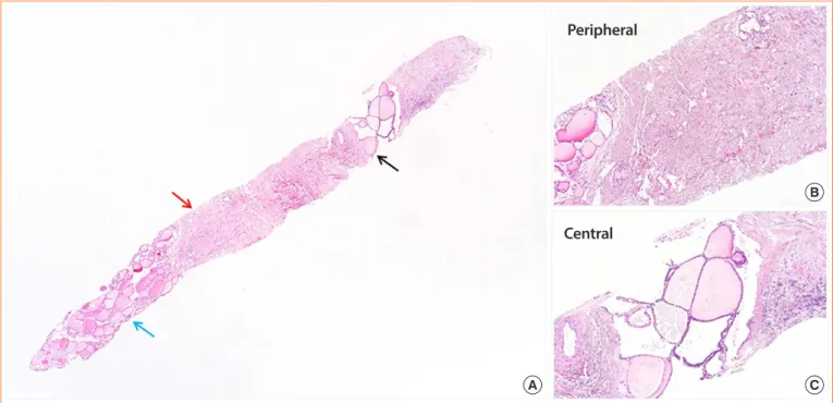
The CNB samples of treated benign thyroid nodules were sub- jected to routine paraffin embedding, followed by hematoxylin and eosin (H&E) staining. For this research, the slides were re- viewed by two pathologists, who assessed various cytomorpho- logical alterations. The location of each alteration was judged on the basis of normal thyroid tissue (Fig. 1A). “Peripheral” lo- cation was defined as any alteration within half distance from the normal thyroid tissue (Fig. 1B) and “central” was defined as more than half distance from the normal thyroid tissue (Fig.
1C). If changes were seen in both “central” and “peripheral,”
the lesion was categorized as “diffuse.” Cytomorphological al- teration without any identifiable follicular thyroid tissue, the presence of only a normal thyroid gland, and samples contain- ing only a few follicular cells that were considered insufficient for reaching a diagnosis, were classified as “not applicable.”
Fig. 1. Orientation of the thyroid core needle biopsy. (A) Normal thyroid tissue is noted on the left lower corner (blue arrow). (B) Less than half near the normal thyroid tissue is defined as ‘peripheral’ (red arrow, A). (C) More than half distance from the normal thyroid tissue is de- fined as ‘central’ (black arrow, A) with focal remaining benign follicular lesion on the central side (H&E stain, ×100).
A C
B
Peripheral
Central
Immunohistochemical staining was performed for galectin-3 (Gal-3, dilution 1:200, BenchMark XT auto-immunostainer, Novo, Ventana, AZ, USA) and Hector Battifora mesothelial cell-1 (HBME-1, dilution 1:200, BenchMark XT auto-immu- nostainer, Dako, Carpinteria, CA, USA), as both are considered good markers of malignancy [29,30]. Galactin-3 staining was limited to the cytoplasm and nucleus, whereas HBME-1 stain- ing was seen along the cell membrane. To determine viability after RFA, immunohistochemical stainings for human mito- chondria, thyroglobulin, thyroid transcription factor-1 (TTF-1), and paired-box gene 8 (PAX-8) were performed on one post- RFA CNB specimen showing slightly viable portion on the H&E slide (Supplemental Table S1).
RESULTS
Five-year follow-up results after RFA
Serum thyroid hormone and thyrotropin levels were within nor- mal range in all patients before RFA, and they were treated for a single benign nodule (nodular hyperplasia, n=7; and nonfunc- tioning benign nodule, n=8). One patient had undergone EA prior to RFA procedure because of a cystic nodule that had in- creased in size after aspiration resulting in a cosmetic problem.
The mean number of RFA sessions was 1.6±0.9 (range, 1 to 4) and mean total energy deposition was 3,234.9±4,915.9 J/mL (range, 384.9 to 19,058.0) (Table 2). The mean nodule volume was 34.3±4.3 mL prior to treatment, which had decreased to
Table 2. Treatment Characteristics (n=16)
Characteristic Initial RFA Additional 2nd RFA Additional 3rd RFA Additional 4th RFA
RF power, W 76.3±22.9
(50–120) 48±23
(10–70) 75±21.2
(60–90) 30
Ablation time, sec 672.1±661.3
(150.0–2,400.0) 355.8±177.2
(120.0–550.0) 591.0±224.9
(432.0–750.0) 492.0
Total energy, J 57,110.0±69,087.7
(15,000.0–216,000.0) 16,662.0±12,362.0
(850.0–27,500.0) 46,710.0±29,401.5
(25,920.0–67,500) 14,760.0
Energy/mL, J 1,639.1±1,332.9
(384.9–4,936.0) 4,970.5±7,903.3
(635.7–19,058.0) 2,858.3±2,300.5
(1,231.6–4,485.0) 1,785.0
No. of RF sessions 13 (4 outside) 7 2 1
Solid componenta
Solid 13 - -
Values are expressed as mean±SD (range).
RFA, radiofrequency ablation; RF, radiofrequency.
aThe solid component is defined as solid (if the solid component is >50%) and predominantly cystic (if the solid component is between 10% and 50%).
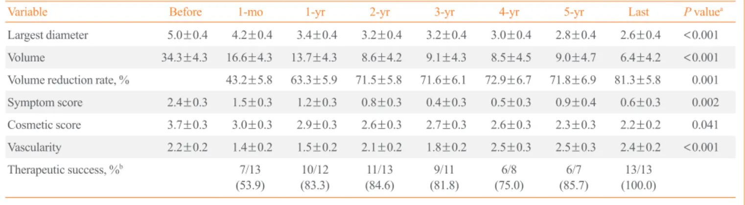
Table 3. Outcomes for the Benign Thyroid Nodules (n=16) after Radiofrequency Ablation
Variable Before 1-mo 1-yr 2-yr 3-yr 4-yr 5-yr Last P valuea
Largest diameter 5.0±0.4 4.2±0.4 3.4±0.4 3.2±0.4 3.2±0.4 3.0±0.4 2.8±0.4 2.6±0.4 <0.001
Volume 34.3±4.3 16.6±4.3 13.7±4.3 8.6±4.2 9.1±4.3 8.5±4.5 9.0±4.7 6.4±4.2 <0.001
Volume reduction rate, % 43.2±5.8 63.3±5.9 71.5±5.8 71.6±6.1 72.9±6.7 71.8±6.9 81.3±5.8 0.001
Symptom score 2.4±0.3 1.5±0.3 1.2±0.3 0.8±0.3 0.4±0.3 0.5±0.3 0.9±0.4 0.6±0.3 0.002
Cosmetic score 3.7±0.3 3.0±0.3 2.9±0.3 2.6±0.3 2.7±0.3 2.6±0.3 2.3±0.3 2.2±0.2 0.041
Vascularity 2.2±0.2 1.4±0.2 1.5±0.2 2.1±0.2 1.8±0.2 2.5±0.3 2.5±0.3 2.4±0.2 <0.001
Therapeutic success, %b 7/13
(53.9) 10/12
(83.3) 11/13
(84.6) 9/11
(81.8) 6/8
(75.0) 6/7
(85.7) 13/13 (100.0) Values are expressed as least-squares mean±standard error or number/total number (%).
aComparison of values before and last year follow-up; bTherapeutic success (volume reduction >50%).
6.0±4.2 mL after a follow-up period of more than 5 years (mean, 5.8 years; range, 38 to 98 months), a volume reduction rate of 81.3%±5.8% (P<0.0001) (Table 3). None of the pa- tients showed any signs of lymph node involvement, distant metastases, or any new suspicious findings on US imaging dur- ing the 5-year follow-up period.
US findings
The nodules were composed of mainly solid portion (n=12) and predominantly solid (n=3). This number does not take into ac- count the four patients who visited our clinic in post-RFA status.
After ablation, color and power Doppler US showed significant reduction of the peripheral and/or intra-nodular vascular signals due to RFA-induced necrosis (P<0.0001). Additional RFA was performed if the results of the treatment did not fulfill the crite- ria for complete ablation. The procedure was performed after confirming that no atypical cells or neoplastic changes were present in the treated benign thyroid nodules. On the last follow- up, we did not observe any nodule larger than its initial size.
Pathological changes
The mean interval between RFA and CNB of benign thyroid nodules was 37.5±22.1 months (range, 1 to 80). On histopatho- logical examinations, we observed acellular hyalinization in 81.2% (13/16), followed by chronic inflammation in 56.2%
(9/16). From the 13 cases showing acellular hyalinization (Fig.
2), diffuse hyalinization was found in eight (61.5%), peripheral hyalinization in one (7.7%), and central hyalinization in one
(7.7%). Chronic inflammation was found diffusely in 55.5%
(5/9), centrally in 22.2% (2/9), and peripherally in 11.1% (1/9).
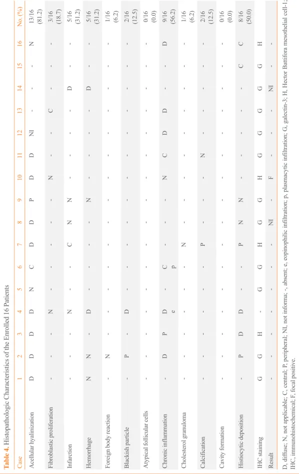
The rest of the findings in order of prevalence were histiocytic deposition, infarction, hemorrhage, fibroblast proliferation, blackish particle, calcification, foreign body reaction, cholesterol granuloma, and cavity formation (Table 4). The procedure had neither altered the thyroid capsule nor changed the thyroid tissue adjacent to the treated area, with maintained follicular architec- ture and benign cytological characteristics. On immunohisto- chemical staining, nine out of 11 cases (81.8%) were negative for Gal-3. The remaining two cases (2/11, 18.1%) were uninfor- mative because of the absence of follicular cells. Three out of four cases (75%) were negative for HMBE-1 and the remaining case (1/4, 25%) was focally positive for HMBE-1, classifying it as benign [31]. In post-RFA samples, one sample with viable ar- eas on H&E stain was positive for TTF-1 and thyroglobulin stainings and negative for human mitochondria antibody. Areas of total infarct were negative for all four markers (human mito- chondria antibody, thyroglobulin, TTF-1, and PAX-8) (Fig. 3).
In all RFA treated nodules in our study, undertreated periph- eral portions were observed. Among them, one patient under- went CNB shortly after RFA (1 month) to assess the presence of any viable tissue, and determine the need for additional treat- ment. An inflammatory response with a few eosinophils was observed in the CNB sample. In six nodules with a volume re- duction rate of more than 90.0%, acellular hyalinization (6/6, 100.0%), and histiocytic deposition (5/6, 83.3%) were predomi- nantly observed. Neither the energy nor the number of RFA ses-
Fig. 2. Treatment effect after radiofrequency ablation (RFA) of a benign follicular nodule. (A) Note acellular dense hyalinization (black ar- row) and the remaining benign follicular lesion (red arrow) (×40). (B) The remaining benign follicular lesion shows mixed benign thyroid follicles with variable sizes and nuclear enlargement without nuclear atypia at higher magnification (H&E stain, ×200).
A B
Table 4. Histopathologic Characteristics of the Enrolled 16 Patients Case12345678910111213141516No. (%) Acellular hyalinizationDDDDNCDDPDDNI---N 13/16 (81.2)
Fibroblastic proliferation---N-----N--C---
3/16 (18.7)
Infarction---N--CNN----D--
5/16 (31.2)
HemorrhageNN-D----N----D--
5/16 (31.2)
Foreign body reaction-N--------------
1/16 (6.2)
Blackish particle-P-D------------
2/16 (12.5)
Atypical follicular cells----------------
0/16 (0.0)
Chronic inflammation-DP
D e
-
C p
---NCDD--D
9/16 (56.2)
Cholesterol granuloma------N---------
1/16 (6.2)
Calcification------P---N-----
2/16 (12.5)
Cavity formation----------------
0/16 (0.0)
Histiocytic deposition-PDD--PNN-----CC
8/16 (50.0)
IHC stainingGGH-GGHGGHGGGGGH Result-------NI-F---NI-- D, diffuse; N, not applicable; C, central; P, peripheral; NI, not informa; -, absent; e, eopinophilic infiltration; p, plasmacytic infiltration; G, galectin-3; H, Hector Battifora mesothelial cell-1; IHC, immunohistochemical; F, focal positive.
sions significantly influenced histopathological alterations. We did not observe any clear association between the energy deliv- ered and the amount of necrotic debris and inflammatory cells in CNB samples (Supplemental Table S2).
DISCUSSION
This is the preliminary study to evaluate whether RFA induces neoplastic transformation. No atypical cells or neoplastic chang- es were detected in the undertreated peripheral portion of be- nign thyroid nodules. We demonstrated that RFA can safely be applied to benign thyroid nodules without any atypical cells or neoplastic changes in the undertreated peripheral portion, which
supports the findings in a previous study [15]. The most com- mon pathologic finding in post-RFA biopsy specimens were acellular hyalinization (81.2%), followed by chronic inflamma- tion (56.2%) and histiocytic deposition (50.0%). RFA neither altered the thyroid capsule nor resulted in neoplastic changes in the thyroid tissue adjacent to the treated area. In 5-year follow- up, RFA significantly reduced the volume of benign thyroid nodules and improved cosmetic and/or symptomatic problems.
Because of the possible regrowth of the undertreated periph- eral portion after RFA of benign thyroid nodules, it is crucial to determine whether RFA promotes any neoplastic transformation and whether the next management should be additional RFA treatment or surgery. Indeed, there is evidence that insufficient Fig. 3. Coexistence of the slightly viable area and totally infarcted area after radiofrequency ablation (RFA). (A) Slightly viable area (black arrow) and totally infarcted area after RFA (red arrow) (H&E stain, ×100). (B) Immunohistochemical (IHC) staining for the human mito- chondria antibody. Both viable and totally infarcted areas are negative for human mitochondria antibody (×100). IHC stains for (C) thyro- globulin and (D) thyroid transcription factor-1 (TTF-1). The totally infarcted area shows loss of expression for thyroglobulin and TTF-1, whereas the slightly viable area is positive for both (×100).
A
C
B
D
RFA facilitates undetected tumor cell progression because sub- optimal heat promotes angiogenesis in non-tumor cells, such as tumor-associated endothelial cells [32,33]. Moreover, in a previ- ous study, Dobrinja et al. [13] observed regrowth of follicular lesions/follicular neoplasm after RFA. Given that regrowth after RFA can be a sign of potential malignancy [14], RFA treated follicular neoplasm underwent surgery and revealed the re- growth to be cancer transformation. Our study supported that RFA treatment of benign thyroid nodules can be safely applied without the need for further pathological confirmation of under- treated peripheral portion.
Most benign nodules grow slowly over time [34], but there is no consensus as to what constitutes significant growth, warrant- ing re-biopsy [34,35]. The American Thyroid Association rec- ommends a repeated biopsy [36] if there is evidence of nodule growth. Recently, CNB has been reported to be a valuable diag- nostic tool that can be used to reduce the incidence of diagnostic surgery [16,17]. Indeed, for patients with benign thyroid nod- ules surgery seems to be an excessive procedure to confirm neoplastic transformation not detected on US. The advantages of a properly obtained CNB sample of thyroid nodules are its ability to sample large amounts of tissue, assess histologic ar- chitecture rather than cytological evaluation and the fact that is not very operator dependent [28]. Previous study revealed that CNB, including the center and periphery of the nodules, provide histologic information regarding degenerative changes [16]. In the present study, the cytolomorphological changes after RFA included both features of tissue damage, such as the presence of histiocytes and hemorrhage, and features of irreversible dam- age, such as infarction, cavity formation, fibroblast prolifera- tion, and acellular hyalinization. No atypical or malignant fol- licular cells were identified in our study. Acellular hyalinization was the most common finding that may cause a long-lasting im- pact in decreasing nodule volume during the follow-up period.
Our pathological findings after RFA for benign nodules may re- flect a process of progressive reabsorption of necrotic material by macrophages and replacement with fibrous scar similar to that seen in LA [11]. Moreover, RFA did not alter the follicular architecture or the tumor capsule, which may be due to retaining the active tip within the nodule throughout the procedure, hence undertreating areas near the thyroid capsule. These findings could explain previous study results [13], which revealed that RFA treated thyroid lesions does not induce perithyroidal fibro- sis, therefore not interfering with later surgery. By contrast, EA can cause perithyroidal fibrosis due to ethanol leakage during the treatment [37].
Sim et al. [38] suggested vital volume tracing as one of the earlier signs of regrowth compared to tracing total volume, which includes ablated volume. They observed regrowth begin- ning 12 months after treatment with a second peak later than 5 years, and suggested additional ablation sessions may be nec- essary to prevent long-term regrowth. Moreover, limitation of RFA approach is that US does not allow detecting very small tu- mor foci within the thyroid or in locoregional lymph nodes.
However, the present study demonstrated the applicability of RFA for treating benign thyroid nodules, with a reduction in volume during the 5-year follow-up, resolution of related symp- toms and cosmetic problems, and no life-threatening complica- tion, sequelae, or unnecessary surgery. Our clinical success might have been due to our use of the moving shot technique, which is well known to ablate the peripheral portion of thyroid nodules and prevent marginal recurrence, which is a major cause of recurrent symptom [23].
There are several limitations of our study: it was a retrospec- tive study, which could cause a selection bias; the follow-up pe- riod was short and our sample size was small which might not be reflective of a larger population. Moreover, there are no de- finitive guidelines on how to use CNB to evaluate RFA treated benign nodules for malignant transformation, resulting in a wide range in the time between RFA and CNB. Nevertheless, we demonstrated that RFA does not induce neoplastic transforma- tion within regrowth of the undertreated peripheral portion on CNB specimens, and does not hinder a subsequent thyroid sur- gery because of the preservation of the thyroid capsule. Finally, we only examined histologic changes in CNB and not surgical specimens. Nonetheless, during monitoring of post-RFA pa- tients with benign thyroid nodules, CNB usage in lieu of sur- gery to observe further histopathological changes provided pos- sible rationale for an additional RFA in the case of regrowth.
In conclusion, this preliminary study demonstrated that RFA is effective in treating benign thyroid nodules without any carci- nogenic effect based on the cytolomorphological changes of follow-up CNBs. RFA did not induce tissue damage in the nor- mal thyroid tissue adjacent to the RFA treated zone, thus not in- terfering with future surgery.
CONFLICTS OF INTEREST
No potential conflict of interest relevant to this article was re- ported.
AUTHOR CONTRIBUTIONS
Conceptualization: J.H.B., D.E.S. Data curation: S.M.H., J.Y.S.
Formal analysis: S.M.H. Methodology: S.M.H., J.Y.S. Valida- tion: S.R.C., Y.J.C., J.H.L. Writing (original draft): S.M.H., J.Y.S. Writing (review & editing): S.M.H., J.Y.S., J.H.B., D.E.S.
ORCID
Su Min Ha https://orcid.org/0000-0002-1833-0919 Jun Young Shin https://orcid.org/0000-0002-4833-9738 Jung Hwan Baek https://orcid.org/0000-0003-0480-4754 Dong Eun Song https://orcid.org/0000-0002-9583-9794 Sae Rom Chung https://orcid.org/0000-0003-4219-7166 Young Jun Choi https://orcid.org/0000-0001-7098-5042 Jeong Hyun Lee https://orcid.org/0000-0002-0021-4477
REFERENCES
1. Tunbridge WM, Evered DC, Hall R, Appleton D, Brewis M, Clark F, et al. The spectrum of thyroid disease in a commu- nity: the Whickham survey. Clin Endocrinol (Oxf) 1977;7:
481-93.
2. Vander JB, Gaston EA, Dawber TR. The significance of nontoxic thyroid nodules. Final report of a 15-year study of the incidence of thyroid malignancy. Ann Intern Med 1968;
69:537-40.
3. Gharib H, Hegedus L, Pacella CM, Baek JH, Papini E. Clin- ical review: nonsurgical, image-guided, minimally invasive therapy for thyroid nodules. J Clin Endocrinol Metab 2013;
98:3949-57.
4. Bernardi S, Dobrinja C, Fabris B, Bazzocchi G, Sabato N, Ulcigrai V, et al. Radiofrequency ablation compared to sur- gery for the treatment of benign thyroid nodules. Int J Endo- crinol 2014;2014:934595.
5. Faggiano A, Ramundo V, Assanti AP, Fonderico F, Macchia PE, Misso C, et al. Thyroid nodules treated with percutane- ous radiofrequency thermal ablation: a comparative study. J Clin Endocrinol Metab 2012;97:4439-45.
6. Hamidi O, Callstrom MR, Lee RA, Dean D, Castro MR, Morris JC, et al. Outcomes of radiofrequency ablation thera- py for large benign thyroid nodules: a mayo clinic case se- ries. Mayo Clin Proc 2018;93:1018-25.
7. Kim JH, Baek JH, Sung JY, Min HS, Kim KW, Hah JH, et al. Radiofrequency ablation of low-risk small papillary thy- roidcarcinoma: preliminary results for patients ineligible for
surgery. Int J Hyperthermia 2017;33:212-9.
8. Bennedbaek FN, Karstrup S, Hegedus L. Percutaneous etha- nol injection therapy in the treatment of thyroid and parathy- roid diseases. Eur J Endocrinol 1997;136:240-50.
9. Crescenzi A, Papini E, Pacella CM, Rinaldi R, Panunzi C, Petrucci L, et al. Morphological changes in a hyperfunction- ing thyroid adenoma after percutaneous ethanol injection:
histological, enzymatic and sub-microscopical alterations. J Endocrinol Invest 1996;19:371-6.
10. Monzani F, Caraccio N, Basolo F, Iacconi P, LiVolsi V, Mic- coli P. Surgical and pathological changes after percutaneous ethanol injection therapy of thyroid nodules. Thyroid 2000;
10:1087-92.
11. Piana S, Riganti F, Froio E, Andrioli M, Pacella CM, Valca- vi R. Pathological findings of thyroid nodules after percuta- neous laser ablation: a series of 22 cases with cyto-histolog- ical correlation. Endocr Pathol 2012;23:94-100.
12. Valcavi R, Piana S, Bortolan GS, Lai R, Barbieri V, Negro R.
Ultrasound-guided percutaneous laser ablation of papillary thyroid microcarcinoma: a feasibility study on three cases with pathological and immunohistochemical evaluation.
Thyroid 2013;23:1578-82.
13. Dobrinja C, Bernardi S, Fabris B, Eramo R, Makovac P, Ba- zzocchi G, et al. Surgical and pathological changes after ra- diofrequency ablation of thyroid nodules. Int J Endocrinol 2015;2015:576576.
14. Kuma K, Matsuzuka F, Kobayashi A, Hirai K, Morita S, Miyauchi A, et al. Outcome of long standing solitary thyroid nodules. World J Surg 1992;16:583-7.
15. Lim HK, Lee JH, Ha EJ, Sung JY, Kim JK, Baek JH. Radio- frequency ablation of benign non-functioning thyroid nod- ules: 4-year follow-up results for 111 patients. Eur Radiol 2013;23:1044-9.
16. Austin PC, Lee DS, D’Agostino RB, Fine JP. Developing points-based risk-scoring systems in the presence of com- peting risks. Stat Med 2016;35:4056-72.
17. Yeon JS, Baek JH, Lim HK, Ha EJ, Kim JK, Song DE, et al.
Thyroid nodules with initially nondiagnostic cytologic re- sults: the role of core-needle biopsy. Radiology 2013;268:
274-80.
18. Na DG, Baek JH, Jung SL, Kim JH, Sung JY, Kim KS, et al.
Core needle biopsy of the thyroid: 2016 consensus state- ment and recommendations from Korean Society of Thyroid Radiology. Korean J Radiol 2017;18:217-37.
19. Jung CK, Baek JH. Recent advances in core needle biopsy for thyroid nodules. Endocrinol Metab (Seoul) 2017;32:407-12.
20. Trimboli P, Giovanella L. The significance of having an ex- cellent patient’s comfort with thyroid core needle biopsy.
Endocrinol Metab (Seoul) 2018;33:53-4.
21. Cibas ES, Ali SZ; NCI Thyroid FNA State of the Science Conference. The Bethesda System for reporting thyroid cy- topathology. Am J Clin Pathol 2009;132:658-65.
22. Jung CK, Min HS, Park HJ, Song DE, Kim JH, Park SY, et al. Pathology reporting of thyroid core needle biopsy: a pro- posal of the Korean Endocrine Pathology Thyroid Core Needle Biopsy Study Group. J Pathol Transl Med 2015;49:
288-99.
23. Jeong WK, Baek JH, Rhim H, Kim YS, Kwak MS, Jeong HJ, et al. Radiofrequency ablation of benign thyroid nod- ules: safety and imaging follow-up in 236 patients. Eur Ra- diol 2008;18:1244-50.
24. Na DG, Lee JH, Jung SL, Kim JH, Sung JY, Shin JH, et al.
Radiofrequency ablation of benign thyroid nodules and re- current thyroid cancers: consensus statement and recom- mendations. Korean J Radiol 2012;13:117-25.
25. Spiezia S, Garberoglio R, Milone F, Ramundo V, Caiazzo C, Assanti AP, et al. Thyroid nodules and related symptoms are stably controlled two years after radiofrequency thermal ab- lation. Thyroid 2009;19:219-25.
26. Lee JH, Kim YS, Lee D, Choi H, Yoo H, Baek JH. Radio- frequency ablation (RFA) of benign thyroid nodules in pa- tients with incompletely resolved clinical problems after ethanol ablation (EA). World J Surg 2010;34:1488-93.
27. Kim YS, Rhim H, Tae K, Park DW, Kim ST. Radiofrequen- cy ablation of benign cold thyroid nodules: initial clinical experience. Thyroid 2006;16:361-7.
28. Na DG, Kim JH, Sung JY, Baek JH, Jung KC, Lee H, et al.
Core-needle biopsy is more useful than repeat fine-needle aspiration in thyroid nodules read as nondiagnostic or atypia of undetermined significance by the Bethesda system for re- porting thyroid cytopathology. Thyroid 2012;22:468-75.
29. Cochand-Priollet B, Dahan H, Laloi-Michelin M, Polivka M, Saada M, Herman P, et al. Immunocytochemistry with cyto- keratin 19 and anti-human mesothelial cell antibody (HBME1) increases the diagnostic accuracy of thyroid fine- needle aspirations: preliminary report of 150 liquid-based
fine-needle aspirations with histological control. Thyroid 2011;21:1067-73.
30. Fadda G, Rossi ED, Raffaelli M, Pontecorvi A, Sioletic S, Morassi F, et al. Follicular thyroid neoplasms can be classi- fied as low- and high-risk according to HBME-1 and galec- tin-3 expression on liquid-based fine-needle cytology. Eur J Endocrinol 2011;165:447-53.
31. Dunderovic D, Lipkovski JM, Boricic I, Soldatovic I, Bozic V, Cvejic D, et al. Defining the value of CD56, CK19, ga- lectin 3 and HBME-1 in diagnosis of follicular cell derived lesions of thyroid with systematic review of literature. Di- agn Pathol 2015;10:196.
32. Ke S, Ding XM, Kong J, Gao J, Wang SH, Cheng Y, et al.
Low temperature of radiofrequency ablation at the target sites can facilitate rapid progression of residual hepatic VX2 carcinoma. J Transl Med 2010;8:73.
33. Kong J, Kong J, Pan B, Ke S, Dong S, Li X, et al. Insuffi- cient radiofrequency ablation promotes angiogenesis of re- sidual hepatocellular carcinoma via HIF-1α/VEGFA. PLoS One 2012;7:e37266.
34. Alexander EK, Hurwitz S, Heering JP, Benson CB, Frates MC, Doubilet PM, et al. Natural history of benign solid and cystic thyroid nodules. Ann Intern Med 2003;138:315-8.
35. Frates MC, Benson CB, Charboneau JW, Cibas ES, Clark OH, Coleman BG, et al. Management of thyroid nodules detected at US: Society of Radiologists in Ultrasound con- sensus conference statement. Radiology 2005;237:794-800.
36. Cooper DS, Doherty GM, Haugen BR, Kloos RT, Lee SL, Mandel SJ, et al. Management guidelines for patients with thyroid nodules and differentiated thyroid cancer. Thyroid 2006;16:109-42.
37. Park HS, Baek JH, Choi YJ, Lee JH. Innovative techniques for image-guided ablation of benign thyroid nodules: com- bined ethanol and radiofrequency ablation. Korean J Radiol 2017;18:461-9.
38. Sim JS, Baek JH, Lee J, Cho W, Jung SI. Radiofrequency ablation of benign thyroid nodules: depicting early sign of regrowth by calculating vital volume. Int J Hyperthermia 2017;33:905-10.