6
Immune Network
Induced by γ-irradiation in Human Endothelial Cells
Eun-Wha Son1, Chul-Koo Cho2 and Suhkneung Pyo1
1Division of Immunopharmacology, College of Pharmacy, Sungkyunkwan University, Suwon, Korea, and
2Department of Radiation Oncology, Korea Cancer Center Hospital, Seoul, Korea
ABSTRACT
Background: Inflammation is a frequent reaction following therapeutic irradiation.
Since the upregulation of adhesion molecules on endothelial cell surface is known to be associated with inflammation, the expression of adhesion molecules is an important therapeutic target. Methods: Treatment of human umbilical endothelial cells (HUVECs) with γ-irradiation (γIR) induces the expression of adhesion proteins such as intercel- lular adhesion molecule-1 (ICAM-1), vascular cell adhesion molecule-1 (VCAM-1), and E-selectin. Changes in the expression of these proteins on γ-irradiated HUVECs which had been treated previously with allicin were measured by ELISA. Results: In the pre- sent study, we demonstrate that allicin inhibits the γIR induced expression of ICAM-1, VCAM-1, and E-selectin on HUVEC in a dose-dependent manner. Allicin was also found to inhibit theγIR induced production of nitric oxide (NO). Conclusion: These data suggest that allicin has a therapeutic potential for the treatment of various inflam- matory disorders associated with increase numbers of endothelial leukocyte adhesion molecules. (Immune Network 2002;2(1):6-11)
Key Words: Allicin, γ-irradiation, endothelial cells, ICAM-1, VCAM-1, E-selectin, NO
Correspondence to: Suhkneung Pyo, Sungkyunkwan University, College of Pharmacy, Suwon 440-746, South Korea. (Tel) +82-31-290-7713, (Fax) +82-31-292-8800, (E-mail) snpyo@skku.
ac.kr
This work was supported by a National Nuclear R & D Program from the Ministry of Science and Technology of Korea.
Introduction
Garlic has been used as a general food and for therapeutic purposes in Oriental for a long time. Pre- vious investigations have shown that garlic plays an important pharmacological role as an anti-microbial (1), anti-thrombotic (2), anti-hypertensive (3,4), anti- hyperglycemic (5), and anti-hyperlipemic (6,7) agent.
It has also been suggested that allium derivatives from garlic regulate nuclear factors involved in the immune and inflammatory functions, as well as proli- feration (8). In addition, it has been recently demon- strated that garlic extracts reduce the migration of leukocyte through the endothelial cell monolayer (9).
Ionizing radiation damage is characterized in part by the generation and the maintenance of an inflam- matory reaction (10). An important event in this in- flammatory response is the localization of leukocytes at the sites of inflammatory lesions through some multistep process. The endothelial cell adhesion mol-
ecules E-selectin, intercellular adhesion molecule-1 (ICAM-1) and vascular cell adhesion molecule-1 (VCAM-1), play an important role in leukocyte adhe- sion and transendothelial migration at sites of inflam- mation. The endothelial expression of these molecule is known to be elevated in both a temporal and spatial association with inflammatory cell infiltrates (11). When activated by inflammatory cytokines, en- dothelial cells exhibit an upregulation of specific adhesion molecules on their surfaces, the ligands for which are borne on circulating leukocyte (12,13). A logical target for new drug development would be the design of compounds that interfere with adhesion molecule interactions. It has been suggested that vari- ous small molecules including glucocorticoids, aspirin and pentoxifylline inhibit the upregulation of adhe- sion protein expression and have a protective effect on inflammatory diseases (14-16). However, no re- sults have been reported on the effects of allicin on the expression of adhesion molecules in human um- bilical endothelial cells.
Nitric oxide (NO) is synthesized by many cell types in various tissues and acts as a vascular relaxing agent, a neurotransmitter and an inhibitor of platelet aggregation (17). In addition, NO is generated during immune and inflammatory responses (17). Moreover, NO may induce toxic reactions against other tissue
of the host and since it is generated at high levels in certain types of inflammation, it has been impli- cated as a pro-inflammatory agent. Equally, NO may act as an anti-inflammatory or immunosuppressive agent via its inhibitory or apoptotic effects on cells.
However, little has been reported upon the roles of NO in γ-irradiation-induced inflammation.
Since allicin, an active principles in garlic has vari- ous related pharmacological effects and the expres- sion of adhesion molecules and NO play an impor- tant role in inflammation, we investigated whether allicin modulates the expression of adhesion proteins and NO release in irradiated-human umbilical vein endothelial cells (HUVECs). The results of the pres- ent study suggest that allicin inhibits the upregulation of γIR-induced adhesion protein expression. We also observed that allicin blocked the production of NO induced by γIR.
Materials and Methods
Reagents. Allicin extract was prepared according to the methods of Prasad et al. (18). Briefly, allicin, diallyl disulfide-oxide, was extracted from a garlicin tablet which contained 2,500μg of allicin per tablet (Madaus Murdock, Inc., 10 Mountain Spring Parkway, Spring- ville, Utah, 84663 USA). The uncoated tablet was uncoated, crushed (0.75 g) and homogenized in test tube containing 5.0 ml of distilled water. The homo- genate was then centrifuged for 10min at 13,000 xg, and the supernatant recentrifuged for 5 min at 13,000 xg and the concentration adjusted to 500μg/ml of allicin.
Anti-ICAM-1 (BBA3), anti-VCAM-1 (BBA6) and anti-E-selectin (BBA1) antibodies were purchased from R&D Systems, USA. Anti-mouse IgG-HRP and p- nitrophenyl phosphate were purchased from Sigma Chemical Co. Fetal bovine serum was purchased from Gibco, USA.
Cells and cell culture. HUVECs were purchased from Clonetics (San Diego, CA) and grown in EGM-2 medium (Clonetics) in gelatin coated tissue culture flasks. For subculturing, the cells were detached using 0.125% of trypsin containing 0.01 M EDTA. Cells used in this study were from the first to the third passage.
γ-irradiation on cells. Cells were irradiated using meth- od previously by Gaugler et al. (19). Just before irradiation, the confluent cell medium was replaced with new medium. Cells were then uniformly irra- diated at room temperature with various doses of a
137Cs γ-source (dose rate of 5.94 Gy/min) (IBL 437 C type H, CIS Biointernational, France). The culture medium was renewed after irradiation. For each dose, control cells were simultaneously exposed to sham irradiation.
Cytotoxicity evaluation. Allicin and γIR at concentra- tions used in the present study were shown to be non-toxic using the following procedure. Viability of HUVECs treated with allicin and/or γIR was deter- mined by using the MTT assay. Irradiated HUVECs were cultured in a gelatin coated 96-well microplate (Costar Products, Cambridge, MA) until confluent.
Cells were treated with or without allicin in qua- druplicate for the indicated time and subsequently 3-(4,5-dimethylthiazol-2-yl)2,5-diphenyl tetrazolium bro- mide (MTT) was added for 4 h. The cells were also inspected visually by trypan blue exclusion staining under an inverted microscope.
ELISA for measurement of adhesion molecules. The cell surface expression of adhesion molecules on endo- thelial monolayers was quantified by ELISA using a modification of the methods described previously (20). After irradiation, HUVECs were seeded at a concentration of 2×10⁴ cells/well in 96-well, flat bottom, gelatin-coated plates (Costar Products, Cam- bridge, MA). Cells were incubated with or without various doses of allicin for the times indicated in the text to measure ICAM-1, VCAM-1 and E-selectin expression. Following incubation, the cells were washed with phosphate buffer saline pH 7.4 (PBS) and fixed with 10% glutaraldehyde for 30 min at 4oC.
Bovine serum albumin (1.0% in PBS) was added to the cells to reduce non-specific binding. Cells were incubated with anti-ICAM-1, anti-VCAM-1 and anti- E-selectin monoclonal antibody or isotype matched control antibody (0.25 g/ml, diluted in blocking buff- er) overnight at 4oC, washed with PBS, and incubated with alkaline phosphatase-conjugated goat anti-mouse secondary antibody (1 g/ml, diluted in PBS). The cells were then washed with PBS and exposed to the peroxidase substrate (p-nitrophenyl phosphate 1 mg/
ml in 0.1 M glycin buffer, pH 10.4 containing 1 mM MgCl2, and 1 mM ZnCl2). Absorbance was deter- mined at 405 nm using a Molecular device microplate reader (Menlo Park, CA). Absorbance values of iso- type matched control antibody were taken as blank and subtracted from the experimental values.
Nitrite determination. Irradiated HUVEC were treated with various doses of allicin for 3days and the accu- mulation of nitrite in culture supernatant was mea- sured using the assay system described by Ding et al.
(21). Briefly, 100μl of supernatant was removed from each well into an empty 96-well plate. After adding of 100μl of Griess reagent to each well, absorbance at 540 nm was measured using a Molecu- lar device microplate reader. Nitrite concentration was calculated from a NaNO2 standard curve. Griess reagent was prepared by mixing one part 0.1% na- phthylethylenediamine dihydrochloride in distilled water plus one part 1% sulfanilamide in 5% concen-
Days after irradiation
Absorbance (405 nm)
0.0 0.1 0.2 0.3 0.4 0.5 0.6
0 Gy 2 Gy 5 Gy 8 Gy 10 Gy
1 2 3 5
**
*
****
*
*
****
4
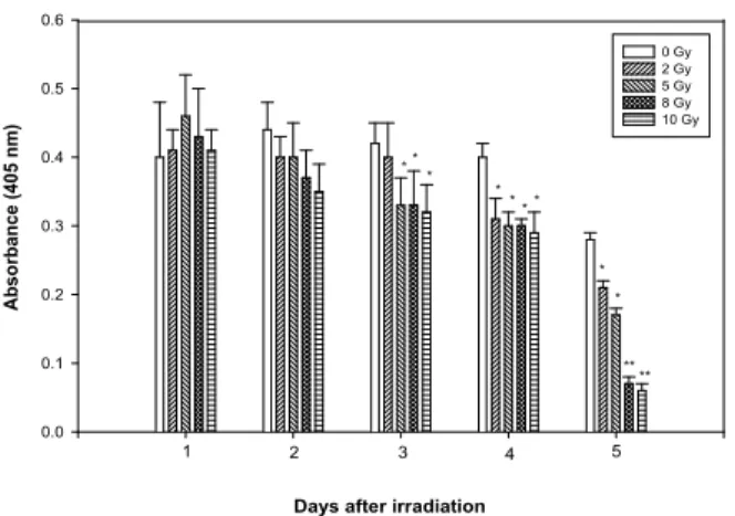
Figure 1. Effect of γ-irradiation on cell viability. Cells were irradiated with various doses of γ-ray for various times. Each experiment was performed in quadruplicate. Results are presented as means±S.E.M of three independent experiments. *p<0.05,
**p<0.01; significantly different from control (sham-irradiated cells).
Allicin (mg/ml)
Control Irradiation 0.01 0.1 1
Absorbance (405 nm)
0 1 2
*
*
**
Figure 2. Dose dependent inhibition of γ-irradiation induced ICAM-1 expression by allicin. Three independent experiments were performed in which confluent HUVEC were sham- or 8 Gy-irradiated with or without the indicated concentrations of allicin and the expression of ICAM-1 was measured by ELISA.
Results are presented as means±S.E.M of three independent experiments. *p<0.05, **p<0.01; significantly different from the irradiation-treated control.
trated H3PO4.
Statistical analysis. Results are presented as means±
S.E.M. Statistical difference between groups was de- termined by one-way analysis of variance (ANOVA) and significant values are indicated with an asterisk (*p<0.05, **p<0.01).
Results
Endothelial cell viability and growth after γIR. Although the effect of radiation on endothelial cells has been extensively reported (19,22,23), we examined the ra- diosensitivity of HUVEC under our culture condi- tions. Viability of the adherent endothelial cells was
>95% at the different times tested after exposure.
However, the number of irradiated cells decreased with time after exposure, whereas the number of non-irradiated cells remained. Three days after 8-Gy of irradiation, the number of irradiated cells was 80%
of that of the control. This percentage decreased to 10% 5 days after irradiation (Fig. 1). In addition, when the cells were treated with various doses of irradiation, the expression of ICAM-1 was maximally induced at 8-Gy (data not shown). Therefore, in subsequent studies 8-Gy was used.
Allicin inhibits γIR-induced ICAM-1 expression on endo- thelial cells in a dose dependent manner. To examine the effect of allicin, HUVEC were incubated without or with various concentrations (0.01, 0.1, 1μg/ml) of allicin for 3 days after γIR. The time of incubation and concentration of allicin used in these experiments had no effect on the viability as determined by trypan blue staining and the morphology of the endothelial cells (data not shown). These concentrations were
based on the concentrations of allicin determined in previous studies (24). As detected by ELISA, ICAM-1 was expressed at low levels on unstimulated endo- thelial cells and was significantly induced by γIR (Fig. 2). Allicin did not effect the basal level of ICAM-1 expression, whereas it led to reduce γIR- induced ICAM-1 expression in a dose dependent manner (Fig. 2).
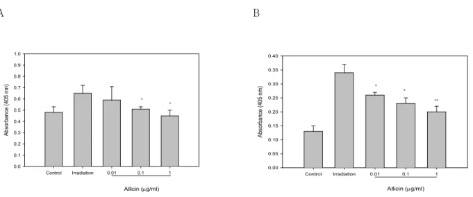
Allicin inhibits γIR-induced VCAM-1 and E-selectin ex- pression. In addition to ICAM-1, γIR also induced VCAM-1 and E-selectin expression in endothelial cells. To examine the effect of allicin on the γIR- induced expression of E-selectin and VCAM-1, HUVECs were incubated with various concentrations of allicin for 6h for in the case of E-selectin and for 3 days for VCAM-1. As measured by ELISA, the unstimulated cells expressed detectable amounts of E-selectin and VCAM-1 (Fig. 3). Upon induction with γIR, a significant increase in the expression of E-selectin was observed. Treatment with allicin inhi- bited slightly, but significantly the expression of VCAM-1 induced by γIR in a dose dependent manner (Fig. 3A). Similarly, γIR-induced E-selectin expression was also inhibited by allicin (Fig. 3B).
Taken together, these results suggest that allicin is effective in blocking the induced levels of expression of ICAM-1, VCAM-1 and E-selectin.
Allicin inhibits γIR-induced NO production. Since NO is known to be an important modulator of inflam- matory response to various stimuli, we determined the effect of allicin on NO production in endothelial
A B
Allicin (mg/ml)
Control Irradiation 0.01 0.1 1
Absorbance (405 nm)
0.0 0.1 0.2 0.3 0.4 0.5 0.6 0.7 0.8 0.9 1.0
*
*
Allicin (mg/ml)
Control Irradiation 0.01 0.1 1
Absorbance (405 nm)
0.00 0.05 0.10 0.15 0.20 0.25 0.30 0.35 0.40
*
*
**
Figure 3. Effect of allicin on γ-irradiation induced VCAM-1 (A) and E-selectin (B) expression by HUVECs. Three independent experiments were performed in which confluent HUVEC were sham- or 8 Gy-irradiated with or without the indicated concentrations of allicin and the expressions of VCAM-1 or E-selectin were measured by ELISA. Results are presented as means±S.E.M of three independent experiments. *p<0.05, **p<0.01; significantly different from the irradiation-treated control.
Allicin (mg/ml)
Control Irradiation 0.01 0.1 1
Nitrite (mM)
0 5 10 15 20 25
* *
**
Figure 4. Effect of allicin on γ-irradiation induced NO pro- duction in HUVEC. Three independent experiments were per- formed in which confluent HUVEC were sham- or 8 Gy- irradiated with or without the indicated concentrations of allicin.
The conditioned media was collected, and nitrite concentration were determined using the Griess reagent. Results are presented as means±S.E.M of three independent experiments. *p<0.05,
**p<0.01; significantly different from irradiation-treated control.
cells. As shown in Fig. 4, treatment of HUVECs with γIR resulted in increased NO release. Moreover, this increased NO production was inhibited by allicin in a dose dependent manner.
Discussion
Garlic extract has been found to promote healing of inflammation in the colon (25). However, although garlic extract has been found to have such anti-
inflammatory properties, very little is known with regard to the effect of allicin, a major component of garlic, on the induction of cell adhesion molecules by γIR. In the present study, allicin was found to block the γIR-induced expression of the leukocyte adhe- sion molecules, ICAM-1, VCAM-1 and E-selectin.
Thus, allicin posseses anti-inflammatory effects on the expression of adhesion protein induced by radiation. This compound also inhibited NO produc- tion in γ-irradiated HUVECs.
Radiation has been shown to induce the expression of a number of genes that participate in the inflam- matory response. These include TNF-α and IL-1 which are known to induce the expression of adhe- sion molecules such as E-selection when added to endothelial cells in culture (26-28). When the effect of γIR was examined on cytokine production in HUVECs, it was found not to induce the production of TNF-α in irradiated cells (data not shown). Re- cently, Hallahan et al. (29) demonstrated that E- selectin gene induction by ionizing radiation is independent of cytokine induction. In accordance with their report, our data confirm that adhesion protein expression did not require cytokine synthesis.
NO is a biologically active gas that is synthesized by a variety of cells, including those of the vascular endothelium, from the guanido group of L-arginine.
Moreover, NO has been invoked as a mediator of vascular phenomena such as arteriolar dilation, plate- let aggregation, and platelet-leukocyte adhesion (17).
It has also been suggested that NO is able to inhibit LPS-induced ICAM-1 expression (30). In addition, Kawachi et al. (31) demonstrated that iNOS-/-
mice injected with TNF-α showed enhanced VCAM- 1 expression in 50% of all tissues compared to the wild-type controls. Based on these findings it is be- lieved that NO inhibits the expressions of adhesion molecules. Our data showed that γIR induces the production of NO and the allicin inhibits NO release.
Recently, it has been shown that UVB radiation acts as a potent stimulator of NO in human endothelial cells and NO is known to be involved in skin ery- thema and inflammation (32). Thus, a role could be proposed for NO either in inhibition or promotion of inflammation. At present the mechanism account- ing for these modulations are unknown. However, it may be possible to override inhibition by enhancing inflammation. Moreover, the involvement of NO in the modulation and regulation of adhesion molecules expressed during in inflammation may be dependent on the source of NO, the cells involved and type of stimulus used to induce the inflammation.
During severe injury, infection, or ischemia and reperfusion damage, spillover of chemoactivators in the systemic circulation results in cellular activation, leading to the release of injurious agents that damage host tissues. These inflammatory mediators can alter the functional integrity of the vascular system, which may be due to the upregulation of the expression of cell adhesion molecules. Thus various strategies, such as monoclonal antibodies against adhesion molecules, soluble receptors, soluble counter-receptors, peptides derived from adhesion molecules to prevent receptor- ligand interactions, and antisense oligonucleotides have been employed to inhibit cell adhesion molecules (33). Flavonoids, glucorticoid, bensothiophene-car- boxamide and vitamin A have been shown to inhibit cytokine-or irradiation-induced cell adhesion molecule expression (14,34-36). Here we demonstrated that allicin effectively blocks the expression of leukocyte adhesion molecules. These studies suggest that allicin may serve as a potential therapeutic tool for radiation- induced inflammation. Further studies are needed to clarify how this modulation occurs and to what extent it occurs in vivo.
References
1. Cellini L, Di Campli E, Masulli M: Inhibition of Helicobacter pylori by garlic extracts (Allium sati- vum). FEMS Immunol Med Microbiol 13;273- 277, 1996
2. Bordia T, Mohammed N, Thomson M: An eva- luation of garlic and onion as antithrombotic agents. Protaglandins Leukot Essent Fatty Acids 54;183-186, 1996
3. Mcmahon FG, Vargas R: Can garlic lower blood pressure? A pilot study. Pharmacotherapy 13;406- 407, 1993
4. Foushee DB, Ruffin J, Banerjee U: Garlic as a natural agent for the treatment of hypertension:
A preliminary report. Cytobios 34;145-152, 1982 5. Chang MLW, Johnson MA: Effect of garlic on
carbohydrate metabolism and lipid synthesis in rats. J Nutr 110;931-936, 1980
6. Yeh YY, Yeh SM: Garlic reduces plasma lipids by inhibiting hepatic cholesterol and triacylgly- cerol synthesis. Lipids 29; 189-193, 1994
7. Eilat S, Oestraicher Y, Rabinkov A: Alteration of lipid profile in hyperlipidemic rabbits by allicin, an active constituent of garlic. Coronay Artery Dis 6;985-990, 1995
8. Pinto JT, Rivlin RS: Antiproliferative effects of allium derivatives from garlic. J Nutr 131;1058S- 1060S, 2001
9. Hobauer R, Frass M, Gmeiner B, Kaye AD, Frost EA: Garlic extract (allium sativum) reduces mi- gration of neutrophils through endothelial cell monolayers. Middle East J Anesthesiol 15;649- 658, 2000
10. Hruza LL, Pentland AP: Mechanisms of UV- induced inflammation. J Invest Dermatol 100;
35S-41S, 1993
11. Carlos T, Harlan JM: Leukocyte-endothelial inte- ractions. Blood 84;17656-1792, 1994
12. Bevilacqua MP, Pober JS, Wheeler ME, Cotran RS, Gimbrone MA Jr: Interleukin 1 acts on cul- tured human vascular endothelium to increase the adhesion of polymorphonuclear leukocytes, mo- nocytes and related cell lines. J Clin Invest 76;
2003-2009, 1985
13. Springer TA: Adhesion receptors of the immune system. Nature 346;425-434, 1990
14. Brostjan C, Anrather J, Csizmadia V, Natrajan G, Winkler H: Glucocorticoids inhibit E-selectin expression by targeting NF-kB and not ATF/
c-jun. J Immunol 158;3836-3844, 1997
15. Neuner P, Klosner G, Pourmojib M, Knobler R, Schwarz T: Pentoxifylline in vivo and in vitro down-regulates the expression of the intercellular adhesion molecule-1 in monocytes. Immunology 90;435-439, 1997
16. Weber C, Erl W, Pietsch A, Weber PC: Aspirin inhibits nuclear factor-kB mobilization and mo- nocyte adhesion in stimulated human endothelial cells. Circulation 91;1914-1917, 1995
17. Moncada, S: The L-arginine:nitric oxide pathway.
Acta Physiol Scand 145;201-227, 1992
18. Prasad K, Laxdal VA, Yu M: Antioxidant activity of allicin, an active principle in garlic. Mol Cell Biochem 148;183-189, 1995
19. Gaugler MH, Squiban C, Van der Meeren A, Bertho JM, Vandamme M, Mouthon MA: Late and persistent up-regulation of intercellular adhe-
sion molecule-1 (ICAM-1) expression by ionizing radiation in human endothelial cells in vitro. Int J Radiat Biol 72;201-209, 1997
20. Gupta B, Ghosh B: Curcuma longa inhibits TNF- α induced expression of adhesion molecules on human umbilical vein endothelial cells. Int J Immunopharmac 21;745-757, 1999
21. Ding AH, Nathan CF, Stuehr DJ: Release of reactive nitrogen intermediates and reactive oxy- gen intermediated from mouse peritoneal macro- phages. Comparison of activating cytokines and evidence for independent production. J Immunol 141; 2407-2412, 1988
22. Rubin P, Johnston CJ, Williams JP, McDonald S, Finkelstein JN: A perpetual cascade of cytokines postirradiation leads to pulmonary fibrosis. Int J Radiat Biol 33;99-109, 1995
23. Eissner G, Kohlhuber F, Grell M, Ueffing M, Scheurich P, Hieke A, Milthoff G, Bornkamm GW, Holler E: Critical involvement of transmem- brane tumor necrosis factor-α in endothelial pro- grammed cell death mediated by ionizing radia- tion and bacterial endotoxin. Blood 86;4184-4193, 1995
24. Kang NS, Moon EY, Cho CG, Pyo S: Immuno- modulating effect of garlic component, allicin on murine peritoneal macrophages. Nutr Res 21;617- 626, 2001
25. Khan I, Ali M: Altered expression of the Na+/H+
exchanger isoform-3 in experimental colitis: effect of garlic. Mol Cell Biochem 200;77-84, 1999 26. Bevilacqua MP, Stengelin S, Gimbrone MA Jr,
Seed B: Endothelial leukocyte adhesion molecule 1: an inducible receptor for neutrophils related to complement regulatory proteins and lectins.
Science 243;1160-1165, 1989
27. Brach MA, Gruss HJ, Kaisho T, Asano Y, Hirano T, Herrmann F: Ionizing radiation induce expres- sion of interleukin 6 by human fibroblasts invol- ving activation of nuclear factor-kappa B. J Biol Chem 268;8466-8472, 1993
28. Sherman ML, Datta R, Hallahan DE, Weichesl- baum RR, Kufe DW: Regulation of tumor necro-
sis factor gene expression by ionizing radiation in human myeloid leukemia cells and peripheral blood monocytes. J Clin Invest 81; 1506-1510, 1991
29. Hallahan D, Clark ET, Kuchibhotla J, Gewertz BL, Collins T: E-selectin gene induction by ioni- zing radiation is independent of cytokine induc- tion. Biochem Biophys Res Commun 217;784- 795, 1995
30. Spiecker M, Darius H, Kaboth K, Hubner F, Liao JK: Differential regulation of endothelial cell ad- hesion molecule expression by nitric oxide do- nors and antioxidants. J Leuk Biol. 63;732-739, 1998
31. Kawachi S, Cockrell A, Laroux FS, Gray L, Granger DN, Van Der Heyde HC, Grisham MB:
Role of inducible nitric oxide synthase in the regulation of VCAM-1 expression in gut inflam- mation. Am J Physiol 277;G572-G576, 1999 32. Deliconstantinos G, Villiotou V, Stavrides JC:
Nitric oxide and peroxynitrite released by ultra- violet B-irradiated human endothelial cells are possibly involved in skin erythema and inflam- mation. Exp Physiol 81;1021-1033, 1996
33. Weiser MR, Gibbs S, Hechtman HB: Strategies to inhibit cellular adhesion molecules. In: Paul LC, Issekutz T eds.: Adhesion molecules in He- alth and Disease. p55-86, New York Marcel Dek- ker Inc., 1997
34. Gerritsen M, Carley W, Ranges GE, Shen C, Phan SA, Ligon G, Perry C: Flavonoid inhibit cytokine-induced endothelial cell adhesion protein gene expression. Am J Pathol 147; 278-292, 1995 35. Cobb RR, Felts KA, Mckenzie TC, Parry G,
Mackman NA: Benxothiophene-carboxamide is a potent inhibitor of IL-1βinduced VCAM-1 gene expression in human endothelial cells. FEBS Lett 382;323-326, 1996
36. Redlich CA, Rockwell S, Chung JC, Sikora AG, Kelley M, Mayne ST: Vitamin A inhibits radia- tion-induced pneumonitis in rats. J Nutr 128;
1661-1664, 1998